

|Articles|September 1, 1996
- ONCOLOGY Vol 10 No 9
- Volume 10
- Issue 9
Commentary (Droz): Prognostic Factors in Low-Stage Nonseminomatous Testicular Cancer
Author(s)Jean-pierre Droz, MD, PhD
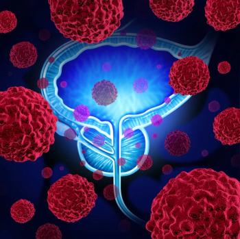
Prognostic Factors in Low-Stage Nonseminomatous Testicular Cancer
Advertisement
Drs. Moul and Heidenreich review prognostic factors for extratesticular subclinical involvement in patients with clinical stage I nonseminomatous germ-cell tumors. There is still no consensus on which prognostic factors are most important in these patients or which treatment strategies should be employed in patients determined to be at high risk.
The most important independent prognostic factors for occult retroperitoneal disease appear to be vascular invasion and the presence of embryonal carcinoma in the primary tumor. According to a study by Klepp et al,[1] vascular invasion also is the main prognostic factor for relapse in pathologic stage I disease.
MRC Studies
The major contributions in this area have come from the Medical Research Council (MRC) Testicular Tumors Subgroup. This group performed a prospective study of surveillance between 1979 and 1983 to assess prognostic factors for relapse.[2] Four prognostic factors were studied: venous invasion, lymphatic invasion, the presence of embryonal carcinoma, and the absence of yolk-sac elements. The prognostic model was validated in a second prospective study between 1984 and 1987,[3] and was then used to enroll patients with a high risk of recurrence to a prospective adjuvant postorchiectomy chemotherapy phase II trial between 1987 and 1994.[4] It is noteworthy that 91 of 126 patients included in a series published by Hoskin et al were also participants in the first MRC study.[5]
Two major practical problems emerge from the MRC findings: (1) the difficulty of making a clear distinction between venous and lymphatic invasion, as pointed out by Hoeltl et al;[6] and (2) the discordance between local pathologists and the central pathology review committee in their assessment of both vascular invasion and the diagnosis of cell types, as demonstrated by Sesterhenn et al.[7] For example, only 54 specimens were considered to have vascular invasion by local pathologists, whereas the central pathologists reported 179 specimens with vascular invasion. Moreover, only the central pathology review committee distinguished between venous and lymphatic invasion. Rates of disagreement between the local and central pathologists were 13% for embryonal carcinoma and 62% for the yolk-sac component.
Moul et al[8] have focused on the percentage of embryonal carcinoma and vascular invasion as prognostic factors. However, insofar as there are difficulties in correctly assessing the cell types, one can question whether it is possible to accurately quantify the percentage of one particular cell type. Moreover, the quantification of the embryonal carcinoma component requires the study of whole paraffin-embedded blocks of tumor, a technique that is particularly time consuming.
In my opinion, risk assessment may be correctly performed by considering only the presence vs absence of vascular invasion (venous and/or lymphatic) and high vs low percentage of embryonal carcinoma (the cut-off point being 25%). Using such a model, as shown in the authors' Table 3, three risk groups are defined: high-, intermediate-, and low-risk groups, with more than 60%, 20% to 60%, and more than 20% rates of occult disease, respectively.
New Prognostic Markers
Moul and Heidenreich carefully review new techniques. However, techniques that may help establish the two most important prognostic factors--factor VIII staining and new embryonal-specific antibodies--also must be developed.
A recent study has attempted to characterize an extremely low-risk cohort of patients with clinical stage I disease[9] by using the volume of embryonal carcinoma and percentage of staining with MIB-1 monoclonal antibody (which identifies the nuclear proliferation-associated antigen Ki-67). Using this model, 41 of 91 patients were predicted to have pathologic stage I disease, and this was confirmed by retroperitoneal lymph node dissection (RPLND) in 40 of the 41. Thus, it seems appropriate to perform studies of new prognostic markers in large series of patients and to validate prognostic models that combine the independent prognostic factors.
Treatment Options for High-Risk Patients
These studies are very important because the results of recent studies of adjuvant postorchiectomy chemotherapy suggest that this treatment may be considered as a third option in high-risk patients, in addition to a wait- and-see approach and surgery. Three studies have demonstrated that chemotherapy is associated with a relapse rate of less than 5% in patients who are at high risk of relapse after orchiectomy only.[4,10,11] Up to now, a randomized trial comparing different treatment strategies did not seem feasible in these patients; the only problem now is to balance the benefits and toxicities of the different treatment options.
A trial conducted by the French Association of Urology/French Federation of Cancer Center will address the question of quality of life, cost-effectiveness, and utility in three cohorts of patients who will be assigned to treatment with surveillance, RPLND, or postorchiectomy adjuvant chemotherapy on the basis of prognostic factor assessment. Studies like this one must be encouraged to better define the treatment strategy that has minimal side effects in this group of clinical stage I patients who are likely to be cured of their disease.
References:
1. Klepp O, Olsson AM, Henrikson H, et al: Prognostic factorsin clinical stage I nonseminomatous germ cell tumors of the testis: Multivariate analysis of a prospective multicenter study. JClin Oncol 8:509-518, 1990.
2. Freedman LS, Jones WG, Peckham MJ, et al: Histopathology inthe prediction of relapse of patients with stage I testicularteratoma treated by orchiectomy alone. Lancet 2:294-298, 1987.
3. Read G, Stenning SP, Cullen MH, et al: Medical Research Councilprospective study of surveillance for stage I testicular teratoma.J Clin Oncol 10:1762-1768, 1992.
4. Cullen MH, Stenning SP, Parkinson MC, et al for the MedicalResearch Council Testicular Tumor Working Party: Short courseadjuvant chemotherapy in high risk stage I nonseminomatous germcell tumors of the testis: A Medical Research Council Report.J Clin Oncol 14:1106-1113, 1996.
5. Hoskin P, Dilly S, Easton D, et al: Prognostic factors in stageI non-seminomatous germ-cell testicular tumors managed by orchiectomyand surveillance: Implications for adjuvant chemotherapy. J ClinOncol 4:1031-1036, 1986.
6. Hoeltl W, Kosak D, Pont J, et al: Testicular cancer: Prognosticimplications of vascular invasion. J Urol 137:683-685, 1996.
7. Sesterhenn IA, Weiss RB, Mostofi FK, et al: Prognosis and otherclinical correlates of pathologic review in stage I and II testicularcarcinoma : A report from the Testicular Cancer Intergroup study.J Clin Oncol 10:69-78, 1992.
8. Moul JW, McCarthy WF, Fernandez EB, et al: Percentage of embryonalcarcinoma and vascular invasion predict pathologic stage in clinicalstage I nonseminomatous testicular. Cancer Res 54:1-3, 1994.
9. Leibovitch I, Nichols CR, Foster RS, et al: Indiana UniversitySchool of Medicine I, IN 46202: Characterization of an extremelylow risk cohort of clinical stage a non seminomatous testicularcancer (NSGCT) ; quantitative immunohistochemistry, histopathologyand radiology. Proc Am Soc Clin Oncol 15:249, 1996.
10. Hoeltl W, Pont J, Kosak D, et al: Treatment decision for stageI non-seminomatous germ-cell tumours based on the risk factor"vascular invasion." Br J Urol 69:83-87, 1992.
11. Klepp O, Swedish-Norvegian Testis Cancer Group (SWENOTECA):Risk-adapted treatment of clinical stage I (CS1) nonseminoma testiscancer (NSTC). Eur J Cancer 31A:S188, 1995.
Articles in this issue
over 29 years ago
Antisense Gene Therapy Trials Underway in Patients With CMLover 29 years ago
BOOK REVIEW: Leukemiaover 29 years ago
Most Terminal AIDS Patients Want to Be Revived if Their Heart Stopsover 29 years ago
Disease Management: State of the Art in Pancreatic Cancerover 29 years ago
How to Better Communicate Cancer Risk to Patientsover 29 years ago
Gemcitabine Shows Promise as Combination Agent in NSCLCover 29 years ago
Data Review Shows Fruits and Vegetables Can Block Major CancersNewsletter
Stay up to date on recent advances in the multidisciplinary approach to cancer.
Advertisement
Related Content
Advertisement


Latest CME


Advertisement
Advertisement
Trending on CancerNetwork
1
Modifiable Risk Factors Suggest Potential for Improving Cancer Prevention
2
Barriers to CAR T-Cell Referral and Center Access in Multiple Myeloma
3
2026 Tandem Meetings: What’s the Latest Research in Multiple Myeloma?
4
Real World Outcomes of CAR T-Cell Therapy in Multiple Myeloma Including Older and Comorbid Patients
5