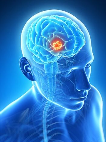
Removing Tissue Surrounding Brain Tumor May Double Survival in Patients with Glioblastoma
The results presented in this study indicated that neurosurgeons may need to change how they approach tumor removal and, when safe, include non-contrast-enhancing tumor during resection to achieve maximal resection.
A study published in JAMA Oncology confirmed an association between maximal resection of contrast-enhanced tumor and overall survival (OS) in patients with glioblastoma across all subgroups.
Additionally, the researchers found that maximal resection of non-contrast-enhanced tumor was associated with longer OS in younger patients, regardless of isocitrate dehydrogenase (IDH) status, and among patients with IDH-wild-type glioblastoma regardless of the methylation status of the promoter region of the DNA repair enzyme O6-methylguanine-DNA methyltransferase.
“Traditionally, the goal of neurosurgeons has been to achieve total resection, the complete removal of contrast-enhancing tumor,” Mitchel Berger, MD, director of the UCSF Brain Tumor Center, said in a press release. “This study shows that we have to recalibrate the way we have been doing things and, when safe, include non-contrast-enhancing tumor to achieve maximal resection.”
In this retrospective, multicenter cohort study, researchers looked at the outcomes of 761 newly diagnosed patients from the University of California, San Francisco diagnosed from January 1, 1997 through December 31, 2017 in a development cohort. The patients were then divided into 4 groups based on age, treatment protocols, and extent of resections of both contrast- and non-contrast-enhanced tumor.
Among those in the development cohort, the authors identified 62 patients whose average survival was 37.3 months (95% CI, 31.6-70.7). These individuals had IDH-mutant tumors or were under 65 with IDH-wild-type tumors and had undergone both radiation and chemotherapy with temozolomide (Temodar)in nearly all cases. Each of the participants had resections with a median of 100% of contrast-enhanced tumor and a median of 90% of non-contrast-enhanced tumor.
Comparatively, 212 participants under 65 who received the same therapies and had IDH-wild-type tumors and reduction of contrast-enhanced tumor but residual non-contrast-enhanced tumors fared worse, with an average survival of 16.5 months (95% CI, 14.7-18.3).
In 2 external cohorts, researchers studied 107 patients from the Mayo Clinic diagnosed from January 1, 2004 through December 31, 2014 and 99 patients from the Cleveland Clinic’s Ohio Brain Tumor Study with data collected from January 1, 2008 through December 1, 2011. Results were validated using the 2 cohorts.
“Although these data show a survival benefit associated with maximal resection, it remains critically important that we do our best to remove tumor in a manner that will not harm the patient,” co-author and neurosurgeon Shawn Hervey-Jumper, MD of the UCSF Brain Tumor Center and of the Weill Institute for Neurosciences, said in a press release.
In an editorial, authors Bryan D. Choi, MD, PhD, Elizabeth R. Gerstner, MD, and William T. Curry Jr., MD, all from Massachusetts General Hospital, indicated that this research leaves room for further investigation.
“Although maximal surgery was found to prolong survival despite IDH mutation status, other molecular characteristics or combinations thereof may ultimately demonstrate the capacity to identify tumors that would specifically benefit from greater extent of resection,” the authors wrote in the editorial. “If so, information obtained through emerging imaging modalities, liquid biopsy, or rapid intraoperative diagnosis could be exploited to help guide clinical decision-making in real time.”
They went on to add that additional work will be necessary to better understand how molecular diagnoses and extent of resection could intersect to potentially influence the role of adjuvant therapies for glioma, specifically in younger patient cohorts.
While maximal resection of both contrast- and non-contrast-enhanced tumor should always be considered, first author Annette Molinaro, PhD, from the UCSF Department of Neurological Surgery and the Department of Epidemiology and Biostatistics explained that we are far from achieving a cure for glioblastoma.
“It’s a complex tumor to treat for a number of reasons,” she said in a press release. “One challenge is that the blood-brain barrier – the network of blood vessels that acts as the brain’s gatekeeper – effectively blocks many cancer agents from reaching their target. Another challenge is that these are heterogenous tumors driven by multiple mutations – if you target one mutation, others will thrive.”
References:
1. Molinaro AM, Hervey-Jumper S, Morshed RA, et al. Association of Maximal Extent of Resection of Contrast-Enhanced and Non-Contrast-Enhanced Tumor With Survival Within Molecular Subgroups of Patients With Newly Diagnosed Glioblastoma. JAMA Oncology. doi:10.1001/jamaoncol.2019.6143.
2. UCSF. Brain Tumor Surgery That Pushes Boundaries Boosts Patients Survival. UCSF website. Published February 4, 2020. ucsf.edu/news/2020/02/416611/brain-tumor-surgery-pushes-boundaries-boosts-patients-survival. Accessed February 7, 2020.
3. Choi BD, Gerstner ER, Curry Jr. WT. A Common Rule for Resection of Glioblastoma in the Molecular Era. JAMA Oncology. doi:10.1001/jamaoncol.2019.6384.
Newsletter
Stay up to date on recent advances in the multidisciplinary approach to cancer.


