

|Articles|June 1, 1996
- ONCOLOGY Vol 10 No 6
- Volume 10
- Issue 6
Role of Thoracoscopic Lymph Node Staging for Lung and Esophageal Cancer
Author(s)Benjamin Movsas, MD, Lawrence R. Coia, MD
Dr. Krasna provides a thoughtful review of thoracoscopy as an emerging technique for the staging of patients with lung and esophageal cancers. In lung cancer, thoracoscopy can be used as a complement to cervical mediastinoscopy in the evaluation of mediastinal and hilar lymph nodes. This is especially true in patients who have left-sided neoplasms with enlarged lymph nodes in the aortico-pulmonary window--a region typically inaccessible to cervical mediastinoscopy.
Advertisement
Dr. Krasna provides a thoughtful review of thoracoscopy as an emerging technique for the staging of patients with lung and esophageal cancers. In lung cancer, thoracoscopy can be used as a complement to cervical mediastinoscopy in the evaluation of mediastinal and hilar lymph nodes. This is especially true in patients who have left-sided neoplasms with enlarged lymph nodes in the aortico-pulmonary window--a region typically inaccessible to cervical mediastinoscopy.
Perhaps the most useful application of thoracoscopy is in the preoperative assessment of patients with pulmonary lesions abutting the mediastinum. Of nine patients with lung cancer in Dr. Krasna's review who underwent thoracoscopic staging, three were found to have mediastinal invasion and thus avoided an unnecessary thoracotomy. Similarly, thoracoscopy enables evaluation of the pleural space, such that the presence of pleural implants or invasion can be assessed preoperatively. As Dr. Krasna points out, however, routine thoracoscopic evaluation of the pleural cavity prior to all thoracotomies for lung cancer is not indicated currently, and this technique should be limited to a study setting at this time.
Potential Benefits in Staging of Esophageal Cancer
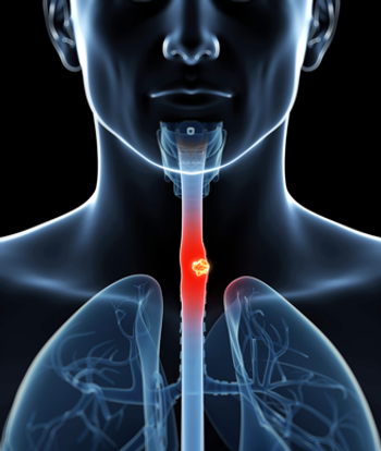
The potential benefits of thoracoscopy in patients with esophageal cancer include improved staging so that an adequate determination of prognosis with present therapies can be made, as well as selection of the appropriate treatment modality for each disease stage. Lymph node involvement occurs in the majority of patients with esophageal cancer, including, in most studies, those undergoing curative surgical resection. Although the accuracy of endoscopic ultrasound for T-staging is 85%, its accuracy for lymph node involvement is only 75%. By contrast, the results reported by Krasna suggest that 93% accuracy for lymph node involvement may be possible with thoracoscopic evaluation.
Information about lymph node status is essential, as the number of lymph nodes involved is predictive of prognosis in patients managed with esophagectomy. Abe et al [1] found no survivors if two or more nodes were involved.
Skinner et al [2] and Sieward [3] observed no survivors if five or more nodes were involved, whereas Ellis et al [4] found that patients with one to four positive nodes had a survival intermediate to that of patients with no involved nodes or more than four positive nodes.
Information regarding the extent of the esophageal tumor at thoracoscopy may also be important for patients initially managed with a nonsurgical approach, such as chemoradiation alone, chemoradiation followed by surgical resection, or chemotherapy followed by surgical resection. At present, patients initially managed with a nonsurgical approach must be staged using the 1983 American Joint Committee on Cancer (AJCC) clinical staging system, or one must rely on the accuracy of ultrasound to appropriately stage patients with the 1988 AJCC criteria. As the volume of tissue irradiated correlates with morbidity, accurate information regarding lymph node involvement provided by video-assisted thoracoscopy may permit the use of tailored radiation fields that conform more closely to the actual regions of involvement, potentially resulting in a reduction in morbidity. Nearly two-thirds of the patients treated with chemoradiation in the landmark study by Herscovic et al [5] developed grade 3 or 4 acute toxicity, including esophagitis and hematologic toxicity, which could be attributed, at least in part, to the use of relatively generous radiation fields, typically encompassing the nodes from the supraclavicular area to the gastroesophageal junction.
Potential problems associated with the use of minimally invasive surgery include delay to the onset of definitive treatment and morbidity associated with the procedure. In experienced hands, the procedure itself may require only 1½ hours to perform; however, when combined with laparoscopy or when performed by surgeons with little experience, a 6-hour procedure is not unusual.
Caveats About Widespread Use of Video-Assisted Surgical Procedures
As with any new technique, there is often a desire to apply such state-of-the art technology to a wide variety of clinical situations. Advances in digital processing and video camera technology have improved the potential of thoracoscopy and led to the emergence of a new set of procedures, termed video-assisted thoracoscopic surgery (VATS). Thoracoscopy has thus recently been used to perform major lung resections, including lobectomies and even pneumonectomies [6]. The rationale for these uses of VATS is its presumed superiority over thoracotomy in reducing morbidity and hospital stay. Yet, Kirby et al challenged this assumption in a randomized trial comparing muscle-sparing thoracotomy and lobectomy to video-assisted lobectomy [7]. Although they reported more postoperative complications in the thoracotomy group (P < .05), the majority of which were prolonged air leaks, they found no significant differences between the two groups with regard to operating time, intraoperative blood loss, duration of chest tube drainage, length of hospital stay, post-thoracotomy pain, or recovery time.
Moreover, VATS exposes the patient to the risk of a major pulmonary resection being performed in an essentially closed chest. Of 1,820 procedures in one series, 439 procedures (24%) were converted to a thoracotomy [7]. Most importantly, long-term follow-up is needed to assess the efficacy of VATS procedures with respect to survival and locoregional control. Lastly, although the exception rather then the rule, an important concern is the potential for tumor implantation or seeding secondary to the VATS procedure itself [8].
Thoracoscopy for wedge resections of lung metastases has rapidly increased in frequency. This technique, however, precludes bimanual palpation of the lung to locate additional lesions not seen on the surface. In one study correlating pathologic and imaging findings in patients undergoing thoracoscopic wedge resections of lung metastases, 22% of patients had fewer lesions at the time of pathologic analysis, as compared with lesions detected on a chest roentgenogram [9]. The inability to adequately palpate the entire lung using the thoracoscope impairs the surgeon's ability to know whether resection of all lesions has been achieved.
Thus, the use of thoracoscopic resection in the management of metastatic disease other than for diagnosis must be reevaluated. By contrast, VATS is rapidly emerging as the procedure of choice in the management of a solitary pulmonary nodule [10].
Exciting Potential Niche for VATS
A possible exciting niche for VATS may be in the management of elderly patients and/or patients with impaired pulmonary function or comorbid medical conditions, who cannot tolerate a standard lung resection. In one study, 30 patients with poor pulmonary function underwent video-assisted thoracoscopic wedge resections of T1 lung cancer [11]. All lesions were successfully resected without converting to thoracotomy, and operative mortality was 3%.
Although long-term follow-up will be required to determine the efficacy of this procedure, this study suggests that video-assisted thoracoscopic wedge resection appears to be a safe procedure in the treatment of peripheral T1 lung cancer in high-risk patients. Such a limited wedge resection via the VATS procedure followed by postoperative radiotherapy to decrease the risk of local failure may yield more promising results then management with radiotherapy alone. This combined-modality approach is currently being examined in a Cancer and Leukemia Group B (CALGB) phase II study for high-operative-risk patients with serious cardiopulmonary dysfunction.
Summary
Now that endoscopic video capabilities have been added to the armamentarium of the thoracic surgeon, the fundamental issue is to determine the appropriate and cost-effective application of this technique. As Dr. Krasna carefully points out, thoracoscopy is a useful staging procedure in selected patients with pulmonary and esophageal tumors. When combined with standard staging procedures, this technique will undoubtedly yield information that will be important in staging and assessing treatment efficacy in clinical trials.
As with laparoscopy, however, initial enthusiasm for VATS must be tempered by the reality of technical challenges and complications, as well as results that have yet to withstand careful scientific scrutiny and the test of time. As Dr. Krasna notes, a randomized, prospective, multi-institutional trial must be conducted to validate the techniques that he helped pioneer in the United States.
References:
1. Abe S, Tachibana M, Shiraishi M, et al: Lymph node metastasis in resectable esophageal cancer. J Thorac Cardiovasc Surg 100:287-291, 1990.
2. Skinner DB, Dowlatshahi KD, DeMeester TR: Potentially curable cancer of the esophagus. Cancer 50:2571-2575, 1982.
3. Sieward JR: Esophageal cancer from the German point of view. Jpn J Surg 19:11-20, 1989.
4. Ellis FH Jr, Watkins E, Krasna MJ, et al: Staging of carcinoma of the esophagus and cardia: A comparison of different staging criteria. J Surg Oncol 55:231-235, 1993.
5. Herskovic A, Marta K, Al-Sarraf M, et al: Combined chemotherapy and radiotherapy compared with radiotherapy alone in patients with cancer of the esophagus. N Engl J Med 326:1593-1598, 1992.
6. Yim AP, Ho JK: Video-assisted thoracoscopic lobectomy: A word of caution. Aust NZ J Surg 65(6):438-441, 1995.
7. Kirby TJ, Mack MJ, Landreneau RJ, et al: Lobectomy--video-assisted thoracic surgery versus muscle-sparing thoracotomy: A randomized trial. J Thorac Cardiovasc Surg 109(5):997-1002, 1995.
8. Walsh GL, Nesbitt JC: Tumor implants after thoracoscopic resection of a metastatic sarcoma. Ann Thorac Surg 59(1):215-216, 1995.
9. McCormack PM, Ginsberg KB, Bains MS, et al: Accuracy of lung imaging in metastases with implications for the role of thoracoscopy. Ann Thorac Surg 56(4):863-866, 1993.
10. Santambrogio L, Nosotti M, Bellaviti N, et al: Videothoracoscopy versus thoracotomy for the diagnosis of the indeterminate solitary pulmonary nodule. Ann Thorac Surg 59(4):868-871, 1995.
11. Shennib HA, Landreneau R, Mulder DS, et al: Video-assisted thoracoscopic wedge resection of T1 lung cancer in high-risk patients. Ann Surg 218(4):555-560, 1993.
Articles in this issue
over 29 years ago
AIDS-related Kaposi's Sarcoma: Options for Today and Tomorrowover 29 years ago
Clinical Manifestations of Kaposi's Sarcomaover 29 years ago
New Developments: A Look to the Futureover 29 years ago
The Epidemiologic Scope of Kaposi's Sarcomaover 29 years ago
Current Therapeutic Options for Kaposi's Sarcomaover 29 years ago
Discussionover 29 years ago
Comments on Bone Marrow Transplantation for Multiple MyelomaNewsletter
Stay up to date on recent advances in the multidisciplinary approach to cancer.
Advertisement
Related Content
Advertisement
Latest CME


Advertisement
Advertisement
Trending on CancerNetwork
1
Modifiable Risk Factors Suggest Potential for Improving Cancer Prevention
2
2026 Tandem Meetings: What’s the Latest Research in Multiple Myeloma?
3
Barriers to CAR T-Cell Referral and Center Access in Multiple Myeloma
4
Dato-DXd Receives Priority Review in Unresectable/Metastatic TNBC
5