
|Articles|October 1, 2005
- ONCOLOGY Vol 19 No 11_Suppl_4
- Volume 19
- Issue 11_Suppl_4
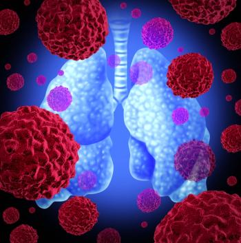
Radiofrequency Ablation in Lung Cancer: Promising Results in Safety and Efficacy
Only about 15% of patients diagnosed with lung carcinoma eachyear are surgical candidates, either due to advanced disease orcomorbidities. The past decade has seen the emergence of minimallyinvasive therapies using thermal energy sources: radiofrequency,cryoablation, focused ultrasound, laser, and microwave; radiofrequencyablation (RFA) is the best developed of these. Radiofrequency ablationis safe and technically highly successful in terms of initial ablation.Long-term local control or complete necrosis rates drop considerablywhen tumors are larger than 3 cm, although repeat ablations can beperformed. Patients with lung metastases tend to fare better with RFlung ablation than those with primary lung carcinoma in terms of localcontrol, but it is unclear if this is related to smaller tumor size at time oftreatment, lesion size uniformity, and sphericity with lung metastases,or to differences in patterns of pathologic spread of disease. The effectsof RFA on quality of life, particularly dyspnea and pain, as well aslong-term outcome studies are generally lacking. Even so, the resultsregarding RF lung ablation are comparable to other therapies currentlyavailable, particularly for the conventionally unresectable or high-risklung cancer population. With refinements in technology, patient selection,clinical applications, and methods of follow-up, RFA will continueto flourish as a potentially viable stand-alone or complementarytherapy for both primary and secondary lung malignancies in standardand high-risk populations.
Advertisement
Only about 15% of patients diagnosed with lung carcinoma each year are surgical candidates, either due to advanced disease or comorbidities. The past decade has seen the emergence of minimally invasive therapies using thermal energy sources: radiofrequency, cryoablation, focused ultrasound, laser, and microwave; radiofrequency ablation (RFA) is the best developed of these. Radiofrequency ablation is safe and technically highly successful in terms of initial ablation. Long-term local control or complete necrosis rates drop considerably when tumors are larger than 3 cm, although repeat ablations can be performed. Patients with lung metastases tend to fare better with RF lung ablation than those with primary lung carcinoma in terms of local control, but it is unclear if this is related to smaller tumor size at time of treatment, lesion size uniformity, and sphericity with lung metastases, or to differences in patterns of pathologic spread of disease. The effects of RFA on quality of life, particularly dyspnea and pain, as well as long-term outcome studies are generally lacking. Even so, the results regarding RF lung ablation are comparable to other therapies currently available, particularly for the conventionally unresectable or high-risk lung cancer population. With refinements in technology, patient selection, clinical applications, and methods of follow-up, RFA will continue to flourish as a potentially viable stand-alone or complementary therapy for both primary and secondary lung malignancies in standard and high-risk populations.
After 52 pack-years of smoking, Don had little to celebrate on his 70th birthday when he was diagnosed with stage IB non-small-cell lung carcinoma. To make matters worse, he was told that, despite his early-stage cancer, he was not a suitable candidate for surgical resection due to his extensive lung disease. Bereft of his most promising option, Don was left without much hope. Although not a candidate for surgery, Don did indeed have other options, many of which would not have been available to him had he been diagnosed a decade earlier. Don did eventually undergo radiofrequency ablation (RFA) of his lung cancer and has just celebrated his 75th birthday. Lung carcinoma remains the leading cause of cancer death in the United States. Over the past decade, lung cancer death rates have more than quadrupled, from 5.4 to 29.4 per 100,000.[1] The American Cancer Society estimates that in 2005 the number of lung cancer deaths will rise to 163,510-90,490 men and 73,020 women-accounting for 28% of all cancer-related deaths. The number of newly diagnosed lung cancers will rise to 172,570, or 93,010 new cases in men and 79,560 in women.[ 2] Nearly 60% of those diagnosed with lung cancer die within 1 year of their diagnosis and nearly 75% within 2 years.[2] Despite recent advances in therapy, the relative 5-year survival rate for all stages of lung cancer has improved only slightly to 15%.[2] For early-stage lung carcinoma, surgical resection confers the best survival option, with 5-year survival rates approaching 80% for stage I disease and 40% for stage II disease.[3] Only about 15% of patients diagnosed with lung carcinoma each year are surgical candidates.[4] Most patients present with advanced or widespread disease at the time of diagnosis and, therefore, are not considered candidates for surgery. Some patients have technically resectable disease but cannot undergo surgery because of comorbid cardiopulmonary disease. This population represents a suitable target for novel, minimally invasive lung-sparing therapies providing local control. Local therapy may also be appropriate in limited metastatic disease. The lung is the second most frequent site of metastatic disease. Surgical resection, or metastasectomy, appears to confer some survival benefits in carefully selected patients.[5] Although it is somewhat uncertain whether this benefit results from total- body tumor burden cytoreduction or a less aggressive natural course of disease in this patient subpopulation, pulmonary metastasectomy is increasingly accepted as treatment in selected patients, with a 10-year survival rate of approximately 25%.[5] At least 90% of the 10-year survivors remain free of disease.[6] Yet the size and number of metastatic nodules frequently preclude attempts at surgical resection. Since the majority of patients develop disease recurrence following metastasectomy, and repeated resections can remove significant amounts of functioning lung, this patient population also represents a suitable target for minimally invasive lung-sparing therapies. Since the early 1990s, an increasing number of minimally invasive techniques have been introduced into clinical practice in the treatment of primary and secondary pulmonary malignancy. Image-guided interventional and video-assisted thoracoscopic approaches have become attractive alternatives to open thoracic surgical resection. The past decade has seen the emergence of minimally invasive therapies using thermal energy sources: radiofrequency, cryoablation, focused ultrasound, laser, and microwave. Radiofrequency ablation is the best developed, secondary to the advent of bipolar and multielectrode and internal tip-cooling RF electrodes as well as advances in computed-tomography (CT) technology. Radiofrequency ablation is a controlled electrosurgical technique that implements high-frequency alternating current to generate localized electromagnetic fields, heating targeted tissues to desiccation, or thermal coagulation. Naturally, cells of targeted tissue die when exposed to high thermal doses. For a variety of reasons, including less efficient heat dissipation, the cells of neoplastic tissues are more sensitive to heat effects than are cells of healthy tissues.[7] Thus, RFinduced hyperthermia exploits this difference in heat sensitivity by creating localized temperature increases in neoplastic tissues to greater than 57C to 60C, while restricting temperatures in healthy tissues to normal ranges.[ 8] Several authors have advocated that lung tumors are well suited to RFA because of the so-called "oven effect," whereby the air (high resistance) surrounding an intraparenchymal tumor (low resistance) affords an insulating effect and traps heat within the targeted tumor.[9] Well established in the treatment of various cardiac and neurologic dysfunctions, RFA has faced a major barrier to further application: the small lesion size created by earlier generation devices and delivery methods. The advent of bipolar and multielectrode and tip-cooled RF electrodes, enabling the creation of larger areas of controlled and reproducible necrosis in animal and human models in vitro and in vivo,[10] has expanded potential clinical applications to include tumor therapy- notably in the treatment of primary and secondary brain and hepatic malignancies.[11,12] The feasibility and safety of percutaneous interstitial thermal ablation of pulmonary tissue have also been investigated. Using a percutaneous CT-guided transthoracic technique, Goldberg et al showed that RFA was not only performed safely in the pulmonary tissue of rabbits but that tissue response to thermal injury was controlled, predictable, and easily monitored.[ 9] Subsequent investigation has been directed to the ablation of abnormal tissue within the pulmonary parenchyma, specifically malignancy. Since 1996, several authors have used CT-guided RF application to successfully treat induced sarcomas within the lungs of rabbit, canine, and ovine models, characterizing tissue changes following lung ablation with at least 95% necrosis to complete eradication of treated tumor nodules at histopathologic analysis and demonstrating the influence of surrounding tissue on ablation outcome.[13-17] Complications recorded during these studies include pneumothorax, pulmonary hemorrhage, and tumor relapse. Patient Selection No solid or strict criteria currently exist regarding patient appropriateness for undergoing RFA. For primary lung carcinoma, much of the current literature focuses on the unresectable or high-risk group.[18-26] These patients have early-stage lung carcinomas that could qualify for surgical resection but are medically inoperable because of comorbid cardiopulmonary disease, particularly severe chronic obstructive pulmonary disease or inability to withstand lung loss. Other relevant populations have limited local recurrence following primary treatment, have refused surgical intervention, seek palliation such as pain control, or desire cytoreduction to render more feasible complementary therapy such as radiation using a smaller field. In any scenario, all imaging-CT, PET, and/or PET/CT-should demonstrate localized disease without hilar or mediastinal nodal and extrathoracic involvement. Radio-graphic staging is limited in its description of the full extent of disease, particularly in groups in whom mediastinoscopy and lymph node sampling cannot be performed due to the high risk of general anesthesia. Even in the best-case scenario, disease is likely to be understaged by imaging alone.[27] Solid or strict criteria are also lacking for tumor characteristics favorable for RFA, although trends are beginning to emerge. "Ideal" lesion features include solitary status. But multiple lesions are considered if they are fewer than five in number, completely intraparenchymal, smaller than 5 cm (more appropriately 3 cm), confined within a single ablation zone, spherical vs irregular, and noncontiguous with the hila and its large airways and pulmonary arteries and veins, or the mediastinum or vital structures within such as the trachea, esophagus, heart, aorta, and great arteries. Akeboshi et al achieved lower rates of complete necrosis in those targeted lesions greater than 3 cm,[22] and Lee et al found that lower rates of control correlated with decreased mean survival rates: 8.7 vs 19.7 months for the complete necrosis group.[21] As with hepatic tumor ablation, tumors close to large arteries and veins are often incompletely ablated, owing to the heatsink effect.[28,29] Tumors close to the hilum likely already have regional nodal involvement. Technique and Delivery Radiofrequency ablation systems approved by the US Food and Drug Administration for coagulation necrosis of soft-tissue tumors have three components: an RF generator, an active electrode, and dispersive electrodes. Radiofrequency energy is introduced into the tissue via the active electrode. As this alternating current moves from the active to the dispersive electrode (ie, electrosurgical return pad) and then back to the active electrode, the ions within the tissue oscillate in an attempt to follow the change in the direction of the alternating current. This movement results in frictional heating of the tissue, and as the temperature within the tissue rises beyond 60C, cells begin to die. This phenomenon creates the region of necrosis surrounding the electrode.[30] Radiofrequency ablation has mostly been performed as an outpatient procedure, usually under conscious sedation. Operators have at times favored deep conscious sedation and even general anesthesia, however,[26] particularly in patients with targeted lesions on the pleura and/or chest wall, and especially in those seeking palliation for pain. As with most interventional procedures, intravenous access is established and blood pressure, heart rate and rhythm, and oxygen saturation are continuously monitored. Because all delivered electrical current must be grounded, RF devices require application of two or four grounding pads to the chest wall or thighs, and proper contact of the electrode gel sometimes necessitates the shaving of body hair. Some authors have advocated prophylactic antibiotics, particularly in the ablation of masses greater than 5 cm, due to the ensuing large volume of necrosis.[22] All intended tumors targeted for RFA should have histopathologic confirmation. Conventional CT with incremental scanning or CT fluoroscopy is used to localize the target tumor. Following standard sterile preparation and draping, 1% lidocaine hydrochloride is administered as local anesthesia intradermally and into the deeper subcutaneous and muscular tissue tract. At this juncture, at least two approaches have been used. Some authors favor placement of a localization needle, such as a 20- or 22-gauge Chiba or spinal needle, with subsequent placement of the RF electrode via tandem needle technique, and others favor direct placement of the RF electrode. The former approach is more practical under conventional scanning.[20] Available Systems Currently, four generator and electrode systems from four different manufacturers are available:
- Boston Scientific (formerly Radiotherapeutics) RF-3000 generator and LeVeen and Concerto multitined expandable needle electrodes
- RITA (RF interstitial tissue ablation) system with 1500X electrosurgical generator and StarBurst SDE, Semi-Flex, XL, and XLi multitined expandable electrodes
- Valleylab (formerly Radionics) Cool-tip RF Ablation System with internally cooled single and clustered needle electrodes, the three-needle array spaced 0.5 cm apart
- Berchtold Elektrotom 106 HiTT with open-perfused electrodes.
In order to optimally deliver a consistent, homogenous, and reproducible area of thermal ablation, Boston Scientific and RITA employ multitined expandable electrode design configurations, while Valleylab opts for single- or clustered-needle electrodes that rely on internal cooling to decrease surrounding tissue impedance, allowing for maximum energy deposition. Similarly, Berchtold uses single- or dual-needle electrodes to deliver RF energy. Although both the Boston Scientific LeVeen and RITA StarBurst XL and XLi expandable array electrodes feature tines that are incrementally deployed to the ablation size required, the LeVeen electrode employs 10 tines in a more horizontal, or "daisy," configuration, while the StarBurst uses a more vertical, or "Christmas tree," configuration. Each manufacturer offers a wide range of electrodes and accessories, such as thinner gauge electrodes with smaller ablation zones achieved with or without tine deployment, larger gauge electrodes with the ability to infuse saline to create a larger ablation zone, and various tine deployment configurations. With the RITA SDE, the tines deploy from the side of the electrode 1 cm from its tip, while the Boston Scientific Concerto uses both end and side tine deployment to create a bipolar device. Side deployment helps deal with difficult tine placement in mobile or extremely dense lesions, termed "push back," whereby the active tines are exposed proximal to the tumor[31] or in close proximity to critical structures. Boston Scientific and RITA market coaxial systems as well. All systems incorporate the principles of temperature, impedance, and time and the feedback of all three to varying degrees to establish ablation end points. Currently, operators determine the RF system and electrode used, based on lesion size, geometry, location, access route, operator familiarity, and device availability. Algorithms for proper and thorough ablation also vary among the four manufacturers. The Boston Scientific system relies heavily on impedance, the RITA system on real-time temperature sensing, and the Valleylab system on time and end temperature. Procedural Considerations
Operators should situate the chosen electrode so as to ensure at least 1-cm margins around the entire target lesion,[31] with multiple and overlapping ablations if needed (Figure 1). Ideally, for tumors measuring between 3 to 5 cm in diameter, six overlapping ablations should be performed-four in the axial plane and two along the z-axis-with all ablations coinciding at the tumor's center.[32] It is critical to have CT documentation, using at least 5-mm collimation or thinner images through target lesion and electrode and tine positions,[33] of electrode placement and, where appropriate, tine deployment with each ablation. Lencioni et al has demonstrated that multiplanar reformations can greatly aid and document accurate tine placement.[34] Depending on the device selected, end points for each ablation are variable, as each system operates on different principles. Each manufacturer provides general algorithms and guidelines, but modification of these guidelines is allowable as the operator gains experience and familiarity with the device(s). The operator determines ablation completion, dependent on adequate margins of coverage, patient condition, and CT imaging end points, which include documentation of electrode position, tine deployment to establish adequate margins, and the presence of groundglass parenchymal change adjacent to areas of ablation and surrounding the targeted tumor in its entirety with 0.5-cm to 1-cm margins.[21,35]
Once ablation of the entire tumor is achieved, the electrode is removed. Tract ablation is recommended but not necessary. A final series of images is obtained to evaluate for any acute complications. If detected, small pneumothoraces may be observed or aspirated, while large pneumothoraces may require placement of an evacuation catheter. Each case should be thoroughly documented, and documentation will vary for each device used. Data forms should specify type of device including electrode used, deployment size, maximum power, average target temperature, number of ablations, tract ablation, and the presence and/or treatment of complications, if any. Immediate postprocedural care involves noninvasive monitoring, pain control, and assessment of potential complications through physician and nurse assessment and postprocedural chest radiographs. Generally, an expiratory chest radiograph should be obtained within the first 2 hours of the procedure, with a second one obtained between 3 and 4 hours after the procedure. Following assessment of the second chest radiograph and examination, the patient may be discharged home. Depending on the patient's clinical course and assessment, the operator will determine whether limited or overnight admission for observation is required. Complications
Both reported complications and complication rates related to RF lung ablation have been rather variable, but overall rates of morbidity and mortality are extremely low.[36] Complications are related to electrode placement and the delivery of RF energy. These include prolonged pain following ablation,[ 20] hemoptysis and pulmonary hemorrhage,[37] pneumonia and abscess,[ 22] pleural effusion,[36] pneumothorax requiring observation and/or evacuation,[36] bronchopleural fistula, cerebral air embolism,[38,39] acute respiratory distress syndrome and death,[19,21,37] inability to retract electrode tines,[40] and electrode tract[41] and pleural[31] tumor seeding. Specific complications related to the delivery of RF energy include dispersive electrode or grounding pad skin burns and interference with coand/ or preexisting medical devices. Lung ablation patients also exhibit a postablation syndrome similar to that described in patients posthepatic tumor ablation,[42] consisting of lowgrade fevers and malaise, with productive cough with brown or rustcolored expectorant and dyspnea, particularly in the severe lung disease population.
Complications have been reported in up to 76% of patients, most of them minor postablation-type symptoms, pneumothoraces, and pleural effusions. Pneumothorax rates have ranged from 4.2% to 53.8%, and those requiring evacuation with pleural catheter or thoracostomy tube from 7.2% to 25%. The occurrence of pleural effusions has been reported as 3.7% to 52.4%. Other complications have been sporadically reported with incidence rates at 10% or less. At least three deaths have been reported, the first due to lethal pulmonary hemorrhage in a patient on a commonly used antiplatelet drug, clopidogrel (Plavix),[37] the second related to Acute Respiratory Distress Syndrome 4 days following the RF procedure,[21] and the third as result of hemoptysis 19 days following RFA of a central tumor.[19] Imaging Follow-Up Reliable imaging is essential for RF ablation to become widely used in the treatment of lung tumors, not only for following tumor regression and postablation coagulation involution but to discern incompletely ablated or recurrent tumor within the ablation zone. Unfortunately, ablation zone size and morphology at conventional CT may not always be useful indices of ablation efficacy. For postablation follow-up, the larger reported patient series have used contrast-enhanced CT, including CT contrast nodule densitometry, positron emission tomography (PET) and PET/CT, and magnetic resonance. Contrast-enhanced CT has been the most widely used and studied. Parameters vary from one series to the next in collimation and timing of image acquisition, rate and volume of administered intravenous contrast, and imaging characteristics suggestive of efficacy. Most authors advocate a pretreatment scan followed by postablation scans at varying time intervals, usually every 3 to 6 months beginning around 1 month and continuing through 12 months. Some authors have scanned postablation patients as early as 1 day and 1 week postprocedure. Immediately following ablation, nonenhanced CT usually shows slightly increased attenuation along the electrode tract and decreased attenuation of the treated tumor, with enveloping ground-glass attenuation. Lee et al considers the thickness and pattern of ground-glass on initial imaging at one day useful in predicting treatment success. All eight tumors in which the ground-glass completely surrounded the treated tumor and extended more than 5 mm beyond the original tumor margins avoided local recurrence at a mean 22.2 months follow-up.[21] During and shortly after ablation, the opacification associated with ablation has been described as light bulb-shaped, surrounding both the electrode and tumor.[43] Gadaleta et al coined the colorful term "cockade phenomenon" to describe the multiple concentric rings with varying densitometric characteristics that appear in the parenchyma surrounding the lesion after 48 to 72 hours, because of their resemblance to a bow made of ribbon historically worn on berets.[24] This group also described clear sectorial hyperemia surrounding the lesion, or conical consolidation with its apex at the hilus lasting 24 to 72 hours.[24] Despite varying CT appearances, ablation zones will be largest immediately or approximately 1 week following ablation and will subsequently decrease over time (Figure 2). Treating metastatic colon carcinoma to the lung, Steinke et al demonstrated that at 1 week almost 100% of treated lesions were larger than baseline, at 1 month 95%, 3 months 76%, and 6 months 47%.[43] Jin et al showed similar decreases over time in the ablation zone, which decreased more than 40% after 1 year of follow-up.[35] Continued enlargement, particularly beyond 6 months, was consistent with partial ablation (Figure 3). Cavitation within the ablation zone seems to be a frequent and uneventful occurrence that may or may not resolve over the course of follow-up.[31] Features of the ablation zone on enhanced CT have also been described. Lee et al defined tumor necrosis as complete when the nonenhancing area at the treatment site had a diameter greater than or equal to that of the initially viable tumor, with no residual portion of the lesion enhancing more than 10 HU after contrast administration.[21] Following RFA of complete and even incompletely ablated tumor, a thin rim, usually less than 5 mm in thickness, can be seen along the circumference of the ablation zone. This benign periablational enhancement,[42] lasting up to 6 months, represents a benign physiologic response to thermal injury, initially from reactive hyperemia and subsequently from fibrosis and giant cell reaction. Both the pattern and appearance of increasing contrast enhancement may serve as useful indicators of incomplete tumor ablation or tumor recurrence within the ablation zone. Irregular peripheral enhancement, referring to scattered, nodular, or eccentric patterns of contrast enhancement within or immediately around the ablation zone, is best seen on delayed imaging during the venous phase (> 180 seconds after contrast injection) and represents residual or recurrent tumor (Figure 4).[42] Irrespective of enhancement pattern, mean contrast uptake substantially decreases within the first 1 to 2 months postablation. In most instances, the appearance of abnormal enhancement first occurs at approximately 3 months and indicates the presence of recurrent disease.[20]
Few authors have incorporated PET or MR into follow-up algorithms. Positron emission tomography studies generally show loss of virtually all fluorodeoxyglucose (FDG) activity (Figure 5), and Akeboshi et al defined complete response as resolution of FDG uptake in previously avid tumors at baseline during the 3-month follow-up, demonstrating 100% sensitivity and specificity at 1 and 3 months postablation.[22] Magnetic resonance, particularly dynamic MR, results appear favorable but do not currently have a robust number of patients and length of follow-up. van- Sonnenberg et al described extensive necrosis as reduced or no enhancement on postprocedural contrast-enhanced MR scans, compared with preprocedure MR.[26] Features that strongly favor disease recurrence on imaging include progressive enlargement of the ablation zone, increasing and/or irregular or nodular enhancement on contrastenhanced CT, recurrent FDG activity on PET, and the appearance of any ancillary findings of disease progression. These include the appearance and/or progression of regional subhilar/ hilar and mediastinal lymphadenopathy, the appearance of satellite or additional pulmonary nodules, and the appearance of sites of new disease outside of the thoracic cavity. Clinical Results
Even though RFA has been used to treat lung tumors in over 500 patients worldwide, further results are required to define its role for pulmonary malignancies. Since clinical RF applications in three patients were first reported by Dupuy et al,[44] the num-ber of published reports has continued to grow. Larger prospective series of treated patients include some with longer than 1-year follow-up. Most of these more current studies report diverse patient populations, however, often mixing primary and secondary lung tumors. Even studies limited to primary lung cancer report heterogeneous patient populations, some of them receiving either prior and/or adjuvant chemotherapy and ranging from stage IA to limited recurrent disease. Most studies are underpowered in terms of consistency and number of diseased individuals. Despite these drawbacks, safety and technical feasibility profiles, the latter approaching 100%, have been established.
Lee et al[21] includes 32 tumors in 30 patients, 4 patients with 5 lung metastases, achieving complete necrosis in 12 of 32 lesions based on imaging parameters. Statistically significant complete necrosis rates were achieved in 6 of 6 (100%) treated tumors smaller than 3 cm vs 6 of 26 (38%) in tumors larger than 3 cm. Additionally, 18 of 32 (60%) patients died within the 21-month follow-up period, with a mean of 6.9 months. Akeboshi et al[22] treated 54 nodules in 31 patients, 13 with primary lung carcinomas. Twenty-five of 36 (69%) treated tumors 3 cm and smaller demonstrated complete necrosis as assessed by imaging vs 7 of 18 (39%) tumors greater than 3 cm. Of the 22 incompletely treated tumors, 13 were retreated, 5 with complete necrosis. No significant statistical difference was found in the 1-year survival rate between patients with complete tumor necrosis and those with residual tumor. Jin et al[35] treated 21 patients, 17 with primary lung carcinoma and 10 of those 17 with stage I disease. Of those 10 with stage I disease, 7 patients (70%) had complete ablation by imaging criteria. Two others with complete response were stage III patients. A total of seven patients were alive at the conclusion of the followup: four of seven complete response stage I disease patients and one partial response stage I disease patient. Three patients with complete response stage I disease died at 6, 11, and 23 months postablation. Yasui et al[23] treated 35 patients with 99 nodules, 3 with early primary lung carcinoma. "Probable" complete coagulation necrosis was achieved in 90 of 99 tumors (91%). Over a 17- month follow-up period in this mostly metastatic disease population, nine local recurrences (9.1%) in six patients were documented. Because of variable length of follow-up and the relatively high attrition rate, the number of patients who survived is indeterminate. Three patients died of extrapulmonary causes. Belfiore et al[25] treated 33 patients with unresectable lung malignancies. At 1 year, 5 patients refused follow-up imaging, 7 were lost to follow- up, 8 had died, 3 had not yet completed follow-up, and the remaining 10 were presumably alive. Six-month cytohistologic analysis from biopsy showed total coagulation necrosis in 7 treated sites and partial necrosis in 12 other sites among 29 patients. At 6 months, clinical improvement in pretreatment symptoms was observed in 12 of 29 (41.3%) patients. Gadaleta et al[24] treated a total of 18 patients, 4 with primary lung carcinoma, and 40 nodules in total. One (5.6%) patient showed local disease relapse, and six (33.3%) patients demonstrated new sites of extrathoracic disease. At 8 months, 17 of 18 patients (94.4%) were alive, 12 (66.7%) without evidence of disease. vanSonnenberg et al[26] treated 30 patients, 18 with primary lung carcinoma. On short-term imaging followup, 26 of 29 patients had necrosis of 90% or more on the basis of postprocedure contrast-enhanced CT or MR studies. Although the longest follow-up was 26 months without recurrence, the duration of follow-up for the remainder of the patients is unclear. Of 11 patients, all 11 (100%), however, had amelioration of their preprocedure pain-4 (36.4%) with complete and 7 (63.6%) with partial improvement. Steinke et al[45] treated 52 colorectal pulmonary metastases in 23 patients. At 1 year, 18 patients were alive with a total of 40 ablation zones: 17 were classified as disappeared, 5 decreased, 4 unchanged, and 14 increased. Five patients died over the course of follow-up from extrapulmonary or widespread disease. In conclusion, 26 of 40 ablation zones followed to 1 year demonstrated local control. Comparative Therapies The spectrum of treatment options for patients with pulmonary malignancies who have poor pulmonary function and/or severe comorbidities is limited. Often, these patients are not considered for surgical resection, or if resection is possible, it is only limited. Most patients thus far treated with RF lung ablation for primary lung carcinoma fall under this category, with excessive risk precluding an invasive operation. Accordingly, RF therapies should be compared with similar alternative techniques for local control, such as limited surgical resection and radiotherapy, and standalone chemotherapy. During the 1980s and 1990s, multiple single-institution nonrandomized studies suggested that early-stage non- small-cell lung cancer treated with wedge or segmental resection conferred substantial risk of disease recurrence.[ 46,47] Reported in 1995 by the Lung Cancer Study Group, the lung cancer recurrence rate was 75% greater in the limited resection group due to a tripling of local tumor recurrence and a 50% increase in the cancer- related death rate.[48] The use of minimally invasive, or thoracoscopic, sublobar resection does not appear to demonstrate better outcomes than procedures via open thoracotomy.[49] More recently, brachytherapy has been used as adjunctive therapy to both limited resection and RFA of non-small-cell lung cancer. Lee et al[50] reported placement of iodine- 125 seeds at the suture line following wedge resection of the lung tumor, achieving a 5-year survival rate of 47%. Jain et al[51] described brachytherapy combined with RF lung ablation in three patients as a promising and minimally invasive alternative approach, especially within the unresectable patient population. Within the subset of patients who either refuse or are medically unfit for surgical resection, a number of studies have evaluated the use of radiation therapy, with survival rates averaging approximately 30% at 5 years.[52,53] In 2001, a meta-analysis[ 54] was performed evaluating 1 randomized and 35 nonrandomized trials by using criteria that required patients to receive > 40 Gy in 20 fractions over 4 weeks or its radiobiological equivalent. In the studies evaluated, the cancer-specific survival rate was 13% to 39% at 5 years. Overall survival at 2 years with continuous hyperfractionated accelerated radiotherapy (37%) was superior to treatment with 60 Gy over 6 weeks (24%). Newer advances in radiation therapy, such as 3D conformal techniques, intensity-modulated radiation therapy, and stereotactic-guided proton beam therapy, show promising results but have not yet gained widespread availability.[55,56] Using stereotactic proton-beam therapy, Bush et al demonstrated 3-year local control and disease-specific survival rates of 74%, and 72%, respectively, and an improvement in local tumor control in T1 vs T2 tumors of 87% vs 49%, respectively, with a trend toward improved survival in patients with higher performance status, female gender, and smaller tumor sizes.[57] Recent studies focused on the role of adjuvant chemotherapy for earlystage lung cancer undergoing resection have shown an improvement in overall survival.[58] In general, however, standard chemotherapy is not used alone for stage I and II non- small-cell lung cancer. Chemotherapy's role as a neoadjuvant to surgery has been evaluated in limited series, with some improvement in survival in patients with stage IIIA disease.[ 59,60] In metastatic disease, combination chemotherapy is used, with response rates of 25% to 30% and no long-term survival.[61,62] New target agents, such as epidermal growth factor inhibitors (ie, erlotinib [Tarceva]) and antiangiogenic compounds (ie, bevacuzimab [Avastin]), have shown improvement in survival rates in patients with advanced disease.[ 63,64] These agents are under investigation in early-stage disease to be used alone or in combination with local therapies. Conclusion Radiofrequency ablation is clearly safe and technically highly successful in terms of initial ablation. Most complications are readily treated by a skilled operator. Long-term local control or complete necrosis rates drop considerably when tumors are larger than 3 cm, although repeat ablations can be performed. Patients with lung metastases tend to fare better with RF lung ablation than those with primary lung carcinoma in terms of local control, but it is unclear if this is related to smaller tumor size at time of treatment, lesion size uniformity, and sphericity with lung metastases, or to differences in patterns of pathologic spread of disease. The effects of RFA on quality of life, particularly dyspnea and pain, as well as long-term outcome studies are generally lacking. Even so, the results regarding RF lung ablation are comparable to other therapies currently available, particularly for the conventionally unresectable or high-risk lung cancer population. Multicenter prospective trials are needed, with more homogeneous and standardized patient populations to gauge true local control rates, the role for cytoreduction, and the occurrence of distant nodal or extrathoracic disease when faced with preprocedural staging with imaging modalities alone, particularly in the high-risk comorbid cardiopulmonary disease group that may be most appropriate for RFA. An argument could be made, however, that members within this high-risk group may not outlive the benefits of having their cancer either controlled or cured due to the severity and poor life expectancy of their comorbid disease. Trials with adjunctive therapies such as chemotherapy, brachytherapy, biopharmacologic agents, and/or radiotherapy can also be envisioned. With refinements in technology, patient selection, clinical applications, and methods of follow-up, RFA will continue to flourish as a potentially viable stand-alone or complementary therapy for both primary and secondary lung malignancies in standard and high-risk populations.
Disclosures:
Dr. Suh has received grants/research support and honoraria from RITA Medical Systems. Dr. Reckamp has received grant/research support from Veterans Affairs and honoraria from Genentech. Dr. Zeidler and Dr. Cameron have no significant financial arrangement or affiliation with any manufacturer of any pharmaceutical or medical device and are not affiliated in any manner with any provider of any commercial medical or health-care professional service.
References:
1. Fry WA, Phillips JL, Menck HR: Ten-year survey of lung cancer treatment and survival in hospitals in the United States: A national cancer data base report. Cancer 86:1867-1876, 1999.
2. American Cancer Society. What are the key statistics for lung cancer? 12 November 2004. http://www.cancer.org/docroot/cri/content/ cri_2_4_1x_what_are_the_key_ statistics_for_lung_cancer_26.asp (8 July 2005).
3. Dienemann H: Principles of surgical treatment in localized non-small cell lung cancer. Lung Cancer 33(suppl 1):S3-S8, 2001.
4. Licker M, Spiliopoulos A, Frey JG, et al: Risk factors for early mortality and major complications following pneumonectomy for nonsmall cell lung carcinoma of the lung. Chest 121:1890-1897, 2002.
5. Freidel G, Pastorino U, Buyse M, et al: Resection of lung metastases: Long-term results and prognostic analysis based on 5206 cases-the International Registry of Lung Metastases. Zentralbl Chir 124:96-103, 1999.
6. Girard P, Baldeyrou P, Le Chevalier T, et al : Surgery for pulmonary metastases. Who are the 10-year survivors? Cancer 74:2791- 2797, 1994.
7. Hall EJ: Hyperthermia in Radiobiology for the Radiologist, 4th ed. Philadelphia, Lippincott, 1994.
8. Duerk JL, Lewin JS, Chung YC: Principles of MR-guided radiofrequency ablation, in Lufkin RB (ed): Interventional MRI. St. Louis, Mosby, 1999.
9. Goldberg SN, Gazelle GS, Compton CC, et al: Radiofrequency tissue ablation in the rabbit lung: Efficacy and complications. Acad Radiol 2:776-784, 1995.
10. McGahan JP, Browning PD, Brock JM, et al: Hepatic ablation using radiofrequency electrocautery. Invest Radiol 25(3):267-270, 1990.
11. Anzai Y, Lufkin R, DeSalles A, et al: Preliminary experience with MR-guided thermal ablation of brain tumors. Am J Neuroradiol 16(1):39-48, 1995.
12. Solbiati L, Ierace T, Goldberg SN, et al: Percutaneous US-guided radio-frequency tissue ablation of liver metastases: Treatment and follow-up in 16 patients. Radiology 202(1):195-203, 1997.
13. Goldberg SN, Gazelle GS, Compton CC, et al: Radio-frequency tissue ablation of VX2 tumor nodules in rabbit lung. Acad Radiol 3(11):929-935, 1996.
14. Miao Y, Ni Y, Bosmans H, et al: Radiofrequency ablation for the eradication of pulmonary tumor in rabbits. J Surg Res 99:265- 271, 2001.
15. Ahrar K, Price RE, Wallace MJ, et al: Percutaneous radiofrequency of lung tumors in a large animal model. J Vasc Interv Radiol 14(8):1037-1043, 2003.
16. Lee JM, Jin GY, Li AL, et al: Percutaneous radiofrequency thermal ablation of lung VX2 tumors in a rabbit model using a cooled tip-electrode: feasibility, safety, and effectiveness. Invest Radiol 38(2):129-139, 2003.
17. Steinke K, Haghighi KS, Wulf S, et al: Effect of vessel diameter on the creation of ovine lung radiofrequency lesions in vivo: Preliminary results. J Surg Res 124(1):85-91, 2005.
18. Nishida T, Inoue K, Kawata Y, et al : Percutaneous radiofrequency ablation of lung neoplasms: A minimally invasive strategy for inoperable patients. J Am Coll Surg 195:426- 430, 2002.
19. Herrera LJ, Fernando HC, Perry Y, et al: Radiofrequency ablation of pulmonary malignant tumors in nonsurgical candidates. J Thorac Cardiovasc Surg 125(4):929-937, 2003.
20. Suh RD, Wallace AB, Sheehan RE, et al: Unresectable pulmonary malignancies: CTguided percutaneous radiofrequency ablationpreliminary results. Radiology 229:821-829, 2003.
21. Lee JM, Jin GY, Goldberg SN, et al: Percutaneous radiofrequency ablation for inoperable non-small cell lung cancer and metastases: Preliminary report. Radiology 230:125-134, 2004.
22. Akeboshi M, Yamakado K, Nakatsuka A, et al: Percutaneous radiofrequency ablation of lung neoplasms: Initial therapeutic response. J Vasc Interv Radiol 15:463-470, 2004.
23. Yasui K, Kanazawa S, Sano Y, et al: Thoracic tumors treated with CT-guided radiofrequency ablation: Initial experience. Radiology231:850-857, 2004.
24. Gadaleta C, Mattioli V, Colucci G, et al: Radiofrequency ablation of 40 lung neoplasms: Preliminary results. Am J Roentgenol 183:361- 368, 2004.
25. Belfiore G, Moggio G, Tedeschi E, et al: CT-guided radiofrequency ablation: a potential complementary therapy for patients with unresectable primary lung cancer-A preliminary report of 33 patients. Am J Roentgenol 183:1003-1011, 2004.
26. vanSonnenberg E, Shankar S, Morrison PR, et al: Radiofrequency ablation of thoracic lesions: Part 2, initial clinical experience- Technical and multidisciplinary considerations in 30 patients. Am J Roentgenol 184:381-390, 2005.
27. Dwamena BA, Ionnad SS, Angobalde TO, et al: Metastases from non-small cell lung cancer: mediastinal staging in the 1990’s: Metaanalytic comparison of PET and CT. Radiology 213:530-536, 1999.
28. Lu DS, Raman SS, Vodopich DJ, et al: Effect of vessel size on creation of hepatic radiofrequency lesions in pigs: Assessment of the “heat sink” effect. Am J Roentgenol 178:47- 51, 2002.
29. Steinke K, Haghighi KS, Wulf S, et al: Effect of vessel diameter on the creation of ovine lung radiofrequency lesions in vivo: Preliminary results. J Surg Res 124(1):85-91, 2005.
30. Curley SA, Izzo F, Delrio P, et al: Radiofrequency ablation of unresectable primary and metastatic hepatic malignancies: Results in 123 patients. Ann Surg 230(1):1-8, 1999.
31. Steinke K, King J, Glenn DW, et al: Percutaneous radiofrequency ablation of lung tumors with expandable needle electrodes: tips from preliminary experience. Am J Roentgenol 183:605-611, 2004.
32. Dodd GD 3rd, Soulen MC, Kane RA, et al: Minimally invasive treatment of malignant hepatic tumors: At the threshold of a major breakthrough. Radiographics 20(1):9- 27, 2000.
33. Antoch G, Kuehl H, Vogt FM, et al: Value of CT volume averaging for optimal placement of radiofrequency ablation probes in liver lesions. J Vasc Interv Radiol 13(11); 1155-1161, 2002.
34. Lencioni R, Crocetti L, Cioni R, et al: Radiofrequency ablation of lung malignancies: Where do we stand? Cardiovasc Intervent Radiol 27(6):581-590, 2004.
35. Jin GY, Lee JM, Lee YC, et al: Primary and secondary lung malignancies treated with percutaneous radiofrequency ablation: Evaluation with follow-up helical CT. Am J Roentgenol 183:1013-1020, 2004.
36. Steinke K, Sewell PE, Dupuy D, et al: Pulmonary radiofrequency ablation-an international study survey. Anticancer Res 24(1):339- 344, 2004.
37. Vaughn C, Mychaskiw G 2nd, Sewell P: Massive hemorrhage during radiofrequency ablation of a pulmonary neoplasm. Anesth Analg 94:1149-1151, 2002.
38. Rose SC, Fotoohi M, Levin DL, et al: Cerebral microembolization during radiofrequency ablation of lung malignancies. J Vasc Interv Radiol 13:1051-1054, 2002.
39. Jin GY, Lee JM, Lee YC, et al: Acute cerebral infarction after radiofrequency ablation of an atypical carcinoid pulmonary tumor. Am J Roentgenol 182:990-992, 2004.
40. Steinke K, King J, Glenn D, et al: Percutaneous radiofrequency of lung tumors: Difficulty withdrawing the hooks resulting in a split needle. Cardiovasc Intervent Radiol 26:583-585, 2003.
41. Yamakado K, Akeboshi M, Nakatsuka A, et al: Tumor seeding following radiofrequency ablation: A case report. Cardiovasc Intervent Radiol 29:2005 (e-published ahead of print).
42. Goldberg SN, Charboneau JW, Dodd GD 3rd, et al: Image-guided ablation: Proposal for standardization of terms and reporting criteria. Radiology 228:335-345, 2003.
43. Steinke K, King J, Glenn D, et al: Radiologic appearance and complications of percutaneous computed tomography-guided radiofrequency-ablated pulmonary metastases from colorectal carcinoma. J Comput Assist Tomogr 27(5):750-757, 2003.
44. Dupuy DE, Zagoria RJ, Akerley W, et al: Percutaneous radiofrequency ablation of malignancies in the lung. Am J Roentgenol 174:57-59, 2000.
45. Steinke K, Glenn D, King J, et al: Percutaneous imaging-guided radiofrequency ablation in patients with colorectal pulmonary metastases: 1-year follow-up. Ann Surg Oncol 11(2):207-212, 2004.
46. Weissberg D, Straehley CJ, Scully NM, et al: Less than lobar resections for bronchogenic carcinoma. Scand Thorac Cardiovasc Surg 27:121-126, 1993.
47. Warren WH, Faber LP: Segmentectomy vs lobectomy in patients with stage I pulmonary carcinoma. Thorac Cardiovasc Surg 107:1087-1094, 1994.
48. Ginsberg RJ, Rubinstein LV: A randomized comparative trial of lobectomy versus limited resection for T1N0 non-small cell lung cancer. Lung Cancer 7:83-88, 1995.
49. McKenna RJ 2nd. Lobectomy by videoassisted thoracic surgery with mediastinal node sampling for lung cancer. Thorac Cardiovasc Surg 107:879-881, 1994.
50. Lee W, Daly BD, DiPetrillo TA, et al: Limited resection for non-small cell lung cancer: observed local control with implantation of I-125 brachytherapy seeds. Ann Thorac Surg 75(1):237-242, 2003.
51. Jain SK, Dupuy DE, Cardarelli GA, et al: Percutaneous radiofrequency ablation of pulmonary malignancies: Combined treatment with brachytherapy. Am J Roentgenol 181:711- 715, 2003.
52. Morita K, Fuwa N, Suzuki Y, et al: Radical radiotherapy for medically inoperable nonsmall cell lung cancer in clinical stage I: A retrospective analysis of 149 patients. Radiother Oncol 42:31-36, 1997.
53. Kupelian PA, Komaki R, Allen P: Prognostic factors in the treatment of node-negative nonsmall cell lung carcinoma with radiotherapy alone. Int J Radiat Oncol Biol Phys 36:607-613, 1996.http://www.chestjournal.org/ c g i / e x t e r n a l _ r e f ? a c c e s s _ n u m = A1996VW12600010&link_type=ISI
54. Rowell NP, Williams CJ: Radical radiotherapy for stage I/II non-small cell lung cancer in patients not sufficiently fit for or declining surgery (medically inoperable): A systematic review. Thorax 56:628-638, 2001.
55. Uematsu M, Shioda A, Suda A, et al: Computed tomography-guided frameless stereotactic radiotherapy for stage I non-small cell lung cancer: A 5-year experience. Int J Radiat Oncol Biol Phys 51:666-670, 2001.http:// www.chestjournal.org/cgi/external_ref?access_ num=000171892800013&link_type=ISI
56. Onishi H, Araki T, Shirato H, et al: Stereotactic hypofractionated high-dose irradiation for stage I non-small cell lung carcinoma: Clinical outcomes in 245 subjects in a Japanese multi-institutional study. Cancer 101(7):1623-1631, 2004.
57. Bush DA, Slater JD, Shin BB, et al: Hypofractionated proton beam radiotherapy for stage I lung cancer. Chest 126(4):1198-1203, 2004.
58. Arrigada R, Bergman B, Dunant A, et al: Cisplatin-based adjuvant chemotherapy in patients with completely resected non-small cell lung cancer. N Engl J Med 350:1713-1721, 2004.
59. Roth JA, Atkinson EN, Fossella F, et al: Long term follow-up of patients enrolled in a randomized trial comparing preoperative chemotherapy and surgery with surgery alone in respectable stage IIIA non-small cell lung cancer. Lung Cancer 21:1-6, 1998.
60. Rosell R, Gomez-Codina J, Camps C, et al: Preresectional chemotherapy in stage IIIA non-small cell lung cancer: A 7-year assessment of a randomized controlled trial. Lung Cancer 26:7-14, 1999.
61. Schiller JH, Harrington D, Belani CP, et al: Comparison of four chemotherapy regimens for advanced non-small cell lung cancer. N Engl J Med 346:92-108, 2002.
62. Kelly K, Crowley J, Bunn PA 2nd, et al: Randomized phase III trial of paclitaxel plus carboplatin versus vinorelbine plus cisplatin in the treatment of patients with advanced non–small-cell lung cancer: A Southwest Oncology Group trial. J Clin Oncol22:3852-3859, 2004.
63. Shepherd FA, Rodrigues Pereira J, Ciuleanu T, et al: National Cancer Institute of Canada Clinical Trials Group. Erlotinib in previously treated non-small-cell lung cancer. N Engl J Med 353(2):123-132, 2005.
64. Johnson DH, Fehrenbacher L, Novotny WF, et al : Randomized phase II trial comparing bevacizumab plus carboplatin and paclitaxel with carboplatin and paclitaxel alone in previously untreated locally advanced or metastatic non-small-cell lung cancer. J Clin Oncol 22:2184-2191, 2004.
Articles in this issue
over 20 years ago
Percutaneous Ablation: Safe, Effective Treatment of Bone Tumorsover 20 years ago
Percutaneous Ablation of Kidney Tumors in Nonsurgical Candidatesover 20 years ago
Tumor Ablation: Treatment and Palliation Using Image-Guided TherapyNewsletter
Stay up to date on recent advances in the multidisciplinary approach to cancer.
Advertisement
Related Content
Advertisement
Latest CME


Advertisement
Advertisement
Trending on CancerNetwork
1
Modifiable Risk Factors Suggest Potential for Improving Cancer Prevention
2
2026 Tandem Meetings: What’s the Latest Research in Multiple Myeloma?
3
Dato-DXd Receives Priority Review in Unresectable/Metastatic TNBC
4
Barriers to CAR T-Cell Referral and Center Access in Multiple Myeloma
5