

|Articles|November 1, 2012
3D Technique Yields Sharper Breast Tumor Images, Using Less Radiation
Author(s)Anne Landry
A US and European team has developed a 3D technique that produces sharper breast images than those available with standard CT scanners, allowing earlier and more accurate detection of breast tumors.
Advertisement
A US and European team has developed a 3D technique that produces sharper breast images than those available with standard CT scanners, allowing earlier and more accurate detection of breast tumors. An added benefit is that the new technique uses less x-ray radiation than a mammogram.
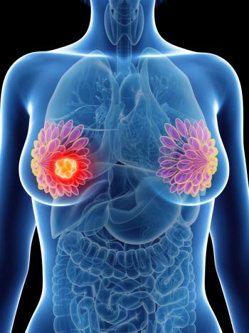
Breast tumor (red) in 3-D. Image courtesy of UCLA.
The study was
The lead author is Yunzhe Zhao, a recent postdoctoral student in the UCLA laboratory of senior author Jianwei (John) Miao, PhD. In their article, Dr. Miao and coinvestigators noted that up to 20% of palpable tumors are not detectable with mammography, which is still the primary imaging tool for screening and diagnosis of human breast cancers; furthermore, they said, only about 40% of biopsied lesions identified using mammography are malignant.
In a news release from UCLA announcing the study findings, Dr. Miao explained that the limitation of mammography “is that it provides only two images of the breast tissue, which can explain why 10% to 20% of breast tumors are not detectable on mammograms.” While CT scans can generate 3D views also, doing this requires a larger radiation dose than a mammogram, he said, posing a risk of tissue damage during breast cancer screening.
For the current study, the European investigators used phase-contrast tomography to x-ray a human breast from multiple angles. They applied a computing algorithm developed by Dr. Miao’s group called equally sloped tomography (EST) to 512 images to produce very high resolution 3D breast images, using less radiation than a mammogram and with fewer tomographic “slices” needed to yield a high-quality image. The tomography method they used measures the difference in x-ray oscillation through normal tissue vs slightly denser tissue such as tumor or bone. Even very small tumors, Dr. Miao said, can change the oscillation of an x-ray significantly to produce a distinct image.
Five independent radiologists from Ludwig Maximilians University performed blind evaluations of the 3D tumor images, ranking them as being sharper and having better contrast and overall quality than 3D breast tissue images created using other standard methods.
Commenting on the study outcomes, the authors wrote that they had “imaged a human breast in three dimensions and identified a malignant cancer with a pixel size of 92 μm and a radiation dose less than that of dual-view mammography,” and that, based on the evaluations of the five radiologists, the new technique “can reduce the radiation dose and acquisition time by [about] 74% relative to conventional phase contrast x-ray tomography, while maintaining high image resolution and image contrast.” They concluded that the results “demonstrate that high-resolution 3D diagnostic imaging of human breast cancers can, in principle, be performed at clinical compatible doses.”
However, senior author Alberto Bravin, PhD, managing physicist of the biomedical research laboratory at the European Synchrotron Radiation Facility, cautioned that this 3D technology is still in the research phase and it will not be available to patients for some time. “A high-quality x-ray source is an absolute requirement for this technique,” Dr. Bravin said in the UCLA news release. “While we can demonstrate the power of our technology, the x-ray source must come from a small enough device for it to become commonly used for breast cancer screening. Many research groups are actively working to develop this smaller x-ray source. Once this hurdle is cleared, our research is poised to make a big impact on society.”
The research was funded by UC Discovery/Tomosoft Technologies; the National Institute of General Medical Sciences, a division of the National Institutes of Health; and the Deutsche Forschungsgemeinschaft-Cluster of Excellence Munich-Centre for Advanced Photonics.
Newsletter
Stay up to date on recent advances in the multidisciplinary approach to cancer.
Advertisement
Related Content
Advertisement


Latest CME


Advertisement
Advertisement
Trending on CancerNetwork
1
Modifiable Risk Factors Suggest Potential for Improving Cancer Prevention
2
Dato-DXd Receives Priority Review in Unresectable/Metastatic TNBC
3
2026 Tandem Meetings: What’s the Latest Research in Multiple Myeloma?
4
Outlining Advances in AI For Breast Cancer Screening/Radiomics
5