
|Articles|July 16, 2019
- ONCOLOGY Vol 33 No 7
- Volume 33
- Issue 7
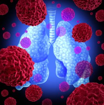
Evolution of Lung Cancer Screening Management
Throughout the last half of the 20th century, despite enormous research efforts, lung cancer remained the most feared as well as lethal cancer across the world. Efforts to update screening management are attempting to change that.
Advertisement
Start of ELCAP
Throughout the last half of the 20th century, despite enormous research efforts, lung cancer remained the most feared as well as lethal cancer across the world. Starting in 1991, a multidisciplinary group of investigators (ELCAP) at Weill Cornell Medical College (WCMC) in New York, mindful of challenge, proposed to leverage the transformative breakthrough in performance of low-dose radiation CT imaging to obtain full thoracic images in a single breath hold to identify early, curable lung cancer. Their research demonstrated that it worked remarkably well.[1] However, they were to find convincing a skeptical world already inured by decades of failed lung cancer detection efforts was to be their greatest challenge.
The research team wanted to learn about previous efforts. For this purpose, they conducted extensive discussions with veterans of the previous, ill-fated National Cancer Institute (NCI)-sponsored randomized trial evaluating the effectiveness of sputum cytology and annual chest x-rays (CXR) as an early detection method.[2] A particularly insightful expert was the chief study statistician, B. Flehinger. Dr. Flehinger had developed a mathematical model to estimate the potential cure rates for lung cancers that were much smaller than were detectable by CXR, but in a range that could be detected by low-dose CT (LDCT).[3,4] Based on the preliminary experience, the investigators hypothesized that the LDCT detection rate of small lung cancer could be as high as 80% of cancers between 4 mm and 10 mm and the Flehinger model suggested that for such cancers, the cure rate could be as high as 80%, much higher than the 18% rate the model predicted for CXR screening.
Based on this preliminary WCMC modeling experience, early detection of curable lung cancer using LDCT seemed feasible. The investigators understood that to compare the quantitative benefit of CT to CXR screening, three probabilities need to be determined for each imaging modality:
1) Probability of detecting a noncalcified nodule (NCN) in a person
2) Probability of diagnosing lung cancer by size and stage
3) Probability of curing all persons diagnosed with lung cancer under screening
They also recognized that, for each imaging modality, these three probabilities need to be determined separately for the baseline round and for all subsequent repeat rounds of screening.[1,5,6] The profound difference between the probabilities for the baseline (“prevalence”) round and the subsequent rounds of repeat screening (“incidence”) had been recognized by Morrison and others.[7] The terms “prevalence” and “incident” had been adapted from acute disease descriptors (eg, myocardial infarction), but any cancer has typically been present but not detectable for a considerable time and thus is a prevalent case, whether detected on baseline or repeat screening. Thus, the ELCAP investigators do not use the terms prevalence or incident cancers, but rather report whether the cancer was detected in the baseline or repeat rounds.
While the probabilities for the baseline round are different from those of repeat rounds of screening, each repeat annual round is a replicate of the other repeat rounds. Thus, a single repeat round would suffice to provide the probabilities, provided the sample size was sufficiently large. The investigators decided that the screening interval should be annual,[5,6] based on analysis of the lung cancer growth rates of lung cancers.[8]
The group’s first study was called the Early Lung Cancer Action Program (ELCAP).[1,6] Its goal was to determine the first two probabilities for LDCT and CXR by recruiting a prospective cohort of 1,000 current and former cigarette smokers, aged 60 years or older with at least 10 pack-years of cigarette smoking, who were asymptomatic for lung cancer. Each ELCAP participant was to receive a baseline LDCT and CXR followed by a repeat screening 12 months later. Upon receiving Institutional Review Board approval in 1992 at WCMC and then at New York University Medical Center, ELCAP recruitment started, thanks to seed philanthropic funding. A grant application was submitted to the NCI, but it was recognized that this would probably take several years to receive a grant award, and as predicted it was awarded in 1995.[5]
During the conduct of the ELCAP trial, participants with noncalcified nodules (NCNs) on LDCTs were presented at weekly multidisciplinary Thoracic Tumor Conferences. Quarterly ELCAP meetings of investigators at both institutions were held to rigorously manage the conduct of this trial. The literature at that time focused on “solitary noncalcified pulmonary nodules,”[9,10] but it was soon recognized that on LDCT, multiple small NCNs were frequently detected in a single participant, and more importantly only few of them represented lung cancers. Thus the focus changed from detecting solitary NCN to detecting one or more NCNs. More importantly, it was recognized that most NCNs remained stable while the lung cancer was identified in rapidly growing LDCT-detected NCNs.[11] Furthermore, standard diagnostic testing and follow-up of NCNs was based on CXR experience, but the ELCAP investigators rapidly moved away from performing a biopsy on all NCNs. Instead for NCN less than 10 mm in size on baseline LDCT, follow-up CT in 3 months was performed to assess growth.[1] If, on follow-up LDCT, the observed NCN growth rate was suggestive of a malignancy, an immediate nodule biopsy was recommended. For larger NCNs (≥ 10 mm) the standard recommendation, which called for biopsy, was followed.
Dr. D. Yankelevitz, an ELCAP investigator, recognized the potential of CT to reliably measure growth rates of small lung cancers on follow-up CTs and led the research projects to confirm that short-term follow-up LDCT scans were very useful in distinguishing between the many small benign NCNs and the few malignant ones.[11-16] This critical innovation was initially supported by philanthropy but then subsequently validated by NCI grant awards,[17] a textbook example of the power of public/private interactions.
The seminal finding of using short-term follow-up LDCT for differentiating lung cancer from benign nodules also highlighted how the efficiency of screening diagnostic workup could be optimized and how it could be integrated into protocols.[18,19] This innovation was rapidly recognized and also integrated by a wide range of other screening research groups.[20,21] In particular, the NELSON trial in the Netherlands and Belgium integrated volume assessment into their protocol.[22]
With the large number of participants with NCNs on LDCT with the need for follow-up scheduling of CT scans, the investigators recognized that data and image management soon became overwhelming, even for a study of 1,000 participants at two institutions. Fortunately, the principal investigator, C. Henschke, was an experienced computer programmer and statistician, and was thus able to rapidly develop and write the needed computer management programs to document, store, and analyze the data for the ELCAP study.
ELCAP Baseline Screening
In 1998, recruitment of 1,000 baseline participants was completed and the baseline results were analyzed and published in the Lancet in 1999.[1] Among the 1,000 participants, NCNs were found by LDCT in 233 (23.3%) and by CXR in 68 (6.8%). Lung cancers were diagnosed in 27 by LDCT and 7 by CXR among the 1,000 participants (2.7% vs 0.7%, P = .0005). Of the 27 lung cancer patients identified by LDCT, 23 (85.1%) were stage I lung cancers while CXR identified only 4, missing 19 (82%) of them. LDCT also found 4 additional cancers, 2 endobronchial cancers, and 2 mediastinal cancers that were not identified by CXR.
Using published long-term survival rates by stage.[23,24] the ELCAP investigators projected a possible cure rate of 80% (a lung cancer fatality rate of 20%) for patients with lung cancer diagnosed in a program of LDCT screening. To robustly estimate the cure rate, however, additional screenings and longer follow-up time were needed.
ELCAP Annual Screening
The annual repeat screenings were completed, analyzed, and submitted in 1999, but only published in Cancer in 2001.[6] As annual rounds are repeated year after year for each participant, while the baseline round is performed only once for each participant, results from annual screening rounds are critical to assess screening performance.
Among the 1,184 annual repeat screening, new or growing NCNs were identified in 30 (2.5%) participants. Seven of the 30 participants were diagnosed with lung cancer, of which 6 (85.7%) had stage I disease. There were far fewer new NCNs on annual repeat screening than on the baseline screening (2.5% vs 23%, P < .0001) and the percentage of screen-diagnosed lung cancers was also different (0.59% vs 2.7%, P < .0001). The percentage of screen-diagnosed stage I disease, however, was similar (85.7% vs 85.1%, P = 1.00), although the median tumor size was smaller in annual rounds of screening (8 mm vs 10 mm, P = .45). Based on this evidence and pathologic findings,[25,26] future protocols for workup in the baseline and annual repeat rounds of screening were different.[18,19,27]
A critical quality assessment parameter for any cancer screening program is the frequency of cancers identified by symptom-prompted workup between the scheduled rounds of repeat screening. Another 2 participants had symptom-prompted workup for pneumonia which led to the diagnosis of lung cancer prior to the first annual round of screening. Both had endobronchial tumors, one stage I small-cell carcinoma and one stage II squamous-cell carcinoma. Thus, the percentage of patients with stage I lung cancer, including screen-diagnosed and symptom-prompted diagnoses, was slightly lower on annual repeat than on baseline screening (77.8% vs 85.1%, P = .63). This was expected as a higher percentage of aggressive cancers are identified in repeat rounds of screening.[26,27]
Prior to 2000, radiologists reviewed the CT images displayed on radiographic films that provided 12 CT images per film.[1,7] Typically, 30 CT images were obtained for each screening participant, using 10-mm slice thickness, although reconstruction at 5-mm overlapping intervals was used to improve the image resolution. With advances in CT technology, particularly with multislice scanners, submillimeter slice thickness acquisitions are now obtained, providing as many as 800 CT images for each participant, and these images are displayed on large computer monitors, which, though expensive, provide far greater detail. Today, in addition to viewing axial images, coronal, sagittal, and maximum intensity projections are routinely provided, further enhancing visualization. These advances have markedly increased identification of tiny NCNs, thus reducing symptom-prompted diagnoses between annual rounds of screening. These marked improvements have also prompted reconsideration of using longer intervals between repeated rounds of screening.[21,28]
Expansion of ELCAP Screening to New York [NY]-ELCAP and International [I]-ELCAP
Immediate international requests for collaborations on LDCT screening followed the publication of the 1999 Lancet article.[1] In response, the ELCAP investigators initiated an International Conference on Screening for Lung Cancer in the fall of 1999 and these conferences have been continued, with the 40th conference held in April 2019.[29,30] At the first conference, participants called for the development of a multi-institutional protocol that, when presented at the next conference 6 months later, was unanimously adopted and then published.[18,19]
The screening protocol was critical, as with the marked advances of CT scanner and viewing technology, radiologists could detect NCNs as small as 2 mm but very few of these NCNs are lung cancers. The exquisite sensitivity of LDCT to identify small lung cancers, compared to CXR, further accentuated the difference in the first two probabilities of the baseline and annual repeat rounds and the need for recognition of these differences in the baseline and repeat protocol.[26,27] These advances also highlighted the need to update the threshold criteria for workup on baseline and on annual repeat screening.[18,31-33] Many additional insights were developed using the enlarging International [I]-ELCAP database, and these recommendations have been incorporated in the updated online protocol.[19] These recommendations are provided for findings made on screening scans of mediastinal masses, emphysema, coronary artery calcifications, aortic valve calcifications, pulmonary artery enlargement, interstitial lung disease, breast density and masses, liver density, and bone. In developing these recommendations, the investigators worked closely with clinicians in different medical specialties and stressed that, different from routine medical practice, these findings were not prompted by symptoms but identified as a result of screening and thus recommendations and workup need to be tailored accordingly. Some of these recommendations have been incorporated, for example, into national guidelines.[34] The importance of using a protocol with detailed recommendations was highlighted by the comparison of I-ELCAP and National Lung Screening Trial (NLST) results,[35] which showed that the use of a regimen of screening markedly decreased unnecessary workup.
In view of the international demand for LDCT screening, A.P. Reeves, a Professor of Electrical Engineering and Computer Science at Cornell University, who had been working with the ELCAP team on tumor growth analyses,[12-18] suggested expansion to a web-based management system that would enable more efficient and comprehensive data and images collection from any site connected to the internet. It would also be able to provide a platform for screening management tools, training, and reporting. Thus, the original ELCAP management system was converted into the updated, web-based ELCAP management system.[36]
This web-based ELCAP management system together with the I-ELCAP protocol made it possible to rapidly expand LDCT screening worldwide.[19] ELCAP investigators developed educational sessions for radiologists and coordinators, and provided the ELCAP management system, free of charge, to all participating institutions. Core features of this system have been widely adopted by other screening data systems. The ELCAP management system has developed a comprehensive, new site onboarding process using a blend of web content, face-to-face interactions at its biannual workshop process, as well as a comprehensive archive of relevant screening publications.[19] An example of the onboarding process is the dual readings service allowing each new participating institution to have the first 200 scans read jointly by the coordinating center with follow-up adjudication discussions and then review of the next 300 scans.[37] Other quality interactions around lung cancer pathology review, assessment of other thoracic detected disease conditions, as well as nursing and navigation measures are available.
By 2000, LDCT screening rapidly expanded throughout New York State as NY-ELCAP and internationally as I-ELCAP programs.[38,39]
I-ELCAP Long-Term Survival for Diagnosed Lung Cancers Under Screening
The expansion of ELCAP provided sufficient data to robustly estimate the probability of cure for patients diagnosed with lung cancer under lung cancer screening (baseline, annual screen-diagnoses as well as symptom-prompted diagnoses rounds).[39] The cure rate, estimated by the 10-year Kaplan-Meier survival rate of 484 patients, was 88% (95% CI, 84%–91%), regardless of the cancer stage or treatment. Patients who were diagnosed with clinical stage I lung cancer and resected within 1 month of diagnosis had a higher estimated cure rate (92% [95% CI, 88%–95%]).
These estimated cure rates for annual LDCT screening for lung cancer have been confirmed at the international conferences, held every 6 months since 1999,[29] including the latest 40th conference held in April 2019, which provided the 20-year follow-up results.
Individualized Benefit of LDCT Screening
The benefit of LDCT screening was individualized as to the age, smoking history, and other pertinent information of each individual considering screening.[40] Using the ELCAP baseline information together with the long-term Kaplan-Meier survival rates, the benefit of a single round of screening was determined based on the person’s age and smoking history. This benefit was due to the increased survival by undergoing the first round of LDCT screening. This approach can be extended to calculate the individual benefit or survival gain from each subsequent annual round of screening.
The ELCAP Management System
The ELCAP management system provided a scalable system to disseminate the standardized data elements needed for consistent, high-quality screening implementation and management. It documented the initial contact of each participant and allowed for all subsequent steps of the screening process, scheduling, reading the LDCT, recommendations for follow-up, and follow-up. This information is needed by coordinators, navigators, nurses, radiologists, and treating physicians on a participant-prompted, timely basis. The system efficiently collected screening data obtained in the process of a clinical screening program and also provided quality control, follow-up management, and reminders. Beyond the clinical needs, the system enabled collection of screening research data, collected in the context of clinical care, for rigorous scrutiny of needed updates and analysis of outcomes.
The ELCAP management system was provided to each participating institution, free of charge. Training for setting up screening programs was provided to each site and is also provided at the international conferences held every 6 months. Training included dual reading by the institution and coordinating center with identification of critical differences as the ELCAP management system provided both the data and images.[37] By the 40th International Conference on Screening held in New York on April 12–13, 2019,[29] the I-ELCAP database has clinical data and images on baseline and repeat rounds on over 82,000 participants throughout the world. The database includes the information on diagnostic work-up, treatment, and follow-up on all participants diagnosed with lung cancer, and also identifies additional diagnoses of metachronous lung cancers during annual follow-up of patients who had been cured of their first lung cancer.
The process of having international meetings every 6 months open to all interested in screening and providing international collaboration for an international health concern has engendered many international collaborations and has enabled an entire family of researchers from many medical, mathematical, engineering, statistics, computing, and other disciplines to work together to continually improve the benefit of LDCT screening for people at risk of lung cancer and other diseases.
The flexibility and utility of the ELCAP cohort design allowed for rapid accumulation of data and thus expansion of knowledge of how to optimize the clinical screening process and the resulting treatment advances.[27,35,41] In fact, considering the latest very low-dose techniques of CT scanners and the findings that can be made on LDCT, LDCT screening should be considered as providing a comprehensive health check of the lungs, heart, and other visualized organs in the LDCT screening,[42] which might prove to be highly cost-effective.[43]
Both the conferences and the management system anticipated the era of “open science, open-source” and have illustrated the power and benefit of an open science approach.[44] The open-science and open-source approach is a formalized approach of the fundamental philosophical underpinning of the Early Diagnosis and Treatment Research Foundation that has provided the ELCAP management system and the international conferences. The ELCAP management system has been made available as an “open source” system for LDCT screening of US veterans who are at high risk of lung cancer and other lung and heart diseases through a grant from the Bristol Myers-Squibb Foundation.[45]
The resulting updated VA-Partnership to increase Access to Lung Screening (VA-PALS)-ELCAP management system is the MUMPS-based software.[46] The ELCAP and updated VA-PALS-ELCAP management systems both use structured forms and radiology reporting, as did the original system, The structured reporting provides the common terminology for characterization of all NCNs, allows for quality assurance assessments, and pooling of data across multi-institutions around the world. In the future, the follow-up recommendations can be automatically generated based on the established screening protocol.
The initial information for each participant is documented in the intake form. Pertinent background information potentially helpful in the interpretation of the LDCT images is collected on the background form, which can be obtained by phone prior to the appointment, at the time the LDCT is performed, or later. As radiologists are responsible for reporting on findings of the entire LDCT, structured reporting of all LDCT findings are documented in the CT evaluation form: NCNs, other lung abnormalities (eg, interstitial lung findings, small airways abnormalities), cardiac abnormalities (eg, coronary artery calcifications and aortic valve calcifications), breast abnormalities (breast density, masses, and other findings), and abdominal abnormalities (eg, liver, adrenal, kidney, esophagus, pancreas, spleen). Input can be done directly or by a dictation template. Upon completion of the CT evaluation form, the written report is automatically generated. The follow-up form is used for documenting whenever the participant returns for a LDCT along with any changes in smoking pattern and diagnosis or hospitalization since the last visit. Biopsy, PET, and treatment forms are available for documenting subsequent diagnostic procedures and interventions.
Advances Stimulated by LDCT Screening
LDCT screening has been disruptive, as its much earlier identification of lung cancers led to updating of recommendations in related disciplines such as smoking cessation, workup of NCNs, recognition of subtypes of NCNs, and pathology criteria of early lung cancer, staging, and treatment.
As a result of the initial ELCAP study, the investigators recognized that LDCT screening enhanced smoking quit rates and did not encourage resumption of smoking.[47,48] These results led to the strong recommendation that smoking cessation programs should be provided with all LDCT screening programs.[49]
Screening led to ever-increasing understanding of small, early lung cancers as to their appearance, growth, and pathologic characteristics that heretofore had not been well-explored due to insufficient data. The impact of CT scanner technology on tumor growth assessment was demonstrated by the limitations due to the tumor appearance and those due to the quantitative measurements of the scanner itself.[50,51] The importance of measurement error in nodule growth assessment was an important factor leading to the formation of the Quantitative Imaging Biomarker Alliance sponsored by the Radiological Society of North America (RSNA).[52] Hopefully, recognition of these limitations will stimulate development of better quantitative tools so that NCN growth can be determined within weeks rather than months, as this will lead to marked improvement of the efficiency of LDCT and to reducing unnecessary biopsies and/or surgery.
Differences in NCN subtypes, solid, part-solid, and nonsolid, were recognized early in the development of screening protocols.[53-56] The terminology was developed by the ELCAP investigators.[56] After review of the I-ELCAP and NLST databases and then of the world literature,[57-62] recommendations for workup of the different subtypes was developed and incorporated into the I-ELCAP protocol.[19] The usefulness of watchful waiting for nonsolid NCNs and concerns about over-diagnosis was addressed by these publications. The key conclusion was that overtreatment should be avoided and that current surgical recommendations for nonsolid NCNs involve watchful waiting as supported by the revised surgical staging classification of the American Joint Committee on Cancer/International Association for the Study of Lung Cancer (AJCC/IASLC) and address the concerns of over-diagnosis.[63-65] These critical findings allowed for watchful waiting, showing that by annual CT follow-up, cancers identified in nonsolid NCNs remained 100% curable even if they developed a solid component during the year.
Other advances were due to the recognition that the LDCT of the chest provides additional information about the lungs as well as the other visualized organs. The Surgeon General reports had documented the impact of active tobacco smoking and involuntary exposure to tobacco smoke on many other organs, including heart disease, chronic pulmonary diseases, and cancers of other organs.[66-69] Secondhand tobacco smoke exposure was shown to increase the risk of lung cancer, emphysema, and cardiovascular disease,[68] which was also assessed in the context of LDCT screening.[70,71] These results suggest that secondhand tobacco smoke should be included as a risk factor. I-ELCAP was also able to demonstrate that women in the United States were found to be more susceptible to lung cancer,[72,73] but when diagnosed with lung cancer, had a lower risk of dying.[73] The lower risk of women dying of lung cancer was confirmed in the NELSON study.[21]
Pathologic criteria for diagnosis of small, early lung cancer needed updating. The ELCAP investigators had developed a pathology protocol and initiated expert pathology panel reviews in 2000,[74] funded in part by the American Cancer Society. Results of these panel reviews of the pathologic specimens available through I-ELCAP have been published.[25,26,75] These reviews led to the new pathologic classification of adenocarcinoma and to the revised World Health Organization classification.[76-79]
When ELCAP started, the 6th staging classification edition was being used.[24] From the beginning, ELCAP and I-ELCAP also documented the modified tumor size categories as many small cancers were identified: < 10 mm, 11–20 mm, and 21–30 mm. These categories have now been incorporated in the 8th staging edition introduced in 2018.[80,81] Different from the 8th classification, however, in I-ELCAP’s modified staging, multiple adenocarcinomas without lymph node metastases are considered as multiple primary stage I cancers, not as higher stages as currently classified in the 8th classification, based on the evidence resulting from analyses of screening results.[75] The 8th edition of staging does not recognize the impact of nodule consistency (solid, nonsolid, part-solid). I-ELCAP investigators have shown in their recent publications that tumor consistency is both an important diagnostic and prognostic factor that needs to be considered in the tumor, node, and metastasis (TNM) staging, particularly for lung cancers < 30 mm in maximum diameter.[82,83] Based on careful documentation and analyses, investigators have found that clinical staging for tumors 30 mm or smaller should be classified based on the tumor size on CT, as neither lymph node enlargement or F-18-fluorodeoxyglucose (FDG)-PET uptake were helpful in identifying lymph node metastases. These publications called for updates of the 8th classification as data for the smallest size category was limited, despite the impressive contribution of the IASLC staging database.
Breast cancer treatment provides an excellent paradigm of the impact of screening on diagnosis and treatment. Prior to screening, mastectomy with radical lymph node dissection was the standard of care, but currently there are many alternative and less radical treatments. The recommendation of lobar resection has not changed in more than 50 years. Two randomized surgical trials started in 2007,[84,85] but their results are not expected until after 2020. In the meantime, new technologies are being developed (eg, robotic surgery, navigational bronchoscopy, percutaneous ablation) that also need assessment.
Lung cancer will follow a similar paradigm as breast cancer. Published cohort studies are providing outcome results while awaiting the results of randomized trials on the extent of surgical resection.[86-91] Quality of life becomes an important consideration in treatment assessment given the high long-term cure rates of screen-diagnosed lung cancer.[92-95]
Clearly, as recognized in the design of ELCAP, it is the curative treatment of early lung cancer that determines whether screening is effective. As new techniques are continuously being introduced, often with limited data for small lung cancers, the Initiative for Early Lung Cancer Research on Treatment (IELCART) was started using a prospective cohort design as previously used for I-ELCAP.[96] The ELCAP management system has been adapted for treatment considerations and also allows for randomization for future innovative randomized trials within its structure. IELCART is envisioned to be as productive as I-ELCAP has been in producing ongoing evidence.
Summary
The low-cost and efficient ELCAP design provided the fundamental clinically relevant information about the benefit of LDCT screening. In the 20 years since our initial ELCAP report in 1999, a vibrant community of LDCT investigators has emerged. Their efforts have ranged from providing the NLST in the United States, the largest randomized screening trial performed, which started in 2002, to the NELSON trial in Europe, which started in 2004.[20,21] The design of the NELSON trial and its use of volumetrics was strongly influenced by extensive discussions of the leading NELSON and ELCAP investigators at I-ELCAP conferences and through personal communications.[22,29] Both the NLST and NELSON trials, when published in 2011 and 2019,[20,21] not only confirmed the ELCAP results reported in 1999, but the NLST also provided a large library of CT images with associated clinical outcomes for research.[97] Important insights of the different screening trial designs and outcome parameters were gained,[98-102] and spinoff screening management optimization approaches were developed.[103-105] Other international trials have reported long-term follow-up of studies with randomized and cohort trial designs across the world that have confirmed the high long-term benefit of CT screening,[28,106-109] thus providing even further assurances as to the robustness of the LDCT screening benefit.
Other screening protocols have emerged since our initial publication in 1999, such as the American College of Radiology Lung Imaging Reporting and Data System (Lung-RADS) and the European Consortium protocols.[103,104] Comparisons of efficiency of these protocols as to the number of workups per diagnosis of lung cancer show that the I-ELCAP is currently the most efficient,[105] but we are excited that each of these efforts provides innovative approaches that benefit all screening participants. As further evidence and technology emerges, it is hoped that protocols will continue to be updated and compared using standardized criteria that measure efficiency and eventually these efforts should lead to further uniform improvements.
Financial Disclosure: Dr. Reeves receives patent royalties from GE, and is president of D4Vision, Inc. Dr. Yankelevitz owns equity in Accumetra and is on the advisory board of GRAIL. Dr. Henschke and Ms. Yip have no significant financial interest in or other relationship with the manufacturer of any product or provider of any service mentioned in this article.
PERSPECTIVE
Low-Dose CT Screening: Where It Began
James L. Mulshine, MD
Innovation is said to be disruptive and the experience in applying low-dose computed tomography (LDCT) to find early lung cancer certainly validated that old bromide. In Dr. Henschke and colleagues’ review, we have the encapsulation of 20 profoundly transformative years in the history of biomedical research. In 1999, lung cancer was a terribly lethal scourge that humbled countless research efforts to improve outcomes. Aside from the courageous interventions of a generation of surgeon generals, a spirited tobacco control community, and a few Japanese epidemiologists, the disease burden of tobacco use was unrelentingly deadly.
The Lancet paper computed tomography (CT) screening report from Dr. Henschke’s team in 1999 was electric and galvanized camps of skeptics and believers alike[1]. This report came at the time of an ongoing and charged reconsideration of screening benefits with breast cancer, as well as prostate cancer. This situation with lung cancer was also colored by memories of initial enthusiasm for chest X-ray screening that did not hold up to more in-depth analysis.
An important, but underappreciated aspect of the New York screening team’s effort was a commitment to continue to pursue research evidence to refine the early lung cancer detection process. This momentum was sustained by a series of workshops that occurred every 6 months for the next 20 years and included advocates, as well as skeptics, from a broad array of medical and scientific disciplines.
Over this time in excess of 300 screening-related manuscripts were published coming from International Early Lung Cancer Action Program (I-ELCAP) collaborative groups in their consortium on aspects from imaging to health policy. Leveraging a remarkably robust informatics capability, the collaboration ultimately accrued over 82,000 screening subjects, including access to over 300,000 thoracic CT scans from participants on five continents. Hundreds of investigators worked with this consortium, including surgical leaders such as Drs. Robert Ginsburg, Nasser Altorki, and Raji Flores. Through coordinated efforts with Drs. William Travis and Adi Gazdar and others from the International Association for the Study of Lung Cancer Pathology Committee, every case of lung cancer detected by ELCAP was reviewed by the pathology panel. That experience led to the changes in international pathology classification and corresponding changes to international staging classifications.
Drs. Harvey Hecht, Matt Cham MD, Joseph Shemesh, and Jagat Narula revealed the dynamic of screen-detected coronary artery disease, while a corresponding revelation of frequent asymptomatic pulmonary parenchymal disease was reported by Javier Zulueta and his Spanish colleagues, principally from the University of Navarra. The list of other new observations is long, from Mary Salvatore team’s findings with interstitial pulmonary fibrosis, to the routine findings of abnormal breast density on screening CTs with Laurie Margolies and colleagues, to recent emerging information about screening for nonalcoholic steatohepatitis (NASH) in the context of low-dose computed tomography screening, with Andrea Branch and colleagues. These activities were thoughtfully acknowledged in Dr. Henshke’s review[2].
An excellent example of the power of this consortial approach was highlighted in a report to the Annals of Internal Medicine on the consequences of selecting the size threshold of detected non-calcified pulmonary nodule relative to the efficiency of a screening management approach. To address this question, Henschke was able to analyze the outcomes of 21, 136 screening cases from a 5-year period of observation to inform the decision as to what threshold would be most appropriate[3] and then further confirm these thresholds using the National Lung Screening Trial Database.[4] Having such robust evidence to refine the efficiency of the screening process reflects how far the I-ELCAP investigators have moved this field.
As Dr. Henschke relates, her group’s work also stimulated competitive and collaborative efforts across the globe with outstanding screening research teams emerging launching the National Lung Screening Trial and the NELSON study, as well as coordinated efforts in Canada, Poland, Spain, Switzerland and many other nations[5-7]. This vibrant experience, while not perfect, did in fact align the contributions of hundreds of talent researchers and advocates to change the world’s perception of and aspirations for lung cancer to the benefit of many.
Financial Disclosure:Dr. Mulshine has no significant financial interest in or other relationship with the manufacturer of any product or provider of any service mentioned in this article.
REFERENCES
1) Henschke C, McCauley D, Yankelevitz D, Naidich D, McGuinness G, Miettinen O, Libby D, Pasmantier M, Koizumi J, Altorki N, and Smith J. Early Lung Cancer Action Project: overall design and findings from baseline screening. Lancet. 1999; 354:99-105.
2) Henschke C, Yankelvitz D, Reeves A, Yip R. Evolution of lung cancer screening management. ONCOLOGY. 2019;____ 33-6.
3) Henschke C, Yip R, Yankelevitz DF, Smith JP for the I-ELCAP Investigators. Definition of a positive test result in computed tomography screening for lung cancer: a cohort study. Annals of Internal Medicine. 2013; 158:246-52 PMID: 23420233
4) Yip R, Henschke CI, Yankelevitz DF, Smith JP. CT screening for lung cancer: alternative definitions of positive test result based on the National Lung Screening Trial and International Early Lung Cancer Action Program databases. Radiology 2014; 273: 591-6. Accompanied by a Radiology podcast. PMID: 24955929
5) National Lung Screening Trial Research Team, Aberle DR, Adams AM, Berg CD, Black WC, Clapp JD, et al. (Reduced lung-cancer mortality with low-dose computed tomographic screening. N Engl J Med. 2011; 365:395-409.
6) De Koning HJ, Van Der Aalst C, Ten Haaf K, et al: Effects of volume CT lung cancer screening: Mortality results of the NELSON randomized-controlled population-based trial. Presented at the 2018 World Conference on Lung Cancer; September 25, 2018; Toronto. Abstract PL02.05.
7) Pyenson B, Henschke C, Yankelevitz DF. Editorial: Population health’s unanimity on lung cancer screening: far ahead of medical advice. Ann Transl Med. 2017; 5:355 PMID: 28936449 PMCID: PMC5599287
References:
1. Henschke C, McCauley D, Yankelevitz D, Naidich D, McGuinness G, Miettinen O, Libby D, Pasmantier M, Koizumi J, Altorki N, and Smith J. Early Lung Cancer Action Project: overall design and findings from baseline screening. Lancet 1999; 354:99-105.
2. Berlin NI, Buncher CR, Fontana RS, et al. The National Cancer Institute Cooperative Early Lung Cancer Detection Program. Am rev Respir Dis 1984; 130:545-549.
3. Flehinger NJ, Kimmel M, Melamed MR. The effect of surgical treatment on survival from early lung cancer. Implications for screening. Chest 1992; 101(4):1013-8.
4. Flehinger BJ, Kimmel M, Polyak T, Melamed MR, Screening for lung cancer. the Mayo Lung Project revisited. Cancer 1993; 72: 1573-80.
5. Early Detection and Diagnosis of Lung Cancer Using CT. National Institutes of Health grant R01-CA-63393. PI: Claudia I. Henschke. NCI grant 1995-1997
6. Henschke CI, Naidich DP, Yankelevitz DF, McGuinness G, McCauley DI, Smith JP, Libby D, Pasmantier M, Vazquez M, Koizumi J, Flieder D, Altorki N, Miettinen OS. Early Lung Cancer Action Project: initial findings on repeat screenings. Cancer 2001; 92:153-9.
7. Morrison A. Screening in chronic disease. 2nd Ed, Monographs in epidemiology and biostatistics. Oxford University Press, New York xiv. 1992.
8. Geddes DM. The natural history of lung cancer: a review based on rates of tumor growth. Br J Dis Chest 1979; 73: 1-17.
9. Lillington GA. Management of solitary pulmonary nodules. Dis Mon 1991; 37: 271-318.
10. Cummings SR, Lillington GA, Richard RJ. Estimating the probability of malignancy in solitary pulmonary nodules. Am Rev Respir Dis 1986; 449-452.
11. Yankelevitz DF, Gupta R, Zhao B, and Henschke CI. Small pulmonary nodules: evaluation with repeat CT--preliminary experience. Radiology 1999; 212:561-6.
12. Yankelevitz DF, Reeves AP, Kostis WJ, Zhao B, and Henschke CI. Small pulmonary nodules: volumetrically determined growth rates based on CT evaluation. Radiology 2000; 217:251-6.
13. Kostis W, Reeves A, Yankelevitz D, and Henschke C. Three-dimensional segmentation and growth-rate estimation of small pulmonary nodules in helical CT images. IEEE Trans Med Imaging 2003; 22:1259-74.
14. Reeves A, Chan A, Yankelevitz D, Henschke C, Kressler B, and Kostis W. On measuring the change in size of pulmonary nodules. IEEE Trans Med Imaging 2006; 25:435-50.
15. Kostis W, Yankelevitz D, Reeves A, Fluture S, and Henschke C. Small pulmonary nodules: reproducibility of three-dimensional volumetric measurement and estimation of time to follow-up CT. Radiology 2004; 231:446-52.
16. Henschke C, Yankelevitz D, Yip R, Reeves A, Farooqi A, Xu D, Smith J, Libby D, Pasmantier M, and Miettinen O. Lung cancers diagnosed at annual CT screening: volume doubling times. Radiology 2012; 263:578-83.
17. Early Repeat CT for Evaluation of Solitary Lung Nodules. National Institutes of Health grant R01-CA-78905. PI: David F. Yankelevitz. NCI grant 1999-2005
18. Henschke C, Yankelevitz D, Smith J, Miettinen O, and ELCAP Group. Screening for lung cancer: the early lung cancer action approach. Lung Cancer 2002; 35:143-8.
19. International Early Lung Cancer Action Program Investigators. International Early Lung Cancer Action Program protocol. Available from: www.IELCAP.org/protocols, Jan 3rd, 2018.
20. National Lung Screening Trial Research Team, Aberle DR, Adams AM, Berg CD, Black WC, Clapp JD, et al. (2011) Reduced lung-cancer mortality with low-dose computed tomographic screening. N Engl J Med 365:395-409.
21. De Koning HJ, Van Der Aalst C, Ten Haaf K, et al: Effects of volume CT lung cancer screening: Mortality results of the NELSON randomized-controlled population based trial. 2018 World Conference on Lung Cancer. Abstract PL02.05. Presented September 25, 2018.
22. van Klaveren RJ, Oudkerk M, Prokop M, Scholten ET, Nackaerts K, Vernbout R, et al. Management of lung nodules detected by volume CT scanning. N Engl J Med 2009; 361:2221-2229.
23. Martini N, Bains MS, Burt ME, Zakowski MF, McCormack P, Rusch VW, Ginsberg RJ. Incidence of local recurrence and second primary tumors in resected stage I lung cancer. J Thorac Cardiovasc Surg 1995; 109(1):120-9.
24. Mountain CF. Revisions in the International System for Lung Cancer. Chest 1997;111:1710-1717.
25. Flieder DB, Vazquez M, Carter D, Brambilla E, Gazdar A, Noguchi M, Travis WD, Kramer A, Yankelevitz DF, Henschke CI. Pathologic findings of lung tumors diagnosed on baseline CT screening. American Journal of Surgical Pathology 2006; 30:606-13
26. Carter D, Vazquez M, Flieder DB, Brambilla E, Gazdar A, Noguchi M, Travis WD, Kramer A, Yip R, Yankelevitz DF, and Henschke CI. Comparison of pathologic findings of baseline and annual repeat cancers diagnosed on CT screening. Lung Cancer 2007; 56:193-9.
27. Henschke CI, Salvatore M, Cham M, Powell CA, DiFabrizio L, Flores R, Kaufman A, Eber C, Yip R, Yankelevitz DF. Baseline and annual repeat rounds of screening: implications for optimal regimens of screening. Eur Radiol 2018; pp 1-10.
28. Pastorino U, Silva M, Sestini S, et al. Prolonged lung cancer screening reduced 10-year mortality in the MILD trial. Ann Oncol. 2019 Apr 1. pii: mdz117. doi: 10.1093/annonc/mdz117.
29. International Early Lung Cancer Action Program Investigators. Program and Consensus statements. International Conferences on Screening for Lung Cancer. Available from:
30. Henschke CI, Boffetta P, Yankelevitz DF, Altorki N. Computed tomography screening: the International Early Lung Cancer Program Experience. Thorac Surg Clin 2015; 25:129-43 PMID:25901557.
31. Henschke CI, Yankelevitz DF, Naidich DP, McCauley DI, McGuinness G, Libby DM, Smith JP, Pasmantier MW, Miettinen OS. CT screening for lung cancer: Suspiciousness of nodules according to size on baseline scans. Radiology 2004; 231:164-8 PMID:14990809
32. Henschke CI, Yip R, Yankelevitz DF, Smith JP for the I-ELCAP Investigators. Definition of a positive test result in computed tomography screening for lung cancer: a cohort study. Annals of Internal Medicine 2013; 158:246-52 PMID: 23420233
33. Yip R, Henschke CI, Yankelevitz DF, Smith JP. CT screening for lung cancer: alternative definitions of positive test result based on the National Lung Screening Trial and International Early Lung Cancer Action Program databases. Radiology 2014; 273: 591-6. Accompanied by a Radiology podcast. PMID: 24955929
34. Hecht HS, Henschke C, Yankelevitz D, Fuster V, Narula J. Combined detection of coronary artery disease and lung cancer. European Heart Journal 2014; 35:2792-6 PMID:25112665
35. International Early Lung Cancer Action Program Investigators (Yip R, Henschke CI, Yankelevitz DF, Boffetta P, Smith JP, writing committee). The impact of the regimen of screening on lung cancer cure: a comparison of I-ELCAP and NLST. European Journal of Cancer Prevention 2015; 24:201-8 PMID:25089376
36. Reeves AP, Kostis WJ, Yankelevitz DF, Henschke CI. A web-based data system for multi-institutional research studies on lung cancer. Scientific Session of the 87 Radiological Society of North America, Chicago IL, November 2001
37. Xu DM, Lee IJ, Zhao S, Yip R, Farooqi A, Cheung EH, Connery CP, Frumiento C, Glassberg RM, Herzog G, Peeke J, Scheinberg P, Shah P, Taylor J, Welch L, Widmann M, Yoder M, Yankelevitz DF, Henschke CI. CT screening for lung cancer: value of expert review of initial baseline screenings. Am J Roentgenol 2015; 204:281-6 PMID: 25349980
38. NY-ELCAP Investigators. CT Screening for lung cancer: diagnoses resulting from the New York Early Lung Cancer Action Project. Radiology 2007; 243:239-49.
39. International Early Lung Cancer Action Program Investigators, Henschke CI, Yankelevitz DF, Libby DM, Pasmantier MW, Smith JP, and Miettinen OS. Survival of patients with stage I lung cancer detected on CT screening. N Engl J Med 2006; 355:1763-71.
40. International Early Lung Cancer Action Program Investigators. CT screening for lung cancer: individualizing the benefit of the screening. European Respiratory Journal 2007; 30:843-7 PMID:17652316
41. Mulshine JL and D’Amico A. Issues with Implementing a High-Quality Lung Cancer Screening Program. CA CANCER J CLIN 2014 doi: 10.3322/caac.21239.PMID: 24976072
42. Mulshine JL. One Screening for Ischemic Heart Disease, Lung Cancer, and Chronic Obstructive Pulmonary Disease: A Systems Biology Bridge for Tobacco and Radiation Exposure. Am J Public Health.2018;108:1294-1295. doi: 10.2105/AJPH.2018.304655. PMID: 30207781
43. Pyenson B, Henschke CI, Yankelevitz DF. Editorial: Population health’s unanimity on lung cancer screening: far ahead of medical advice. Ann Transl Med 2017: 5: 355 PMID: 28936449 PMCID: PMC5599287
44. National Academies of Sciences, Engineering, and Medicine. 2018. Open Science by Design: Realizing a Vision for 21st Century Research. Washington DC:
Washington, DC: The National Academies Press. doi:
45. Office of Public and Intergovernmental Affairs. VA to increase access to safe and effective lung screening programs. [Press release]. August 31, 2017. Retrieved from
46. Henschke CI, Reeves AP, Avila R, Moghanaki D, Jirapatnakul A, Yankelevitz DF. Open source CT lung screening management system. World Lung Cancer Conference, Barcelona 2019.
47. Ostroff J, Buckshee N, Mancuso C, Yankelevitz D, and Henschke C. Smoking cessation following CT screening for early detection of lung cancer. Prev Med 2001; 33:613-21.
48. Anderson CM, Yip R, Henschke CI, Yankelevitz DF, Ostroff JS, and Burns DM. Smoking cessation and relapse during a lung cancer screening program. Cancer Epidemiol Biomarkers Prev 2009; 18:3476-83.
49. Warren GW, Ostroff JS, Goffin JR. Lung cancer screening, cancer treatment, and addressing the continuum of health risks caused by tobacco. Am Soc Clin Oncol Educ Book. 2016;35:223-
50. Zhang L, Yankelevitz DF, Henschke CI, Jirapatnakul AC, Reeves AP, and Carter D. Zone of transition: a potential source of error in tumor volume estimation. Radiology 2010; 256:633-9.
51. Henschke C, Yankelevitz D, Yip R, Archer V, Zahlmann G, Krishnan K, Helba B, and Avila R. Tumor volume measurement error using computed tomography (CT) imaging in a Phase II clinical trial in lung cancer. Journal of Medical Imaging 2016; 3:035505
52. Radiologic Society of North America Quantitative Imaging Biomarkers Alliance (QIBA) Calculator. (2017)
53. Mirtcheva RM, Vazquez M, Yankelevitz DF, Henschke CI. Bronchioloalveolar carcinoma and adenocarcinoma with bronchioloalveolar features presenting as ground-glass opacities on CT. Clin Imaging 2002; 26:95-100 PMID:11852215
54. Henschke CI, Shaham D, Yankelevitz DF, Kramer A, Kostis WJ, Reeves AP, Vazquez M, Koizumi J, and Miettinen OS. CT screening for lung cancer: significance of diagnoses in its baseline cycle. Clin Imaging 2006; 30:11-5.
55. Henschke CI, Wisnivesky JP, Yankelevitz DF, Miettinen OS. Screen-diagnosed small stage I cancers of the lung: genuineness and curability. Lung Cancer 2003; 39:327-30 PMID:12609571
56. Henschke C, Yankelevitz D, Mirtcheva R, McGuinness G, McCauley D, and Miettinen O. CT screening for lung cancer: frequency and significance of part-solid and nonsolid nodules. AJR Am J Roentgenol 2002; 178:1053-7.
57. Yankelevitz DF, Yip R, Smith JP, Liang M, Liu Y, Xu DM, Salvatore MM, Wolf AS, Flores RM, Henschke CI, and International Early Lung Cancer Action Program Investigators Group. CT Screening for Lung Cancer: Nonsolid Nodules in Baseline and Annual Repeat Rounds. Radiology 2015; 277:555-64.
58. Henschke CI, Yip R, Smith JP, Wolf AS, Flores RM, Liang M, Salvatore MM, Liu Y, Xu DM, Yankelevitz DF, and International Early Lung Cancer Action Program I. CT Screening for Lung Cancer: Part-Solid Nodules in Baseline and Annual Repeat Rounds. AJR Am J Roentgenol 2016; 207:1176-1184.
59. Yip R, Yankelevitz DF, Hu M, Li K, Xu DM, Jirapatnakul A, and Henschke CI. Lung Cancer Deaths in the National Lung Screening Trial Attributed to Nonsolid Nodules. Radiology 2016; 281:589-596.
60. Yip R, Henschke C, Xu D, Li K, Jirapatnakul A, and Yankelevitz D. Lung cancer manifesting as part-solid nodules in the National Lung Screening Trial. AJR Am J Roentgenol 2016; 208: 1011-1021. doi: 10.2214/ajr.16.16930
61. Yip R, Wolf A, Tam K, Taioli E, Olkin I, Flores R, Yankelevitz D, and Henschke C. Outcomes of lung cancers manifesting as nonsolid nodules. Lung Cancer 2016; 97:35-42.
62. Yip R, Li K, Liu L, Xu D, Tam K, Yankelevitz DF, Taioli E, Becker B, and Henschke CI. Controversies on lung cancers manifesting as part-solid nodules. Eur Radiol 2018; 28:747-759.
63. Travis WD, Asamura H, Bankier A, et al. The IASLC Lung Cancer Staging Project: proposals for coding T categories for subsolid nodules and assessment of tumor size in part-solid tumors in the forthcoming eighth edition of the TNM classification of lung cancer. J Thorac Oncol. 2016; 11:1204-1223.
64. Yankelevitz DF. Chan C, Henschke CI. Overdiagnosis: a malformed concept. J Thorac Imag 2019; 34(3): 151-153.
65. Yankelevitz D, Henschke C, Altorki N, and I-ElCAP Investigators. Balancing curability and unnecessary surgery in the context of computed tomography screening for lung cancer. J Thorac Cardiovasc Surg 2014; 147:1619-26.
66. U.S. Department of Health and Human Services. Women and smoking: A Report of the Surgeon General. Atlanta, GA: U.S. Department of Health and Human Services, Centers for Disease Control and Prevention, National Center for Chronic Disease Prevention and Health Promotion, Office on Smoking and Health, 2001.
67. U.S. Department of Health and Human Services. The Health Consequences of Smoking: A Report of the Surgeon General. Atlanta, GA: U.S. Department of Health and Human Services, Centers for Disease Control and Prevention, National Center for Chronic Disease Prevention and Health Promotion, Office on Smoking and Health, 2004.
68. U.S. Department of Health and Human Services. The Health Consequences of Involuntary Exposure to Tobacco Smoke: A Report of the Surgeon General. Atlanta, GA: U.S. Department of Health and Human Services, Centers for Disease Control and Prevention, National Center for Chronic Disease Prevention and Health Promotion, Office on Smoking and Health, 2006.
69. U.S. Department of Health and Human Services. The Health Consequences of Smoking: 50 years of progress: A Report of the Surgeon General. Atlanta, GA: U.S. Department of Health and Human Services, Centers for Disease Control and Prevention, National Center for Chronic Disease Prevention and Health Promotion, Office on Smoking and Health, 2014.
70. Yankelevitz DF, Cham MD, Hecht HS, Yip R, Shemesh S, Narula J, Henschke CI. The association of secondhand tobacco smoke and CT angiography-verified coronary atherosclerosis. JACC Cardiovasc Imaging 2017; 10: 652-659. PMID: 27852512
71. Henschke CI, Yip R, Boffetta P, Markowitz S, Miller A, Hanaoka T, Zulueta JJ, Yankelevitz DF. CT screening for lung cancer: importance of emphysema for never smokers and smokers. Lung Cancer 2015; 88:42-7 PMID:25698134
72. Henschke CI, Miettinen OS. Women’s susceptibility to tobacco carcinogens. Lung Cancer. 2004; 43:1-5 PMID:14698531
73. International Early Lung Cancer Action Program Investigators (Henschke CI, Principal Investigator). Women’s susceptibility to tobacco carcinogens and survival after diagnosis of lung cancer. JAMA 2006; 296:180-4 PMID:16835423.
74. Vazquez M, Flieder D, Travis W, Carter D, Yankelevitz DF, Miettinen OS, and Henschke CI. Early lung cancer action project pathology protocol. Lung Cancer 2003; 39:231-2.
75. Vazquez M, Carter D, Brambilla E, Gazdar A, Noguchi M, Travis WD, Huang Y, Zhang L, Yip R, Yankelevitz DF, and Henschke CI. Solitary and multiple resected adenocarcinomas after CT screening for lung cancer: histopathologic features and their prognostic implications. Lung Cancer 2009; 64:148-54.
76. Travis WD, Brambilla E, Noguchi M, Nicholson AG, Geisinger K, Yatabe Y, Powell CA, Beer D, Riely G, Garg K, Austin JH, Rusch VW, Hirsch FR, Jett J, Yang PC, Gould M, and Society American Thoracic. International Association for the Study of Lung Cancer/American Thoracic Society/European Respiratory Society: international multidisciplinary classification of lung adenocarcinoma: executive summary. Proc Am Thorac Soc 2011; 8:381-5.
77. Travis W, Brambilla E, Noguchi M, Nicholson A, Geisinger K, Yatabe Y, Beer D, Powell C, Riely G, Van Schil P, Garg K, Austin J, Asamura H, Rusch V, Hirsch F, Scagliotti G, Mitsudomi T, Huber R, Ishikawa Y, Jett J, Sanchez-Cespedes M, Sculier J, Takahashi T, Tsuboi M, Vansteenkiste J, Wistuba I, Yang P, Aberle D, Brambilla C, Flieder D, Franklin W, Gazdar A, Gould M, Hasleton P, Henderson D, Johnson B, Johnson D, Kerr K, Kuriyama K, Lee J, Miller V, Petersen I, Roggli V, Rosell R, Saijo N, Thunnissen E, Tsao M, and Yankelevitz D. International Association for the Study of Lung Cancer/American Thoracic Society/European Respiratory Society international multidisciplinary classification of lung adenocarcinoma. J Thorac Oncol 2011; 6:244-85.
78. Travis WD BE, Burke AP, Marx A, Nicholson AG, WHO Classification of Tumours of the Lung, Pleura, Thymus and Heart. 2015, Lyon: International Agency for Research on Cancer.
79. Travis W, Brambilla E, Burke A, Marx A, and Nicholson A. Introduction to the 2015 World Health Organization classification of tumors of the lung, pleura, thymus and heart. Journal of Thoracic Oncology 2015; 10:1240-2.
80. Detterbeck FC, Boffa DJ, Kim AW, and Tanoue LT. The Eighth Edition Lung Cancer Stage Classification. Chest 2017; 151:193-203.
81. Rami-Porta R, Bolejack V, Crowley J, Ball D, Kim J, Lyons G, Rice T, Suzuki K, Thomas Jr CF, Travis WD, Wu YL, IASLC Staging and Prognostic Factors Committee, Advisory Boards, and Participating Institutions. The IASLC Lung Cancer Staging Project: Proposals for the Revisions of the T Descriptors in the Forthcoming Eighth Edition of the TNM Classification for Lung Cancer. J Thorac Oncol 2015; 10:990-1003.
82. Yip R, Ma T, Yankelevitz DF, Henschke CI for the International Early Lung Cancer Action Program Investigators. Survival with Parenchymal and Pleural Invasion of Non-small-cell Lung Cancers less than 30 mm. J Thorac Oncology. 2019; 14:890-902.
83. Wang Y, Yip R, Zhu Y, Fevrier E, Ma T, Taioli E, Yankelevitz DF, Flores R, Henschke CI. Importance of staging of lung cancers, 30 mm or less, separately for subsolid and solid nodules. IASLC Abstract, Barcelona 2019.
84. Nakamura K, Saji H, Nakajima R, Okada M, Asamura H, Shibata T, Nakamura S, Tada H, Tsuboi M. A phase III randomized trial of lobectomy versus limited resection for small-sized peripheral non-small cell lung cancer (JCOG0802/WJOG4607L), Jpn J Clin Oncol. 2010 Mar; 40(3): 271-4. doi: 10.1093/jjco/hyp156.
85. CALGB 140503: A phase III randomized trial of lobectomy versus sublobar resection for small (≤2cm) peripheral non-small cell lung cancer (NCT00499330). Available from: http://www.cancer.gov/about-cancer/treatment/clinical-trials/search/view?cdrid=555324
86. Altorki N, Yip R, Hanaoka T, Bauer T, Aye R, Kohman L, Sheppard B, Thurer R, Andaz S, Smith M, Mayfield W, Grannis F, Korst R, Pass H, Straznicka M, Flores R, and Henschke C. Sublobar resection is equivalent to lobectomy for clinical stage 1A lung cancer in solid nodules. J Thorac Cardiovasc Surg 2014; 147:754-62; Discussion 762-4.
87. Buckstein M, Yip R, Yankelevitz D, Henschke C, Rosenzweig K, and International Early Lung Cancer Action Program Investigators. Radiation therapy for stage I lung cancer detected on computed tomography screening: results from the international early lung cancer action program. Journal of Radiation Oncology 2014; 3:153-7.
88. Flores RM, Nicastri D, Bauer T, Aye R, Andaz S, Kohman L, Sheppard B, Mayfield W, Thurer R, Korst R, Straznicka M, Grannis F, Pass H, Connery C, Yip R, Smith JP, Yankelevitz DF, Henschke CI, Altorki NK, and Writing committee for the IEI. Computed Tomography Screening for Lung Cancer: Mediastinal Lymph Node Resection in Stage IA Nonsmall Cell Lung Cancer Manifesting as Subsolid and Solid Nodules. Ann Surg 2017; 265:1025-1033.
89. Barta JA, Henschke CI, Flores RM, Yip R, Yankelevitz DF, Powell CA. Lung Cancer Diagnosis by Fine Needle Aspiration is Associated with Reduction in Resection of Nonmalignant Lung Nodules. Ann Thorac Surg. 2017; 103: 1795-1801 PMID: 28274517.
90. Wolf AS, Swanson SJ, Yip R, Liu B, Tarras ES, Yankelevitz DF, Henschke CI, Taioli E, Flores RM. The Impact of Margins on Outcomes after Wedge Resection for Stage 1 Non-Small Cell Lung Cancer. Ann Thor Surg 2017; 104:1171-1178 PMID: 28669499
91. Berlin E, Buckstein M, Yip M. Yankelevitz DF, Henschke CI, Rosenzweig K. Definitive Radiation for Stage I lung cancer in a screened population: Results from the I-ELCAP. Int J Radiat Oncol Biol Phys. 2019;104:122-126.
92. Schwartz RM, Yip R, Flores RM, Olkin I, Taioli E, Henschke C; I-ELCAP Investigators. The impact of resection method and patient factors on quality of life among stage IA non-small cell lung cancer surgical patients. J Surg Oncol 2017; 115:173-180. PMID:27790715
93. Schwartz R, Yip R, Olkin I, Sikavi D, Taioli E, Henschke CI for the I-ELCAP Investigators. Impact of Surgery for Stage 1A Non-small Cell Lung Cancer on Quality of Life. J Community Support Oncol 2016; 14:37-44 PMID:26870841
94. Schwartz RM, Gorbenko K, Kerath SM, Flores R, Ross S, Taylor TN, Taioli E, Henschke C. Thoracic surgeon and patient focus groups on decision-making in early stage lung cancer surgery. Future Oncology 2018; 14:151-163. PMID: 29231095
95. Schwartz RM, Gorbenko K, Kerath SM, Flores R, Ross S, Taylor TN, Taioli E, Henschke C. Thoracic surgeon and patient focus groups on decision-making in early stage lung cancer surgery. Future Oncology 2018; 14:151-163. PMID: 29231095
96. Flores R, Taioli E, Yankelevitz D, Becker BJ, Jirapatnakul A, Reeves A., Yip R. Fevrier E., Tam K, Steiger B, Henschke CI, Initiative for Early Lung Cancer Research on Treatment Investigators. Initiative for Early Lung Cancer Research on Treatment: Development of Study Design and Pilot Implementation. Journal of Thoracic Oncology 2018; 13:946-957. PMID:29578108
97. Clark KW, Gierada DS, Marquez G, Moore SM, Maffitt DR, Moulton JD, et al. Collecting 48,000 CT exams for the lung screening study of the National Lung Screening Trial. J Digit Imaging. 2009;22:667-80. https://www.ncbi.nlm.nih.gov/pubmed/18777192
98. Yankelevitz DF, Smith JP. Understanding the core result of the National Lung Screening Trial. N Engl J Med. 2013;368:1460-1.
99. Henschke CI, Yankelevitz DF, Kostis WJ. CT screening for lung cancer. Semin Ultrasound, CT and MRI 2003; 24:23-32 PMID:12708641
100. I-ELCAP Investigators (PI: C. Henschke). An Update of CT screening for lung cancer. Seminars in Ultrasound, CT and MR. 2005; 26:438-56 PMID:16274003
101. Henschke CI, Boffetta P, Gorlova OY, Yip R, Delancey J, Foy M. Assessment of Lung-cancer Mortality Reduction from CT Screening. Lung Cancer 2011; 71:328-32
102. Foy M, Yip R, Chen X, Kimmel M, Gorlova OY, Henschke CI. Modeling the mortality reduction due to CT screening for lung cancer. Cancer 2011; 117:2703-8. PMID:21656748 PMCID:PMC3138899
103. American College of Radiology (ACR). (2014) Lung CT screening reporting & data system (Lung-RADS Version 1.0).
104. Oudkerk M, Devaraj A, Vliegenthart R, Henzler T, Prosch H, Heussel CP, et al. (2017) European position statement on lung cancer screening. Lancet Oncology 18: e754-e76
105. Henschke CI, Yip R, Ma T, Aguayo SM, Zulueta J, Yankelevitz DF, for the I-ELCAP Investigators. CT screening for lung cancer: comparison of three baseline screening protocols. Eur Radiol 2018; 29:3321-3322. PMID: 30511179
106. Sone S, Nakayama T, Honda T, et al. Long-term follow-up study of a population-based 1996-1998 mass screening programme for lung cancer using mobile low-dose spiral computed tomography. Lung Cancer 2007; 58:329-341
107. Nawa T, Fukui K, Nakayama T, et al. A population–based cohort study to evaluate the effectiveness of lung cancer screening using low-dose CT in Hitachi city, Japan. Jpn J Clin Oncol. 2019; 49:130-136.
108. Nawa, T., et al., Long-term prognosis of patients with lung cancer detected on low-dose chest computed tomography screening. Lung Cancer, 2012. 75(2): p. 197-202.
109. Nawa T, Nakagawa T, Mizoue T, et al. A decrease in lung cancer mortality following the introduction of low-dose chest CT screening in Hitachi, Japan. Lung Cancer 2012; l5:197-202.
Articles in this issue
over 6 years ago
Addressing the Disparities in Prostate Cancer Care and Outcomesover 6 years ago
ASCO Highlights 2019over 6 years ago
Multiple Primary Tumors Over a Lifetimeover 6 years ago
Diffuse Hepatic Infiltration by Metastatic Melanomaover 6 years ago
Overview: FDA Approval of Olaparib Maintenance for BRCA-MutatedNewsletter
Stay up to date on recent advances in the multidisciplinary approach to cancer.
Advertisement
Related Content
Advertisement
Latest CME


Advertisement
Advertisement
Trending on CancerNetwork
1
Modifiable Risk Factors Suggest Potential for Improving Cancer Prevention
2
2026 Tandem Meetings: What’s the Latest Research in Multiple Myeloma?
3
Dato-DXd Receives Priority Review in Unresectable/Metastatic TNBC
4
Barriers to CAR T-Cell Referral and Center Access in Multiple Myeloma
5