

|Articles|August 1, 2003
- ONCOLOGY Vol 17 No 8
- Volume 17
- Issue 8
The Multidisciplinary Management of Paragangliomas of the Head and Neck, Part 2
Author(s)Kenneth Hu, MD, Mark S. Persky, MD
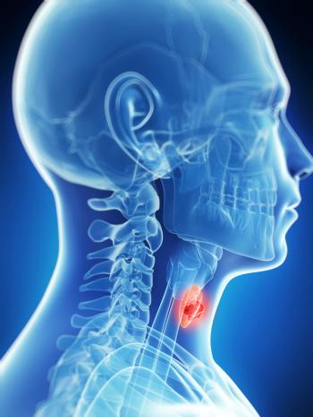
Paragangliomas most commonly occur in the carotid body, jugulotympanicarea, and vagus nerve but have also been reported in otherareas of the head and neck. These tumors are highly vascular andcharacteristically have early blood vessel and neural involvement,making their treatment particularly challenging. Surgery has traditionallybeen the preferred method of treatment, especially in light of recentadvances in technique. However, compared to radiation therapy, it canresult in a higher incidence of cranial nerve dysfunction. Radiationtherapy has the advantage of avoiding the increased morbidity ofsurgery while offering an equal possibility of cure. Part 2 of this articlediscusses radiation therapy as primary treatment of patients who areineligible for surgery and the elderly and infirm. Results with radiotherapyare comparable to those achieved with surgery. The efficacy ofsalvage therapy with either surgery or radiation is discussed, and atreatment algorithm for these tumors is proposed.
Advertisement
ABSTRACT: Paragangliomas most commonly occur in the carotid body, jugulotympanic area, and vagus nerve but have also been reported in other areas of the head and neck. These tumors are highly vascular and characteristically have early blood vessel and neural involvement, making their treatment particularly challenging. Surgery has traditionally been the preferred method of treatment, especially in light of recent advances in technique. However, compared to radiation therapy, it can result in a higher incidence of cranial nerve dysfunction. Radiation therapy has the advantage of avoiding the increased morbidity of surgery while offering an equal possibility of cure. Part 2 of this article discusses radiation therapy as primary treatment of patients who are ineligible for surgery and the elderly and infirm. Results with radiotherapy are comparable to those achieved with surgery. The efficacy of salvage therapy with either surgery or radiation is discussed, and a treatment algorithm for these tumors is proposed.
Part 1 of this two-part series focused on techniques for diagnosing paragangliomas-highly vascular tumors that arise from extra-adrenal portions of the paraganglion system-and the use of surgery as primary treatment. Part 2 reviews radiation therapy as primary treatment of patients with jugular paragangliomas and occasionally carotid body and vagal paragangliomas. We will also address salvage therapy with either surgery or radiation and, finally, will describe a treatment algorithm that we are currently studying.
Radiation Therapy in the Management of Paragangliomas
Radiotherapy has traditionally been relegated to the treatment of unresectable paragangliomas (eg, extensive skull-base involvement or intracranial extension) or of tumors in the medically infirm and elderly. Some physicians have advocated surgery as the only curative option, based on the assumption that paragangliomas are radioresistant and will ultimately progress following radiotherapy. However, the risk of perioperative morbidity and long-term postoperative complications, including cranial nerve dysfunction, can be considerable.
Certainly, surgery may be the preferred treatment of resectable paragangliomas when complete resection entails acceptable morbidity. With technical advances in preoperative embolization, skull-base surgery, and reconstruction, a greater proportion of cases can now be effectively resected. However, if surgery results in significant debility requiring longterm rehabilitation, other effective options should be considered.[1,2]
Observation of these slow-growing tumors may be considered in patients with small and moderate-sized, asymptomatic paragangliomas who can be closely monitored. Alternatively, radiotherapy has proven to be an effective therapeutic option over the past several decades and should be considered as a form of primary treatment. Therapeutic options must always be weighed judiciously, and the choice of treatment is dependent on the physician's experience, the clinical findings, the patient's preference, and the risks of morbidity.
Outcomes With Radiotherapy
Local Control
Both conventionally fractionated radiotherapy and, more recently, stereotactic radiosurgery have been used primarily to treat jugular paragangliomas of the temporal bone, and significantly less frequently for treatment of carotid body and vagal paragangliomas. Successful radiotherapeutic treatment of paragangliomas must be defined because total resolution of the tumor after treatment is rare. Thus, "local control" usually means stability (or regression) of tumor size and no progression (or improvement) of neurologic symptoms.
The first major review to demonstrate the efficacy of radiotherapy was reported in 1962 by Alford and Guildford, who analyzed data from 106 cases of paraganglioma collected from the literature.[3] Although more advanced lesions were treated with radiation, the local control rate was comparable to that of surgery (72% vs 78%). Unfortunately, follow-up was relatively short for a disease with a long natural history.
TABLE 1
Radiotherapy for Head and Neck Paragangliomas
Since then, a multitude of reports with as much as 35 years of follow-up have demonstrated the effectiveness of radiation therapy in controlling head and neck paragangliomas (see Tables 1 and 2).[1,2,4-39] The accumulated data represent a wide variety of dose levels (30 to 70 Gy), radiation techniques (electrons, photons, radium implant), and beam energies (superficial, cobalt, 4 to 6 megavoltage). As shown in Table 1, local control rates range from 65% to 100%, with a mean of 90%, based on more than 1,000 tumors from 35 published series (crude local control = 907/1,009).[1,2,4-36] No obvious difference in local control is detectable between tumors that primarily involve the temporal bone vs those that involve the carotid body and vagus nerve (local control = 97%, as shown in Table 2).[4,10,21,23,37,38] Mean or median follow-up was at least 10 years[4,6-8,11,39,14,22,24,32,35] in many series, with a maximum follow-up of more than 30 years in some.[4,8,10,15,22,35]
TABLE 2
Local Control After Radiotherapy for Carotid Body Tumors and Vagal Paragangliomas
Kim et al reported a local control rate of 90% for 225 temporal bone tumors from 11 radiotherapy series published between 1965 and 1980.[27] Schild et al updated the analysis by including studies published from 1965 to 1992.[9] Following radiotherapy, 91% of patients (337/370) achieved local control. In their report, Kim et al demonstrated a dose-response relationship between local control and the total radiotherapy dose delivered.[27] The optimal dose appeared to be greater than 40 Gy. The local failure rate was 1% vs 25% in patients who received > 40 Gy (n = 142) vs < 40 Gy (n = 83), respectively.
FIGURE 1
Three-Dimensional Treatment Plan
In general, a total dose of 45 Gy delivered over 5 weeks is recommended (Figure 1). However, a schedule of 35 Gy delivered over 3 weeks has also been shown to be effective in a Canadian study.[24] The majority of lesions remain stable in size or show modest regression on radiographic evaluation after the completion of radiotherapy.[ 14,27,40] Paralleling the high rates of local control are diseasespecific survival rates ranging from 65% to 100%, with the majority of series reporting rates of 90% to 100% and overall survival rates of 65% to 100% (Table 1).[1,2,4-39]
The mechanisms by which radiation prevents tumor growth and reduces symptoms are not completely understood because the chief cells can remain long after irradiation.[1,41-43] Radiation most likely induces an obliterative endarteritis with resultant fibrosis, which possibly prevents regrowth in addition to promoting partial tumor involution.[2,44] Whether radiation exerts a primary effect on chief cells is difficult to determine because morphogically intact but persistent cells may lose the ability to reproduce.[45]
Neurologic Results
Limited data demonstrate improvement in symptoms after radiotherapy. The most frequent symptoms associated with temporal bone paragangliomas are hearing loss and tinnitus, which manifest in 55% to 58% and 56% to 66% of patients, respectively.[6,10,13,14,21,24] Following radiotherapy, tinnitus commonly improves or resolves in up to 88% of cases,[10,24] although one report cited improvement of hearing impairment in 35%, with complete resolution in 5%.[24] The incidence of other lower cranial neuropathy at presentation is usually about 40% for temporal bone tumors but may range from 8% to 60%.[6,8-11,14,21]
TABLE 3
Cranial Neuropathy Response After Definitive Radiotherapy
Often, the presenting cranial nerve symptoms are long-standing, with only a modest possibility of improvement after radiotherapy. The reported rates of improvement vary from 0% to 83%, with an average of 35% for all series that document responses (Table 3).[6,9,10,12,14,16,19,21,23, 24,26] Complete restoration of cranial nerve function can occur in about 10% (range: 8% to 20%); however, the majority show no change. Notably, only about 1% of cranial neuropathies worsen after radiotherapy.[10,19,23]
Radiotherapy vs Surgery
Extraction of meaningful data from retrospective studies is inherently fraught with difficulty. Single-institution retrospective series often report conflicting results based on small patient numbers and selection bias. For example, Ogura et al reported a cure rate of 80% (39/49) after surgery alone for jugular paragangliomas vs 25% (3/12) after radiotherapy alone and 64% after combined surgery and radiation.[46] In this series and those from Glasscock et al, surgery was recommended as the treatment of choice for resectable tumors, and radiotherapy was proposed for large, inoperable tumors.[47]
Moore et al reported successful control in 88% (7/8) of tympanic paragangliomas and 25% (1/4) of petrosal/ extrapetrosal tumors treated with surgery alone vs 100% (5/5) of similar tumors treated with radiotherapy alone and 88% of tumors treated with surgery followed by radiotherapy.[48] Other series have demonstrated no obvious differences in response rates following surgery vs radiotherapy.[ 44,49] Carrasco and Rosenman reported a large series showing similar recurrence rates after surgery (7%, 19/283) as after radiotherapy (8%, 24/299).[49] The mortality rate from progressive disease was low following treatment with either modality- 2.5% (7/283) with surgery vs 6% (18/299) with radiation. Due to the retrospective nature of the data, however, the impact of selection bias on this analysis cannot be determined.
TABLE 4
Local Control After Primary Radiotherapy vs Surgery vs Surgery Plus Radiotherapy in Temporal Bone Paragangliomas
• Common Staging System-In an effort to extract meaningful data, outcomes may be compared based on a common staging system. Few studies report their results based on a staging system that can be used for crossmodality and interinstitutional comparison. However, several series used the McCabe/Fletcher staging system (see Table 4 in part 1 of this article, in the June 2003 issue of ONCOLOGY), which categorizes temporal bone paragangliomas as tympanic, tympanomastoid, and petrosal/extrapetrosal (Table 4, above).[4,8,14,15,27] Although this staging system relies on outdated radiographic modalities such as plain x-ray films and retrograde jugulography, it is similar in principle to the Fisch and Glasscock/Jackson systems (Table 4, part 1), which are currently preferred.
A combined analysis of five studies reporting the results of surgical or radiotherapeutic treatment of temporal bone tumors staged according to the McCabe/Fletcher system is presented in Table 4.[4,8,14,15,27] Patients were treated with radiotherapy alone (n = 94), surgery alone (n = 45), or surgery and radiation (n = 39). Patients in the last cohort received radiation, usually in the postoperative setting, for gross residual disease. Median follow-up ranged from 11 to 16 years. The average local control rates were 93%, 78%, and 85% for patients treated with radiotherapy, surgery, or a combination of surgery and radiotherapy, respectively.
Despite a selection bias against the radiotherapy group (65% were group III paragangliomas) compared to the surgery alone (13% were group III) or combined surgery and radiation group (51% were group III), local control appeared to be at least as good after radiotherapy alone as after surgery alone or the combined treatment. Local control for group III tumors was better after definitive radiotherapy (92%) or combined-modality therapy (95%) compared to surgery alone (17%). Patients with early- or intermediate- group tumors did well regardless of the treatment approach.
McCabe and Fletcher recommend surgery alone for tympanic tumors, combined treatment for tympanomastoid tumors, and radiotherapy alone for petrosal or extrapetrosal tumors.[ 22] However, the combined analysis suggests that surgery or radiotherapy alone are equally effective alternatives for group I and II tumors (local control: 87% and 88% after surgery for group I and II tumors vs 93% and 100% after radiation therapy) and that radiotherapy is the preferred treatment for advanced tumors (local control: 92% for group III tumors; see Table 4).[4,8,14,15,27]
Moreover, a combined approach does not appear to improve local control compared to radiotherapy alone. Kim et al also emphasized that there is no obvious benefit to debulking subtotal resection in conjunction with definitive radiotherapy.[27] The pooled analysis encompasses retrospective data from 174 patients and is subject to selection/reporting bias. Nevertheless, it represents a comparison based on a common staging system, with reasonably long-term follow- up. Unfortunately, the respective morbidities of the different treatment groups were not detailed in these studies.
Toxicity of Fractionated Radiotherapy
TABLE 5
Severe Complications After Radiotherapy for Paraganglioma
Severe complications after radiation therapy have been reported for 30 series involving a total of 795 patients and are summarized in Table 5.[1,4-16,18,19,21-33, 35,36] This review spans 37 years of literature (1964-2001) and a wide variety of radiation techniques and doses. The incidence of severe complications is 6% (49/795) and of treatment-related mortality, 0.6% (5/795). Of the five reported deaths, four were the result of brain complications including necrosis and abscess. The majority of such complications were related to the use of excessive doses, outdated treatment techniques (orthovoltage- which is preferentially absorbed in the bone or radium implant), or reirradiation. Of importance is the low incidence (0.4%, 3/795) of radiationinduced secondary malignancy, including two fibrosarcomas occurring 15 and 25 years posttreatment, and one osteosarcoma occurring 5 years later (Table 5).[1,4-16,18,19,21-33,35,36]
With modern treatment planning and appropriate dosing, definitive radiotherapy for paragangliomas should be well tolerated. In the acute setting, patients may experience dermatitis, alopecia, mucositis, epilation, and alterations in taste and saliva along with either otitis externa or media. The potential complications vary according to the radiation technique and site of treatment. At a standard total dose of 45 Gy delivered over 25 fractions, most of these side effects are transient, and the incidence of treatmentrelated mortality is exceedingly low. Chronic toxicities such as osteoradionecrosis and brain complications should also be negligible. Care must be taken to minimize dose inhomogeneity and volume of treated normal tissue, especially brain tissue (Figure 1).
Stereotactic Radiosurgery
TABLE 6
Experience With Stereotactic Radiosurgery in the Treatment of Paragangliomas
Stereotactic radiosurgery (SRS) has been explored in the primary treatment of paragangliomas. The major advantage of stereotactic radiation is that one large dose of radiation (usually 12 to 18 Gy) is delivered to a precisely defined area. The rationale for delivering one large dose is to effectively treat tumors that may be radioresistant while minimizing the amount of normal tissue in the treatment field. A small number of series have demonstrated success using this approach to treat jugular paragangliomas, with no reported local failures except in one series (Table 6).[40,50-54] Treatment appears to be well tolerated with no reported severe chronic toxicities. Although initial results appear promising, follow-up has been short (median: 20 to 38 months), and more experience is needed to define the role of SRS vis vis conventionally fractionated radiotherapy and surgery.
The limitations of SRS include eligibility restrictions, lesion size and location, and a greater risk of geographic miss due to the sharp dose gradient.[50] Tumors may be no greater than 4 cm (to achieve tight dose conformality), and only lesions located above the stereotactic frame may be treated (due to treatment planning constraints). Therefore, SRS may primarily be used to treat skull-based lesions because lesions originating from or extending into the upper neck may not be adequately covered.
Salvage Therapy
Late recurrences have been reported up to 19 years after radiotherapy,[ 4] with the majority recurring within 10 years of radiotherapy (Table 1).[1,2,4-36] Thus, the need for continued long-term surveillance remains important.
Carrasco and Rosenman found that salvage rates are high when local failure occurs after definitive radiotherapy or surgery, based on a review of 24 series comprising 582 patients treated with surgery and/or radiation from 1964 to 1987.[49] Overall, 4% (25/582) died of disease (2.5% [7/283] of surgery patients and 6% [18/299] of radiation patients), and salvage was achieved in 88%. Of the 62 patients with a recurrence after initial resection, 95% (56/59) were salvaged with definitive radiation and 100% (3/3) with additional surgery, for an overall salvage rate of 95% (59/62). Salvage of radiotherapy failures with surgery or additional radiotherapy was successful in 95% (19/ 20) and 71% (25/35), respectively, for an overall rate of 80% (44/55). Thus, the use of either surgery or radiotherapy as initial treatment does not preclude the success of salvage surgery or radiotherapy in tumor recurrence.
Treatment Algorithm
FIGURE 2
Management of Carotid Body Tumors FIGURE 3
Management of Paragangliomas
Based on the outcomes and considerations discussed here, we propose a treatment algorithm (shown in Figures 2 and 3), which we are currently studying on a prospective basis. Patients presenting with the signs and symptoms of paraganglioma should begin with a thorough history and physical examination to evaluate neural function. A positive family history may be strongly indicative of multiple tumors, which should be considered even in sporadic cases. Computed tomography scanning and magnetic resonance imaging are used to evaluate the extent of tumor, especially skull-base or intracranial involvement. These imaging studies will usually identify the presence of unsuspected multiple paragangliomas or the possibility of malignant metastatic regional disease. If necessary, a fine-needle aspiration biopsy can be performed safely to establish a diagnosis. If surgery is contemplated, angiography is indicated to identify tumor blood supply, visualize possible vessel invasion, and evaluate bilateral cerebral blood supply.
Refinements in surgery, interventional radiology, and medical care have resulted in acceptable surgical mortality rates, even among patients with extensive tumors. However, surgical treatment of paragangliomas- especially those with a natural history of early skull-base (eg, jugular paragangliomas) and cranial nerve involvement (eg, vagal paragangliomas)- results in a higher incidence of cranial nerve dysfunction, compared to radiation therapy. Indeed, evidence suggests occasional improvement in cranial nerve function in paragangliomas treated with radiation therapy.
Certainly, lower cranial nerve dysfunction (cranial nerves VII, IX-XII) represents a potentially significant debility, especially if multiple nerves are affected. Although rehabilitative techniques can improve the patient's ability to cope with these problems, it is still better to avoid these debilities if cure can be achieved. Radiation therapy has the advantage of avoiding the increased morbidity of surgery while offering an equal possibility of cure.[4-6] The morbidity of radiation therapy must be considered, but proper treatment planning with appropriate dosing (4,500 cGy over 5 weeks for fractional therapy) can result in minimal side effects.
• Indications for Surgery-Because surgery and radiation therapy demonstrate equal cure rates,[4] as indicated above, the focus should be on the associated morbidity and mortality of each treatment. Primary surgery should be considered for patients with small to moderate-sized carotid body tumors and tympanic paragangliomas without cranial nerve dysfunction. These tumors can usually be totally excised without significant morbidity. Primary surgery is also indicated for patients with jugular or vagal paragangliomas in the presence of lower cranial dysfunction (cranial nerves VII, IX-XII), whereby resection of the tumor would not result in additional significant functional deficits, especially in younger and middle- aged patients without coexisting medical problems. If a diagnosis of malignant paraganglioma is established, tumor resection with a neck dissection is indicated, assuming that there is no evidence of systemic disease. Postoperative radiation therapy is recommended.
• Indications for Radiation Therapy-Primary radiation therapy is currently the accepted treatment for elderly, poor-risk patients with multiple or severe medical conditions, and patients with extensive skull-base or intracranial involvement. Radiation therapy is also indicated in patients with jugular or vagal paragangliomas and no evidence of lower cranial nerve dysfunction and in patients with multiple or bilateral tumors with the potential for severe postoperative debility from cranial nerve dysfunction.[52,55-60] Observation without treatment is also an option for older patients who can be followed closely.[61] Salvage therapy- either surgery for previously irradiated patients or radiation therapy for previously resected patients-has also proven effective in controlling local disease.
Conclusions
As is evident by the complexities associated with the management of paragangliomas, multidisciplinary cooperation is crucial for achieving cure with minimal treatment-related morbidity. These tumors are highly curable, and the majority of patients live for several decades after their initial diagnosis.
References:
1.
Hatfield PM, James AE, Schulz MD:Chemodectomas of the glomus jugulare. Cancer30:1164-1168, 1972.
2.
Spector GJ, Compagno J, Perez CA, etal: Glomus jugulare tumors: Effects of radiotherapy.Cancer 35:1316-1321,1975.
3.
Alford BR, Guildford FR: A comprehensivestudy of tumours of the glomus jugulare.Laryngoscope 72:765-787, 1962.
4.
Hinerman RW, Mendenhall WM, AmdurJ, et al: Definitive radiotherapy in the managementof chemodectomas arising in the temporalbone, carotid body, and glomus vagale.Head Neck 23:363-371, 2001.
5.
Evenson LJ, Mendenhall WM, ParsonsJT, et al: Radiotherapy in the management ofchemodectomas of the carotid body and glomusvagale. Head Neck 20:609-613, 1998.
6.
Mumber MP, Greven KM: Control ofadvanced chemodectomas of the head and neckwith irradiation. Am J Clin Oncol 18:389-391,1995.
7.
Cole JM, Beiler D: Long-term results oftreatment for glomus jugulare and glomus vagaletumors with radiotherapy. Laryngoscope104:1461-1465, 1994.
8.
Larner JM, Hahn SS, Spaulding CA, etal: Glomus jugulare tumors. Long-term controlby radiation therapy. Cancer 69:1813-1817, 1992.
9.
Schild SE, Foote RL, Buskirk SJ, et al:Results of radiotherapy for chemodectomas.Mayo Clin Proc 67:537-540, 1992.
10.
Powell S, Peters N, Harmer C: Chemodectomaof the head and neck: Results of treatmentin 84 patients. Int J Radiat Oncol BiolPhys 22:919-924, 1992.
11.
Verniers DA, Keus RB, SchouwenburgPF, et al: Radiation therapy, an important modeof treatment for head and neck chemodectomas.Eur J Cancer 28A:1028-1033, 1992.
12.
Skolyszewski J, Korzeniowski S, PszonJ: Results of radiotherapy in chemodectoma ofthe temporal bone. Acta Oncol 30:847-849,1991.
13.
Boyle JO, Shimm DS, Coulthard SW:Radiation therapy for paragangliomas of thetemporal bone. Laryngoscope 100:896-901,1990.
14.
Pryzant RM, Chou JL, Easley JD: Twenty-year experience with radiation therapy fortemporal bone chemodectomas. Int J RadiatOncol Biol Phys 17:1303-1307, 1989.
15.
Wang ML, Hussey DH, Doombos JF, etal: Chemodectoma of the temporal bone: Acomparison of surgical and radiotherapeuticresults. Int J Radiat Oncol Biol Phys 14:643-648, 1988.
16.
Friedland JL, et al: Chemodectomasarising in temporal bone structures. Head NeckSurg 10:S53-S55, 1988.
17.
Hansen HS, Thomsen KA: Radiotherapyin glomus tumours (paragangliomas). A 25year-review. Acta Otolaryngol 449(suppl):151-154, 1988.
18.
Konefal JB, Pilepach MW, Spector GJ,et al: Radiation therapy in the treatment ofchemodectomas. Laryngoscope 97:1331-1335,1987.
19.
Dawes PJ, Fillippou M, Welch AR, etal: The management of glomus jugulare tumours.Clin Otolaryngol 12:15-24, 1987.
20.
Zinreich ES, Lee DJ: Radiotherapy forthe treatment of paragangliomas in the temporalbone. Ear Nose Throat J 65:181-184,1986.
21.
Mitchell DC, Clyne CA: Chemodectomasof the neck: The response to radiotherapy.Br J Surg 72:903-905, 1985.
22.
Sharma PD, Johnson AP, Whitton AC:Radiotherapy for jugulo-tympanic paragangliomas(glomus jugulare tumours). J LaryngolOtol 98:621-629, 1984.
23.
Lybeert ML, van Andel JG, EijkenboomWM, et al: Radiotherapy of paragangliomas.Clin Otolaryngol 9:105-109, 1984.
24.
Cummings BJ, Beale FA, Garrett PG, etal: The treatment of glomus tumors in thetemporal bone by megavoltage radiation. Cancer53:2635-2640, 1984.
25.
Reddy EK, Mansfield CM, HartmanGV: Chemodectoma of glomus jugulare. Cancer52:337-340, 1983.
26.
Dickens WJ, Million RR, Cassisi NJ,et al: Chemodectomas arising in temporalbone structures. Laryngoscope 92:188-191,1982.
27.
Kim JA, Elkon D, Lim ML, et al: Optimumdose of radiotherapy for chemodectomasof the middle ear. Int J Radiat Oncol Biol Phys6:815-819, 1980.
28.
Wang CC: Paraganglioma of the headand neck, in Wang CC (ed): Radiation Therapyfor Head and Neck Neoplasms. Chicago,Year Book Medical Publishers, 1990.
29.
Gibbin KP, Henk JM: Glomus jugularetumours in South Wales-a twenty-year review.Clin Radiol 29:607-609, 1978.
30.
Simko TG, Griffin TW, Gerdes AJ, etal: The role of radiation therapy in the treatmentof glomus jugulare tumors. Cancer42:104-106, 1978.
31.
Arthur K: Radiotherapy in chemodectomaof the glomus jugulare. Clin Radiol 28:415-417, 1977.
32.
Tidwell TJ, Montague ED: Chemodectomasinvolving the temporal bone. Radiology116:147-149, 1975.
33.
Newman H, Rowe JF Jr, Phillips TL:Radiation therapy of the glomus jugulare tumor.Am J Roentgenol Radium Ther Nucl Med118:663-669, 1973.
34.
Maruyama Y: Radiotherapy of tympanojugularchemodectomas. Radiology105:659-663, 1972.
35.
Fuller AM, Brown HA, Harrison EG Jr,et al: Chemodectomas of the glomus jugularetumors. Laryngoscope 77:218-238, 1967.
36.
Grubb WB, Lampe I: The role of radiationtherapy in the treatment of chemodectomasof the glomus jugulare. Laryngoscope75:1861-1871, 1964.
37.
Valdagni R, Amichetti M: Radiationtherapy of carotid body tumors. Am J ClinOncol 13:45-48, 1990.
38.
Mendenhall WM, Million RR, ParsonsJT, et al: Chemodectoma of the carotid bodyand ganglion nodosum treated with radiationtherapy. Int J Radiat Oncol Biol Phys 12:2175-2178, 1986.
39.
Hoogenhout J, et al: Surgery and radiotherapyin the management of glomus bodytumors, p 67. Proceedings of the EuropeanSociety of Therapeutic Radiology and Oncology,1990.
40.
Jordan JA, Roland PS, McManus C,et al: Stereotactic radiosurgery for glomusjugulare tumors. Laryngoscope 110:35-38,2000.
41.
Glasscock ME 3rd, Jackson CG, DickinsJR, et al: The surgical management ofglomus tumors. Laryngoscope 89:1640-1651,1979.
42.
Myers EN, Newman J, Kaseff L, et al:Glomus jugulare tumor: A radiographic-histologiccorrelation. Laryngoscope 81:1838-1851,1971.
43.
Gardner G, Cocke EW JR, RobertsonJT, et al: Combined approach surgery for removalof glomus jugulare tumors. Laryngoscope87:665-688, 1977.
44.
Van Miert PJ: The treatment of chemodectomasby radiotherapy. Proc R Soc Med57:946-951, 1964.
45.
Suit HD, Gallager HS: Intact tumor cellsin irradiated tissue. Arch Pathol 78:648, 1964.
46.
Ogura JH, Spector GJ, Gado M: Glomusjugulare and vagale. Ann Otol 87 (5 pt1):622-629, 1978.
47.
Glasscock ME, Haris PF, Newsome G:Glomus tumours: Diagnosis and treatment.Laryngoscope 84:2006-2032, 1974.
48.
Moore G, Yarington CT Jr, ManghamCA Jr: Vagal body tumors: Diagnosis and treatment.Laryngoscope 96:533-536, 1986.
49.
Carrasco V, Rosenman J: Radiation therapyof glomus jugulare tumors. Laryngoscope103(suppl 60):23-27, 1993.
50.
Feigenberg SJ, Mendenhall WM, HinermanRW, et al: Radiosurgery for paragangliomaof the temporal bone. Head Neck24:384-389, 2002.
51.
Liscak R, Vladyka V, Simonova G, etal: Leksell gamma knife radiosurgery of thetumor glomus jugulare and tympanicum. StereotactFunct Neurosurg 70(suppl 1):152-160,1998.
52.
Eustacchio S, Leber K, Trummer M, etal: Gamma knife radiosurgery for glomus jugularetumours. Acta Neurochir 8:811-818,1999.
53.
Foote RL, Coffey RJ, Gorman DA, etal: Stereotactic radiosurgery for glomus jugularetumors: A preliminary report. Int J RadiatOncol Biol Phys 38:491-495, 1997.
54.
Foote RL, Pollock BE, Gorman DA, etal: Glomus jugulare tumor: Tumor control andcomplications after stereotactic radiosurgery.Head Neck 24:332-339, 2002.
55.
Netterville JL, Reilly KM, RobertsonD, et al: Carotid body tumors: A review of 30patients with 46 tumors. Laryngoscope105:115-126, 1995.
56.
Maier W, Marangos N, Laszig R:Paraganglioma as a systemic syndrome: Pitfallsand strategies. J Laryngol Otol 113:978-982, 1999.
57.
Urquhart AC, Johnson JT, Myers EN, etal: Glomus vagale: Paraganglioma of the vagusnerve. Laryngoscope 104:440-445, 1994.
58.
Gardner P, Dalsing MA, Weisberger E,et al: Carotid body tumors, inheritance, and ahigh incidence of associated cervical paragangliomas.Am J Surg 172:196-199, 1996.
59.
Gardner G, Cocke EW Jr, RobertsonJH, et al: Skull base surgery for glomus jugularetumors. Am J Otology (suppl):126-134,1985.
60.
Sillars HA, Fagan PA: The managementof multiple paraganglioma of the headand neck. J Laryngol Otol 107:538-542, 1993.
61.
Glasscock ME 3rd: The history of glomustumors: A personal perspective. Laryngoscope103(suppl 60):3-6, 1993.
Articles in this issue
over 22 years ago
Ovarian Cancer in Elderly Womenover 22 years ago
Commentary (Bryan/Berek): Ovarian Cancer in Elderly Womenover 22 years ago
Recent Advances in Hormonal Therapy for Advanced Prostate Cancerover 22 years ago
Commentary (Balducci): Ovarian Cancer in Elderly Womenover 22 years ago
Treatment of Complications After Breast-Conservation Therapyover 22 years ago
Medicare to Add PET Coverage for Some Thyroid Cancer PatientsNewsletter
Stay up to date on recent advances in the multidisciplinary approach to cancer.
Advertisement
Related Content
Advertisement

Latest CME


Advertisement
Advertisement
Trending on CancerNetwork
1
Modifiable Risk Factors Suggest Potential for Improving Cancer Prevention
2
2026 Tandem Meetings: What’s the Latest Research in Multiple Myeloma?
3
Dato-DXd Receives Priority Review in Unresectable/Metastatic TNBC
4
Barriers to CAR T-Cell Referral and Center Access in Multiple Myeloma
5