

|Articles|August 1, 1997
- ONCOLOGY Vol 11 No 8
- Volume 11
- Issue 8
Parotid Gland Cancer Surgical Practice Guidelines
The Society of Surgical Oncology surgical practice guidelines focus on the signs and symptoms of primary cancer, timely evaluation of the symptomatic patient, appropriate preoperative evaluation for extent of disease, and role of the surgeon in
Advertisement
The Society of Surgical Oncology surgical practice guidelines focuson the signs and symptoms of primary cancer, timely evaluation of the symptomaticpatient, appropriate preoperative evaluation for extent of disease, androle of the surgeon in diagnosis and treatment. Separate sections on adjuvanttherapy, follow-up programs, or management of recurrent cancer have beenintentionally omitted. Where appropriate, perioperative adjuvant combined-modalitytherapy is discussed under surgical management. Each guideline is presentedin minimal outline form as a delineation of therapeutic options.
Since the development of treatment protocols was not the specific aimof the Society, the extensive development cycle necessary to produce evidence-basedpractice guidelines did not apply. We used the broad experience residingin the membership of the Society, under the direction of Alfred M. Cohen,MD, Chief, Colorectal Service, Memorial-Sloan Kettering Cancer Center,to produce guidelines that were not likely to result in significant controversy.
Following each guideline is a brief narrative highlighting and expandingon selected sections of the guideline document, with a few relevant references.The current staging system for the site and approximate 5-year survivaldata are also included.
The Society does not suggest that these guidelines replace good medicaljudgment. That always comes first. We do believe that the family physician,as well as the health maintenance organization director, will appreciatethe provision of these guidelines as a reference for better patient care.
Symptoms and Signs Early-stage disease
- Asymptomatic
- Lump in the parotid region
Advanced-stage disease
- Enlarged cervical lymph nodes
- Rapidly enlarging mass in the parotid region
- Mass in the parotid region that has been present for a long time,withrecent rapid growth
- Facial weakness
- Pressure symptoms in the ear
- Involvement of the skin by a parotid mass
- Pain
Evaluation of the Symptomatic Patient Work-up
- Clinical examination and thorough head and neck examination
- Fine-needle aspiration of the parotid mass in selected patients
- CT scan for large tumors
Appropriate timeliness of surgical referral
- A lump in the parotid region should be considered a parotid tumor unlessproven otherwise.
- If the patient's general condition is satisfactory, all parotid massesshould be surgically removed both for diagnostic and therapeutic purposes.
Preoperative Evaluation for Extent of Disease Physical examination Chest x-ray
CT scan
- Indicated in selected patients to evaluate the extent of disease andthe presence of nodal metastasis
Role of the Surgeon in Initial Management Surgical considerations
- The surgeon's responsibilities include:making a standard parotid incisionand being prepared to do a superficial parotidectomy with identificationand preservation of the facial nerve.
- For most standard masses in the parotid region, surgical therapy includessuperficial parotidectomy with identification and preservation of the facialnerve. In HIV-positive patients, local excision of a lymphoepithelial cystmay be considered.
- If the tumor shows a high-grade malignancy, the deep jugular lymphnodes should be evaluated and performance of a supraomohyoidneck dissectionshould be considered if there are no suspicious nodes.
- If the nodes are clinically apparent, a comprehensive neck dissectionshould be considered.
- If the facial nerve is functioning preoperatively, every attempt shouldbe made to preserve it or, if the tumor is involving the nerve, to graftit.
- If the facial nerve is paralyzed preoperatively, a radical parotidectomyshould be considered. Immediate nerve repair with the greater auricularnerve or sural nerve should be considered.
These guidelines are copyrighted by the Society of Surgical Oncology(SSO). All rights reserved. These guidelines may not be reproduced in anyform without the express written permission of SSO. Requests for reprintsshould be sent to: James R. Slawny, Executive Director, Society of SurgicalOncology, 85 West Algonquin Road, Arlington Heights, IL 60005.
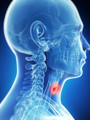
The major salivary glands include the parotid, submandibular, and sublingualglands. In addition, there are approximately 600 to 700 minor salivaryglands distributed throughout the upper aerodigestive tract.
Although salivary gland tumors are not very common, they represent aninteresting clinical entity for which early diagnosis and appropriate treatmentafford the best chance of cure. The incidence of salivary tumors is estimatedat 40 cases per million people. Approximately 75% to 80% of these tumorsinvolve the parotid gland.
The vast majority (80%) of parotid masses are benign, while only 20%are malignant. In contrast, 50% of submandibular salivary tumors and 80%of minor salivary gland tumors are malignant. Of parotid tumors, 90% originatein the superficial lobe of the parotid and only 10% arise from the deeplobe itself
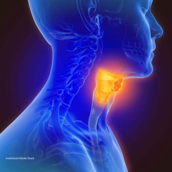
The diagnosis of a parotid gland tumor is based primarily on clinicalexamination. A lump in the parotid region should be considered a parotidtumor unless proven otherwise.
Ancillary diagnostic tests include CT scanning and fine-needle aspirationbiopsy. Computed tomography is very helpful in evaluating the extent ofthe tumor when clinical evaluation of involvement of the deep lobe is difficult.
In general, fine-needle aspiration is unnecessary for superficial parotidtumors. However, if there is a clinical dilemma regarding the extent ofdisease or whether a lesion is of salivary or nonsalivary pathology, fine-needleaspiration is of considerable help. Fine-needle aspiration is also usefulin differentiating a mass in the tail of the parotid from an enlarged lymphnode. It is vitally important to distinguish between a high neck mass andparotid tumor.
The diagnostic accuracy of fine-needle aspiration exceeds 80%. However,the fine-needle aspiration biopsy findings should be critically evaluatedin view of the clinical judgment. Other investigations, such as sialographyand CT sialography, are not commonly used.
The operating surgeon should be prepared to make the appropriate incisionfor parotid exposure and be sufficiently skilled to find the facial nerveand preserve it. Evaluation of facial nerve function is very critical preoperativelyand postoperatively. A functioning facial nerve rarely needs to be sacrificed.
The most common benign tumor is pleomorphic adenoma, which can be easilymanaged by superficial parotidectomy. Other benign tumors, such as lymphoepithelialcysts and oncocytomas, are occasionally seen and are also managed by superficialparotidectomy.
The most common malignant tumor of the parotid is mucoepidermoid carcinoma,which can be either high or low grade. Adenoid cystic carcinoma is a specifichistologic entity with a high incidence of local recurrence and systemicmetastasis.
Several histologic classifications of parotid tumors have been proposed.The simple method devised by Spiro at Memorial Sloan-Kettering Cancer Centeris shown in Table 1, and a more elaborateclassification method is shown in Table 2.
The TNM staging of salivary gland tumors is shown in Table3 on , along with approximate 5-year survival rates by stage. T-stagingof parotid tumors relates directly to the size of the tumor. Each T-stageis subdivided further depending on whether or not there is local extension.
The incidence of lymph node metastasis in all parotid tumors is 15%to 25%. Overall survival is considerably poorer in the presence of lymphnode metastasis.
Rates of 10- and 20-year survival are 90% and 85%, respectively, inpatients with stage I disease and 60% and 55%, respectively, in those withstage II disease. In patients with stage III or IV tumors, the 10-yearsurvival rate is approximately 25% and the 20-year rate is approximately15%.
Survival is also related to tumor grade. Survival is excellent for patientswith low-grade tumors, ranging from 80% to 90% for 10 years. For thosewith high-grade tumors, survival is approximately 25% for 10-year follow-up.
Spiro and colleagues at Memorial Sloan-Kettering have described a differentstaging system that includes the presence of facial nerve dysfunction asan important factor. Facial nerve palsy in patients with parotid tumorsindicates an overall poor prognosis.
The mainstay of treatment of parotid tumors is adequate and appropriatesurgery, which generally includes superficial parotidectomy. If a parotidtumor is locally excised or incised, the likelihood of local recurrenceis high.
Lymphoepithelial cysts are common in HIV-positive patients, and localexcision is adequate in such patients. If the tumor originates in the deeplobe of the parotid, appropriate therapy consists of a superficial parotidectomyand removal of the tumor with preservation of the facial nerve. Preservationof facial nerve function may be very difficult in patients with deep lobeparotid tumors.
If the facial nerve or its branches are already involved with tumorand the patient has facial nerve weakness, the nerve should be sacrificedand reconstruction may be considered. If the proximal stump of thefacial nerve is available and the distal branches can be identified, anerve graft may be used. The greater auricular nerve is readily availablefor grafting. Prior to the surgical intervention, the likelihood of facialnerve weakness must be carefully explained to the patient.
Elective neck dissection is generally not considered. Only 15% to 25%of patients present with lymph node metastasis either at the time of initialdiagnosis or during follow-up. In patients with high-grade, high-stageparotid tumors, supraomohyoid neck dissection may be considered as a stagingprocedure.
Postoperative radiation therapy has been used more commonly recently,except for small parotid tumors. The indications for postoperative radiationtherapy include: high-grade, high stage tumor; lymph node metastasis; skininvolvement. Locoregional recurrence has been significantly reducedby aggressive postoperative radiation therapy.
Specialized centers have used neutron therapy for advanced or recurrentparotid tumors, with satisfactory results.
References:
Armstrong JG, Harrison LB, Spiro RH, et al: Malignant tumors of majorsalivary gland origin: A matched pair analysis of the role of combinedsurgery and postoperative radiotherapy. Otolaryngol Head Neck Surg116:290-293, 1990.
Duncan W, Orr JA, Arnott SJ, et al: Neutron therapy for malignant tumorsof the salivary glands: A report of the Edinburgh experience. RadiotherOncol 8:97-104, 1987.
Fu KK, Leiber SA, Levine ML, Friedlander LM, et al: Carcinoma of themajor and minor salivary glands. Cancer 40:2882-2890, 1977.
Spiro RH: Salivary neoplasms: Overview of a 35 year experience with2807 patients. Head Neck Surg 8:177-184, 1986.
Spiro RH, Huvos AH, Strong EW: Cancer of the parotid gland, a clinicopathologicstudy of 288 primary cases. Am J Surg 130:452-459, 1975.
Articles in this issue
over 28 years ago
Can We Make Low-Fat Foods More Palatable?over 28 years ago
The Prostate Cancer Intervention Versus Observation Trial (PIVOT)over 28 years ago
Practical Tips for Caring for HIV/AIDS Patientsover 28 years ago
The Role of Exercise in the Prevention and Treatment of Cancerover 28 years ago
Pap Smear RefinedNewsletter
Stay up to date on recent advances in the multidisciplinary approach to cancer.
Advertisement
Related Content
Advertisement

Latest CME


Advertisement
Advertisement
Trending on CancerNetwork
1
Modifiable Risk Factors Suggest Potential for Improving Cancer Prevention
2
2026 Tandem Meetings: What’s the Latest Research in Multiple Myeloma?
3
Dato-DXd Receives Priority Review in Unresectable/Metastatic TNBC
4
Barriers to CAR T-Cell Referral and Center Access in Multiple Myeloma
5