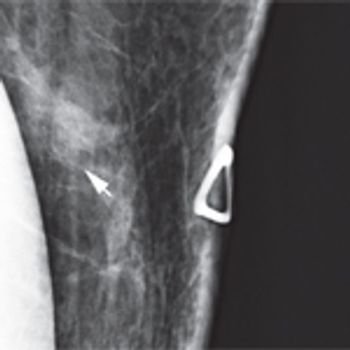
A 46-year-old woman had a routine screening mammogram that showed new calcifications in the posterior left breast. A diagnostic mammogram showed several small punctate calcifications, and a 6-month interval follow-up was recommended.
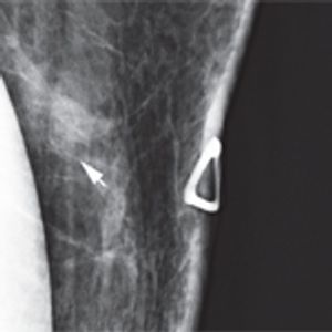
A 46-year-old woman had a routine screening mammogram that showed new calcifications in the posterior left breast. A diagnostic mammogram showed several small punctate calcifications, and a 6-month interval follow-up was recommended.

The Case: A 48-year-old perimenopausal woman noted a lump in her left breast. She had had a mammogram 9 months earlier without abnormality. After ultrasound imaging confirmed a solitary mass measuring about 1.5 cm, a core needle biopsy demonstrated a poorly differentiated mammary carcinoma with chondroid features.
July 15th 2017