|Poll|October 14, 2013
A 51-Year-Old Patient Develops Scaly Plaque on the Right Foot
Author(s)Ted Rosen, MD
Advertisement
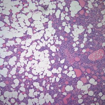
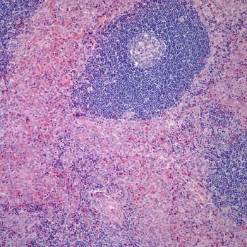
A 51-year-old man slowly developed a 15 cm × 8 cm erythematous, notably indurated, and variably scaly plaque located on the medial and dorsal aspect of the right foot. By the time he sought medical attention, this minimally pruritic lesion had been present about 2.5 years. The rest of the cutaneous examination was unremarkable. Ipsilateral inguinal adenopathy was not detected. Several skin biopsies showed a similar pattern, consisting of hyperkeratosis and acanthosis accompanied by an intraepidermal clonal infiltrate of CD8+ atypical lymphocytes with pleomorphic nuclei, and a dense upper dermal infiltrate composed of more benign-appearing lymphocytes.
This clinical and pathological picture is most consistent with what disease entity?
Advertisement
Related Content
Advertisement


Advertisement
Advertisement
Trending on CancerNetwork
1
Pirtobrutinib Earns Positive CHMP Opinion Across All CLL Treatment Lines
2
Generic Enzalutamide Receives Tentative FDA Approval in Prostate Cancer
3
40 An AI Digital Twin Compares Favorably to Radiologists for Landmark Identification and Measurement in Early-Stage Breast Cancer: A Retrospective, Multi-Institution Clinical Study
4
Cellular Therapy and Antibody-Drug Conjugates Shape Day 1 of ASCO Breakthrough 2026
5