|Articles|October 1, 1997
- ONCOLOGY Vol 11 No 10
- Volume 11
- Issue 10
The Biodistribution and Pharmacokinetics of Stealth Liposomes in Patients with Solid Tumors
Author(s)Simon Stewart, MD, Kevin J. Harrington, MD
Liposomes have been proposed as potential vehicles for drug therapy targeted to solid tumors, in particular, because of the increased permeability of these tumors to macromolecules. The recent development of Stealth
Advertisement
ABSTRACT: Liposomes have been proposed as potential vehicles for drug therapy targeted to solid tumors, in particular, because of the increased permeability of these tumors to macromolecules. The recent development of Stealth liposomes has renewed clinicians interest in liposomal-targeted therapy due to their ability to evade the reticuloendothelial system. Thus they remain in circulation longer with a concomitant increase in duration of exposure to the tumors. We examined the biodistribution of indium 111-labeled Stealth liposomes in 17 patients with a variety of selected malignancies. The median half-life of 111In-labeled liposomes was 55 hours, with a pattern of biodistribution in the liver, spleen, and bone marrow consistent with uptake by the reticuloendothelial system. The high levels of 111In activity seen in some of the tumors suggest that liposomes may also be used to carry therapeutic doses of radiopharmaceuticals. Liposomes have the potential to significantly improve tumor targeting in oncology, and new liposomal drug formulations may advance drug delivery with increased tumor response rates and decreased toxicity. [ONCOLOGY 11(Suppl 11:)33-37, 1997]
Liposomes are colloidal structures composed of a lipid bilayer surrounding an aqueous medium.[1] They can be used to carry a wide spectrum of different compounds and have been postulated as potential vehicles for targeted drug therapy.[2]
Solid tumors, with their increased permeability to macromolecules, colloids, and liposomes, may be particularly susceptible to this form of therapy. These larger molecules can more easily enter tumor blood vessels because of defects between the gap junctions of the vessels endothelial cells.[3,4] Furthermore, the clearance of these larger molecules from the perivascular space is reduced due to both the absence of organized lymphatics and the relatively high interstitial pressure found in tumors. Liposomes trapped in the perivascular space are degraded slowly, gradually releasing their contents to the surrounding environment. Thus they deliver more chemotherapy to tumor cells in the immediately surrounding tissue than to normal cells in the more distant, healthy tissue.
One of the main disadvantages of conventional liposomes is that their lipid bilayer attracts plasma proteins and other opsonins onto their surfaces, leading to structural instability and leakage of their contents into the circulation. Protein-coated liposomes are rapidly cleared by the reticuloendothelial system,[5] and the plasma half-life of conventional liposomes is approximately six hoursan insufficient amount of time to efficiently access the tumor vasculature.
Stealth liposomes were devised to correct these instability problems and thus their development has renewed clinicians interest in liposomal-targeted therapy.[6-8] The lipid bilayer of Stealth liposomes is coated with linear segments of polyethylene glycol, a hydrophilic polymer. This polymer causes a hydrated shell to form around the external surface of the lipid bilayer, thus creating a steric barrier against interactions with plasma proteins, opsonins, and cell surface receptors. Consequently, Stealth liposomes are able to evade the reticuloendothelial system and remain in the circulation longer. Their reported plasma half-lives of about 40 to 60 hours afford them a greater opportunity to interact with the tumor vasculature.[9]
Abundant evidence supports the concept of Stealth technology. Liposome accumulation has been demonstrated in a variety of xenograft tumors in nude mice.[4] Both doxorubicin and cisplatin are found to be considerably more cytotoxic in xenograft models when given as liposomal formulations than as free drugs.[10]
In this article, we discuss clinical studies performed with two liposomal anthracycline formulations: Dauno-Xomedaunorubicin in a conventional liposome, and Doxildoxorubicin in a stealth liposome. Both anthracycline formulations are active against HIV-related Kaposis sarcoma and have been compared with the combination of doxorubicin (Adriamycin), bleomycin, (Blenoxane), and vincristine (ABV) chemotherapy regimens in separate phase III trials. Liposomal daunorubicin, at an intravenous dose of 40 mg/m2 every two weeks, was shown to be approximately as effective as ABV (25% vs 28%, respectively) with significantly less toxicity.[11]
Pegylated liposomal doxorubicin (PEG-LD), at a dose of only 20 mg/m2 every three weeks, was twice as effective as the ABV combination chemotherapy regimen (response rate of 58% vs 23%).[12] Similar results were observed in a European phase III study where PEG-LD was compared with a regimen of bleomycin and vincristine (BV).[13] The activity of PEG-LD against solid tumors has, thus far, only been described in preliminary reports in metastatic breast carcinoma[14] and relapsed ovarian cancer.[15]
Designing clinical studies is difficult due to the paucity of data on the biodistribution of liposomes in patients with solid tumors. Tumor targeting in human cancers has not yet been confirmed, let alone quantified. In addition, it is not known which tumor sites or histological subtypes possess the leaky vasculature necessary for successful liposomal localization or over what time period this localization occurs.
The little we know about biodistribution in humans comes from studies using conventional liposomes; even less information exists on the distribution of the more promising Stealth liposomes. In order to answer these questions, we have conducted a preliminary study to examine the biodistribution of indium 111-labeled Stealth liposomes in patients with selected malignancies.
Patient Population
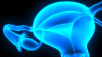
Seventeen patients with a variety of locally advanced biopsy-confirmed malignancies, and who were referred for radiotherapy, were entered into this study. The sites of malignancy were as follows: head and neck cancer (5 patients), breast cancer (5 patients), non-small-cell lung cancer (4 patients), high-grade glioma (2 patients), and carcinoma of the cervix (1 patient).
111In-Labeled Stealth Liposomes
Stealth liposomes containing diethylenetriaminepentacetic acid (DTPA) were supplied by Sequus Pharmaceuticals Inc., Menlo Park, CA and stored at -20°C. These liposomes were of the same composition as those used to encapsulate doxorubicin for the Doxil formulation. They measured approximately 90 nm in diameter and contained approximately 55% soybean phosphatidylcholine, 40% cholesterol, 5% polyethyleneglycol derivatized distearoylphosphatidylethanolamine, and a small quantity of a-tocopherol.
A 20 mL solution of these liposomes was thawed to room temperature and then radiolabeled by the addition of 2 mL of 111In oxine solution for a 15-minute incubation period. During the incubation, the DTPA inside the liposomes chelated and trapped 111In diffusing across the lipid bilayer. Any free 111In left in solution outside the liposomes was in turn chelated by the addition of 3.8 mg of ethylenediaminetetraacetic acid (EDTA) in a 5% solution of dextrose. This step ensured that any injected 111In not encapsulated by liposomes would be chelated by EDTA and rapidly excreted in the urine. The labeling efficiency was assayed before injection by separating a 10 µL aliquot of the liposome solution on a Sephadex G50 column.
Pharmacokinetics
Patients received between 65 and 107 MBq of 111In-labeled Stealth liposomes, diluted to 500 mL in 5% dextrose, as a 45-minute intravenous infusion. Blood was taken for 111In activity at 30 minutes, and again at 4, 24, 48, 72, 96, and 240 hours. Serial 24-hour urine collections were taken to monitor 111In excretion over the first 96 hours.
Scintigraphy
Gamma camera scintigraphy was performed on an MS-2 dual-headed gamma camera using high resolution, medium energy collimators. Whole body images were acquired at 6 cm per minute. Before the injection of 111In-labeled liposomes, the tissue attenuation of gamma radiation was compensated by performinga whole body transmission scan using a 57Co source. Whole body scans were undertaken at 0.5, 4, 24, 48, 73, 96, and 240 hours after injection. Single-photon emission computed tomography (SPECT) and static imaging of regions of interest were performed when appropriate. The tumor and normal organ 111In content was estimated using a validated regions of interest method from the scintigraphy images.
Pharmacokinetics
The half-life of 111In-labeled liposomes ranged from 40 to 103 hours (median, 60 hours). The free 111In chelated to EDTA was rapidly excreted in the urine in the first 4 hours. Following this elimination, there was a gradual excretion of 111In with a range of 4% to 28% of the injected dose being excreted within the first 96 hours.
Normal Tissue Biodistribution
Sequential whole body gamma camera scintigrams revealed a consistent pattern of biodistribution (Figure 1). An early blood pool image was seen at 30 minutes by which time the EDTA-bound 111In was clearly seen in the urinary tract and bladder. The blood pool 111In activity then declined slowly in keeping with the long biological retention time of Stealth liposomes in the circulation. Over the next 72 hours, prominent 111In activity was noted in the liver, spleen, and bone marrow, but no residual activity in the urinary system was seen. This pattern of biodistribution was consistent with uptake by the reticuloendothelial system.
Tumor Biodistribution
Twelve of the 17 tumors were imaged on whole body gamma camera scintigraphy, and using SPECT, three tumors were seen 72 hours after injection. The tumors were best visualized 48 to 96 hours postinjection when the blood pool was decreasing but the tumor localization was still increasing (Figure 2).
A wide heterogeneity was apparent in 111In concentration among the 15 different tumors, with 111In ranging from 0.5% to 4% of the injected dose per tumor. All five patients with head and neck tumors had strikingly positive scans (Figure 1), whereas patients with breast tumors tended to have less 111In concentration. The 111In activity in head and neck tumors was high enough for these lesions to still be detected on plain scintigraphy 10 days after injection.
This study supports the concept of using liposomal-targeted therapy for treatment of solid tumors. The liposomes used in this study were retained in the circulation for many days despite some retention by the reticuloendothelial system. This long plasma half-life permitted extended access to the tumor vasculature and thus tumor localization was seen in 15 of the 17 tumors. The data from this study indicate that tumor targeting occurs for several days after injection, with tumor localization evident for up to 10 days after injection. The inability to detect tumor localization at later times in this study was due to loss of signal from the radioactive decay of 111In.
The use of Stealth liposomes as vehicles for drug delivery offers exciting avenues for further research. The present study indicates that liposomes may deliver encapsulated cytotoxic drugs to solid tumors at higher concentrations than to the surrounding normal tissue. It is expected that an increase in response rate and a decrease in systemic toxicity accompany the increased tumor exposure to the contained cytotoxic agents.
The increased exposure of tumor to liposomal chemotherapy would explain the impressive response rates seen in HIV-related Kaposis sarcoma.[16] However, active uptake by the reticuloendothelial system exists, and myelosuppression might still be a dose-limiting toxicity. The uptake by the reticuloendothelial system probably accounts for the increased myelosuppression seen when PEG-LD was compared with the BV regimen.[13]
This study demonstrated considerable heterogeneity in the concentration of liposomes in a variety of tumor types. While one can speculate that tumors able to concentrate liposomes would be more amenable to liposomal chemotherapy than tumors with poor liposome localization, an exact correlation between the concentration of liposomes in tumors and the clinical efficacy of liposomal chemotherapy is not yet known.
A subgroup of tumors with poor or non-leaky vasculature, unable to concentrate liposomes, would account for our inability to demonstrate liposome targeting in two of our patients. Tumors without vascular access would not be expected to respond to liposomal chemotherapy because the active drug remains in the liposomes in the circulatory and reticuloendothelial systems, unable to reach the tumor tissue. This feature may have important implications in the design of future clinical studies.
In addition to carrying conventional cytotoxic drugs, Stealth liposomes have the potential to deliver a variety of different agents to solid tumors. One conceivable application to exploit the impressive local concentration of liposomes in tumors is to use them to carry radiosensitizing agents during radiotherapy. Radiosensitizing agents, such as the halogenated pyrimidines (BUdR and IUdR), are known to be effective radiation sensitizers but fail in clinical practice because they also sensitize the surrounding normal mucosa. Stealth liposomes may allow for the delivery of these and other radiosensitizing agents selectively to tumors without the concomitant sensitization of the surrounding normal tissue. This concept of differential radiosensitization is clearly attainable as illustrated in Figure 1 and Figure 2 where both tumors are specifically targeted when compared with the surrounding normal tissue.
The high levels of 111In activity seen in some of the tumors studied herein also suggest that liposomes may be used to carry therapeutic doses of radiopharmaceuticals as an enhancement to radiation when combined with external beam radiotherapy. Preliminary calculations suggest that it may be feasible to deliver a radiation dose of about 10 Gy to some tumors with minimal radiation dose to the surrounding normal tissue.
In conclusion, this study and others suggest that liposomes are on the threshold of making major contributions to tumor targeting in oncology. New liposomal drug formulations may improve drug delivery with increased tumor response rates and decreased toxicity. Future liposomal therapy may include their use in the delivery and enhancement of radiotherapy.
References:
1. Lasic DD, Papahadjopoulis D: Liposomes revisited. Science 267:1275-1276, 1995.
2. Gregoriades G, Swain CP, Wills EJ, et al: Drug-carrier potential of liposomes in cancer chemotherapy. Lancet 1:1313-1316, 1974.
3. Huang SK, Lee K-D, Hong K, et al: Microscopic localization of sterically stabilized liposomes in colon carcinoma-bearing mice. Cancer Res 52:5135-5143, 1992.
4. Huang SK, Martin FJ, Jay G, et al: Extravasation and transcytosis of liposomes in Kaposis Sarcoma-like dermal lesions of transgenic mice bearing the HIV tat gene. Am J Pathol 143:1-14, 1993.
5. Gabizon AA: Liposomal anthracyclines. Hematol Oncol Clin North Am 8:431-450, 1994.
6. Allen TM, Chonn A: Large unilamellar liposomes with low uptake by the reticulo-endothelial system. FEBS Lett 223:42-46, 1987.
7. Woking PK, Newman MS, Huang SK et al: Pharmacokinetics, biodistribution and therapeutic efficacy of doxorubicin encapsulated in STEALTH liposomes (Doxil). Liposome Res 4:667-687, 1994.
8. Papahadjopoulos D, Allen TM, Gabizon A, et al: Sterically stabilized liposomes: Improvements in pharmacokinetics and anti-tumor therapeutic efficiency. Proc Natl Acad Sci USA 85:6949-6953, 1988.
9. Gabizon A, Castane R, Uziely B, et al: Prolonged circulation time and enhanced accumulation in malignant exudates of doxorubicin encapsulated in polyethylene-glycol coated liposomes. Cancer Res 54:987-992, 1994.
10. Huang SK, Mayhew E, Giliani S, et al: Pharmacokinetics and therapeutics of sterically stabilized liposomes in mice bearing C 26 colon carcinoma. Cancer Res 52: 6774-6781, 1992.
11. Gill PS, Scadden DT, Cohen P et al: Randomized phase III trial of liposomal daunorubicin versus doxorubicin, bleomycin, and vincristine in AIDS-related Kaposis Sarcoma. J Clin Oncol 14:2353-2364, 1996.
12. Northfelt DW: Liposomal anthracycline chemotherapy in the treatment of AIDS-related Kaposis sarcoma, page 21 (this issue).
13. Stewart S, Jablonowski H, Goebel FD, et al: A randomized comparative trial of liposomal doxorubicin versus bleomycin and vincristine in the treatment of AIDS-related Kaposis sarcoma. J Clin Oncol, 1996 (in press).
14. Ransom M, OByrne K, Carmichael J et al: Phase II dose-finding trial of Dox-SL (Stealth Liposomal Doxorubicin Hcl) in the treatment of advanced breast cancer (abstract). Proc Amer Soc Clin Oncol 15:a161, 1996.
15. Muggia F, Hainsworth S, Jeffers S, et al: Liposomal doxorubicin (Doxil) is active against refractory ovarian cancer (abstract). Proc Amer Soc Clin Oncol 15:a781, 1996.
16. Harrison M, Tomlinson ND, Stewart S: Liposomal entrapped doxorubicin: An active agent in AIDS related Kaposis sarcoma. J Clin Oncol 13:914-920, 1995.
Articles in this issue
over 28 years ago
Vinorelbine in Non-Small-Cell Lung Cancerover 28 years ago
Paclitaxel and Vinorelbine in Non-Small-Cell Lung Cancerover 28 years ago
Safety Data From North American Trials of Vinorelbineover 28 years ago
Cisplatin Alone vs Cisplatin Plus Vinorelbine in Stage IV NSCLCover 28 years ago
Current Management of Unresectable Non-Small-Cell Lung Cancerover 28 years ago
The Economics of Prostate Cancer ScreeningAdvertisement
Related Content
Advertisement


Advertisement
Advertisement
Trending on CancerNetwork
1
FDA Approves Afami-cel in Metastatic Synovial Sarcoma
2
The Moonlight Shift: Dr Joshua Richter on Myeloma, Myths, and the C-Word
3
Current Treatment Landscape of HER2-Positive Metastatic Breast Cancer and Phase 3 Trial Overview
4
How Are Key Lymphoma Data From ASCO and EHA 2026 Shaping Treatment?
5