
|Articles|November 1, 2006
- ONCOLOGY Vol 20 No 12
- Volume 20
- Issue 12
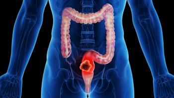
Cancer of the Cervix: Current Management and New Approaches: Review 3
Author(s)Beth Erickson, MD
This article summarizes the current management of patients with newly diagnosed cervical cancer. The topics range from the management of early-stage disease to the phase III randomized studies that have established the current standard of care for patients with locally advanced cancer of the cervix. New approaches to combined-modality therapy with the goal of improving outcomes and decreasing complications are also described.
Advertisement
I have read with interest the paper entitled, "Cancer of the Cervix: Current Management and New Approaches," in this issue of ONCOLOGY. Shivnani et al have written a nice review of contemporary management of cervical cancer, focusing in large part on radiation therapy. Their consideration of evolving technologies and practice patterns give rise to a number of questions.
Chemotherapy Issues
Although chemotherapy has become standard therapy since 1999 for women with cervical cancer, its use and efficacy in both early and advanced disease remain in question. The five US trials discussed by Shivnani et al were diverse in terms of patient entry.[1-5] The study by Rose et al offered the only evidence of benefit of combined therapy in women with stage III or IVA cancer.[4,6] Morris et al found no survival benefit with chemoradiation for patients with stage III or IVA disease.[2] The only US trial to include stage IB/IIA disease treated with chemoradiation without surgery was Radiation Therapy Oncology Group (RTOG) 9001.[2] These patients had positive pelvic lymph nodes or tumors greater than 5 cm in diameter and appeared to benefit from the addition of chemotherapy. That said, we still need to question the role of chemotherapy with small-volume node-negative IB/IIA tumors.
Use of chemotherapy appears to increase survival but also toxicity. More focused selection criteria need to be determined to exclude patients who will not benefit. The doses of chemotherapy given with radiation are meant specifically to enhance the effect of radiation, rather than truly being systemic doses. If a patient has nonbulky pelvic disease, this may not be needed. In contrast, patients with bulky disease need chemotherapy both as a radiation sensitizer and as a systemic agent. The absence of systemic doses to address micrometastases may explain why there was no improvement in survival in stage III and IV disease in RTOG 9001.
Role of Imaging
In the five randomized cervical cancer studies that proved the efficacy of chemotherapy, eligible patients were required to have either surgical staging or a lymphangiogram to rule out occult para-aortic metastases. Unfortunately, off study, clinicians have been using computed tomography (CT) alone as proof or lack thereof of para-aortic disease. CT has a sensitivity of 34% and a specificity of 96%, whereas lymphangiography has a sensitivity of 79% and specificity of 73%.[7] Morris et al used lymphangiography or surgical staging to rule out para-aortic disease, but few institutions have the ability to perform and interpret these studies.[2] Surgical staging is probably the most rigorous method to rule out nodal disease if a sufficient number of nodes are submitted and sectioned properly.
Implementation of positron-emission tomography (PET) scanning prior to therapy may be the best imaging test for detecting para-aortic disease.[8-12] Grigsby et al have proven that the accuracy of PET in detecting subclinical para-aortic as well as supraclavicular disease was much greater than that of CT. With PET, we can identify patients with para-aortic disease and offer para-aortic irradiation rather than submitting them to upfront surgery and/or concurrent chemoradiation to the pelvis alone. For para-aortic positive patients, sequencing with chemotherapy remains important, as does dose escalation with intensity-modulated radiation therapy.[13-16] Future research will need to define a more effective paradigm for treating patients with para-aortic disease to improve survival-without excessive late toxicity. Radioprotection with agents such as amifostine (Ethyol) may also help.
Surgery vs Radiation
The choice of surgery vs radiation in early cervical cancer remains very physician-specific. Our surgical colleagues are not inclined to use imaging prior to pelvic exploration. Data on magnetic resonance imaging (MRI) demonstrates a much better ability to quantify tumor bulk and spread into the parametrium than physical examination alone.[17-20] PET can identify patients with occult lymph node disease, perhaps avoiding the delay of an aborted hysterectomy for these patients and allowing them to proceed with more appropriate chemoradiation.
Brachytherapy practice patterns are shifting in the United States. Cesium sources are becoming more difficult to buy due to decreased production. The Patterns of Care studies have shown that low-dose-rate (LDR) rather than high-dose-rate (HDR) brachytherapy has predominated in the US to date. The most recent (1996-1999) Patterns of Care study demonstrated that 27.8% of patients were treated with LDR, 13.3% with HDR, and 0.9% with a combination of LDR and HDR therapy. These statistics are similar to what was seen in the 1992-1994 survey.[21,22] Now that LDR options are decreasing, more radiation oncologists will need to consider either pulsed-dose-rate or HDR brachytherapy techniques. Despite the frequent use of this modality in Asia, India, and Europe, there is little consensus as to the optimum dose fractionation scheme. The Gynecologic Oncology Group and RTOG are now allowing HDR techniques for national protocols. There is hope that this will lead to a more in-depth analysis of outcome for HDR in the United States.
As HDR becomes more pervasive, it will be even more important to understand the dose distribution in normal organs as well as in the tumor during brachytherapy. Image-guided brachytherapy has been a long time coming for gynecologic cancer (unlike its established use in prostate cancer). MRI is especially helpful during brachytherapy due to its soft-tissue resolution revealing tumor and its extensions as well as the critical normal organs. Use of MRI for brachytherapy imaging has been difficult because of applicator and software issues. After years of delay, these factors are finally being resolved. With the advent of MRI simulators, it will be much more likely that image-guided therapy with each insertion will be feasible.
Image-Based Treatment Planning
Guidelines for image-based treatment planning have been carefully developed by the GEC-ESTRO Gynecologic Working Group.[23,24] These have been shared with investigators in the US, leading to the formation of the Transatlantic Image-Guided Brachytherapy Group. Further collaborative efforts will produce the necessary data to validate these guidelines. The Vienna Group has already demonstrated an increase in local control for bulky tumors and a decrease in complications with better coverage of tumor and exclusion of normal tissues.[23,25]
Appropriate imaging for specific problems in cervical cancer can give us important pivotal information that can change patient outcome. Though tradition is rich in the treatment of cervical cancer, it is important to step into the future with new approaches that can enhance care and quality of life.
-Beth A. Erickson, MD
References:
1. Keys H, Bundy B, Stehman F, et al: Cisplatin, radiation, and adjuvant hysterectomy compared with radiation and adjuvant hysterectomy for bulky stage IB cervical carcinoma. N Engl J Med 340:1154-1161, 1999.
2. Morris M, Eifel P, Lu J, et al: Pelvic radiation with concurrent chemotherapy compared with pelvic and para-aortic radiation for high risk cervical cancer. N Engl J Med 340:1137-1143, 1999.
3. Peters III W, Liu P, Barrett II R, et al: Concurrent chemotherapy and pelvic radiation therapy compared with pelvic radiation therapy alone as adjuvant therapy after radical surgery in high risk early-stage cancer of the cervix. J Clin Oncol 18:1606-1613, 2000.
4. Rose P, Bundy B, Watkins E, et al: Concurrent cisplatin-based radiotherapy and chemotherapy for locally advanced cervical cancer. N Engl J Med 340:1144-1153, 1999.
5. Whitney C, Sause W, Bundy B, et al: Randomized comparison of fluorouracil plus cisplatin vs. hydroxyurea as an adjunct to radiation therapy in stage II B-IVA carcinoma of the cervix with negative para-aortic lymph nodes: A Gynecologic Oncology Group and Southwest Oncology Group study. J Clin Oncol 17:1339-1348, 1999.
6. Rose P, Eifel P: Combined radiation therapy and chemotherapy for carcinoma of the cervix. Cancer J 7:86-94, 2001.
7. Heller P, Malfetano J, Bundy B, et al: Clinical-pathologic study of stage IIB, III, IVA carcinoma of the cervix: Extended diagnostic evaluation for paraaortic node metastasis- A Gynecologic Oncology Group study. Gynecol Oncol 38:425-430, 1990.
8. Grigsby P, Heydon K, Mutch D, et al: Long-term follow-up of RTOG 92-10: Cervical cancer with positive para-aortic lymph nodes. Int J Radiat Oncol Biol Phys 51:982-987, 2001.
9. Grigsby P, Siegel B, Dehdashti F, et al: Lymph node staging by positron emission tomography in patients with carcinoma of the cervix. J Clin Oncol 19:3745-3749, 2001.
10. Rose P, Adler L, Rodriguez M, et al: Position emission tomography for evaluating para-aortic nodal metastasis in locally advanced cervical cancer before surgical staging: A surgicopathologic study. J Clin Oncol 17:41-45, 1999.
11. Lin W, Hung Y, Yeh L, et al: Usefulness of 18F-fluorodeoxyglucose positron emission tomography to detect para-aortic lymph nodal metastasis in advanced cervical cancer with negative computed tomography findings. Gynecologic Oncology 89:73-76, 2003.
12. Tsai CS, Chang TC, Lai CH, et al: Preliminary report of using FDG-PET to detect extrapelvic lesions in cervical cancer patients with enlarged pelvic lymph nodes on MRI/CT. Int J Radiat Oncol Biol Phys 58:1506-1512, 2004.
13. Kavanagh B, Schefter T, Wu Q, et al: Clinical application of intensity-modulated radiotherapy for locally advanced cervical cancer. Semin Radiat Oncol 12:260-271, 2002.
14. Portlance L, Chao C, Grigsby P, et al: Intensity-modulated radiation therapy (IMRT) reduces small bowel, rectum, and bladder doses in patients with cervical cancer receiving pelvic and para-aortoc irradiation. Int J Radiat Oncol Biol Phys 51:261-266, 2001
15. Ahmed R, Kim R, Duan J, et al: IMRT dose escalation for positive para-aortic lymph nodes in patients with locally advanced cervical cancer while reducing dose to bone marrow and other organs at risk. Int J Radiat Oncol Biol Phys 60:505-512, 2004.
16. Mutic S, Malyapa R, Grigsby P, et al: PET-guided IMRT for cervical carcinoma with positive para-aortic lymph nodes-a dose ecalation treatment planning study. Int J Radiat Oncol Biol Phys 55:28-35, 2003.
17. Hricak H, Lacey C, Sandles L, et al: Invasive cervical carcinoma: Comparison of MR Imaging and surgical findings. Radiology 166:623-631, 1988.
18. Hricak H, Yu K: Radiology in invasive cervical carcinoma. AJR Am J Roentgenol 167:1101-1108, 1996.
19. Rubens D, Thornbury J, Angel C, et al: Stage IB cervical carcinoma: Comparison of clinical, MR, and pathologic staging. AJR Am J Roentgenol 150:135-138, 1988.
20. Greco A, Mason P, Leung AWL, et al: Staging of carcinoma of the uterine cervix: MRI-surgical correlation. Clin Radiol 40:401-405, 1989.
21. Erickson B, Eifel P, Moughan J, et al: Patterns of brachytherapy practice for patients with carcinoma of he cervix (1996-1999): A patterns of care study. Int J Radiat Oncol Biol Phys 63:1083-1092, 2005.
22. Eifel P, Moughan J, Owen J, et al: Patterns of radiotherapy practice for patients with squamous carcinoma of the uterine cervix: Patterns of care study. Int J Radiat Oncol Biol Phys 43:351-358, 1999.
23. Haie-Meder C, Potter R, Van
Limbergen E, et al: Recommendations from gynaecological (GYN) GEC-ESTRO Working Group (I): Concepts and terms in 3D image-based 3D treatment planning in cervix cancer brachytherapy with emphasis on MRI assessment of GTV and CTV. Radiother Oncol 44:235-245, 2005.
24. Potter R, Haie-Meder C, Van
Limbergen E, et al: Recommendations from gynaecological (GYN) GEC ESTRO Working Group (II): Concepts and terms in 3D image-based treatment planning in cervix brachytherapy-3D dose volume parameters and aspects of 3D image-based anatomy, radiation physics, radiobiology. Radiother Oncol 78:67-77, 2006.
25. Potter R, Dimopoulos P, Georg S, et al: Impact of systemic MRI assisted 3D treatment planning on local control and morbidity in cervix cancer: Vienna experience in 145 patients treated by intracavitary +/- interstitial HDR brachytherapy from 1998-2003. Radiother Oncol 81(suppl 1):S109, 2006.
Articles in this issue
over 19 years ago
Outliers in Testicular Cancer Managementover 19 years ago
Further Thoughts on a Rare Entityover 19 years ago
'DES Daughters' at Higher Risk for Breast Cancerover 19 years ago
FDA Approves Bevacizumab Plus Chemotherapy to Treat NSCLCover 19 years ago
First Oral Liquid Formulation of Tamoxifen Launchedover 19 years ago
Pulmonary Carcinoid Tumors: The Need for Tailored Assessmentover 19 years ago
Management of Difficult Germ-Cell Tumors: Review 2Advertisement
Related Content
Advertisement


Advertisement
Advertisement
Trending on CancerNetwork
1
Risvutatug Rezetecan Improves Overall Survival in Relapsed SCLC Trial
2
Dostarlimab Yields Sustained Complete Responses in dMMR/MSI-H Rectal Cancer
3
FDA Accepts NDA for Mezigdomide Combo in R/R Multiple Myeloma
4
Clinical Scenario - Triple-Negative Breast Cancer
5