In the United States, cancer of the pancreas accounts for nearly 40,000 deaths annually and is the fourth leading cause of cancer-related mortality. The vast majority of patients present with metastatic or unresectable disease. Only 20% of patients are candidates for surgery, and therefore curable. The 5-year survival rate for patients with pancreatic adenocarcinoma is only 6%, with surgical resection being essential for long-term survival. Recent research has identified a precise subset of patients with borderline resectable pancreatic cancer for whom resection yielding durable survival is possible. This population is being actively studied to identify optimal treatment strategies for long-term survival. In this article we will discuss the definitions of resectability, describe the current diagnostic tests for pancreatic cancer, and review strategies for maximizing treatment outcomes in patients with resectable pancreatic cancer.
Introduction
Pancreatic ductal adenocarcinoma persists as a highly lethal disease. Multidisciplinary care that includes surgical resection is the only treatment associated with long-term survival; however, only approximately 20% of patients present with surgically resectable disease.[1,2] The ability of neoadjuvant therapy to reduce both tumor size and the extent of locally advanced disease has improved the treatment outcomes associated with many solid malignancies; this approach has been studied over the past 20 years in patients with pancreatic cancer, and is now being actively investigated. Until the early 2000s, pancreatic adenocarcinomas were defined as being resectable, locally advanced, or metastatic.[3] However, in the past decade, along with recent advances in pancreatic imaging, surgical technique, and-perhaps most importantly-multimodality treatments for pancreatic cancer, a distinct subset of patients has emerged: those with borderline resectable pancreatic cancer.[4] Unlike most other solid malignancies, this distinct category of pancreatic cancer is defined not only by anatomic parameters, but also by the biologic and physiologic state (eg, comorbidities) of the individual patient.
The identification of borderline resectable pancreatic cancer as a distinct clinical entity serves to clarify a divergence along the continuum between technically resectable and locally advanced unresectable cancers. It separates out the subgroup of patients for whom a margin-negative (R0) resection is difficult to achieve without major vascular resection, but who are not truly unresectable from anatomic and biologic standpoints. Patients with resection margin–positive pancreatic tumors have poor overall survival and do not benefit from chemoradiation therapy.[5,6] Multiple studies have demonstrated the association of margin-positive resection with early recurrence and more aggressive pancreatic disease. Therefore, categorizing the resectability of a pancreatic adenocarcinoma is a critical goal of the initial patient evaluation.[7]
As we will discuss, there is currently no uniform definition of borderline resectable pancreatic cancer, and consensus on this diagnostic entity continues to evolve. Many retrospective studies and clinical trials continue to group patients with borderline resectable disease together with patients who have locally advanced disease. Future studies will likely separate the two groups and define them as distinct entities of pancreatic adenocarcinoma. The goal of combining systemic chemotherapy, local radiation therapy (RT), and other local ablative therapies is to optimize clinical outcomes for patients with borderline resectable pancreatic cancer, by achieving negative surgical margins and ultimately improving overall survival. In recent years, biologic and physiologic parameters have been incorporated into the pancreatic cancer staging system, due to the efforts of forward-thinking leaders in the field of pancreatic cancer and the growing recognition of high rates of distant metastatic recurrence after surgical resection of early-stage pancreatic adenocarcinoma.[8,9]
Defining Borderline Resectable Pancreatic Cancer
The current ambiguity of the anatomic definition of borderline resectable pancreatic cancer is evident from the four major published definitions of resectability, developed separately by the National Comprehensive Cancer Network (NCCN), MD Anderson Cancer Center (MDACC), the Americas Hepato-Pancreato-Biliary Association (AHPBA)/Society of Surgical Oncology (SSO)/Society for Surgery of the Alimentary Tract (SSAT), and the Alliance for Clinical Trials in Oncology (Table 1). Therefore, in interpreting results of clinical studies of pancreatic cancer management published in the medical literature, one must be mindful of which guidelines have been followed. As an example, compared with the other three sets of guidelines, the AHPBA/SSO/SSAT criteria are the most conservative, with “borderline” disease classified as any abutment of the superior mesenteric vein (SMV), whereas the NCCN guidelines require the presence of SMV distortion rather than just contact. A patient classified under the AHPBA/SSO/SSAT guidelines as having “borderline” disease may, in fact, be resectable based on the NCCN guidelines. Thus, the existence of differences in the current guidelines on resectability of pancreatic cancer is important to bear in mind until one unified classification system is developed that enables identical resectability criteria to be used in all investigational trials.
Borderline tumors in the pancreatic head and/or uncinate process are defined as those with any of the following:
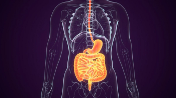
• Tumor abutment to the superior mesenteric artery (Figure 1).
• Short segment abutment or encasement of the common hepatic artery.
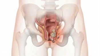
• Occlusion or distortion of the SMV–portal vein confluence (Figure 2), with a vein of the appropriate caliber (internal diameter) above and below the occlusio to allow for vascular reconstruction.
Tumors involving the body and tail of the pancreas are characterized as borderline if they abut the celiac axis by 180 degrees or less (ie, ≤ 50% of contact) or by more than 180 degrees without involvement of the aorta and with an intact and uninvolved gastroduodenal artery. The details of the specific degree of contact or distortion based on the different criteria for left-sided vs right-sided pancreatic cancers are outlined in Table 1.
In an effort to identify patients with borderline resectable pancreatic cancer based on biology and physiology, developers of the MDACC guidelines proposed two additional useful and prognostic categories to consider: type A and type B borderline resectable tumors. Type A is based on the aforementioned anatomic considerations. Patients with type B tumors have clinical findings that are suspicious but not diagnostic for metastatic disease; these may include radiographically indeterminate liver lesions, suspicious but not biopsy-proven distant lymph nodes, a biopsy-proven regional lymph node, or cancer antigen (CA) 19-9 levels greater than 1,000 units/mL (in the presence of normal levels of bilirubin).[4] These findings have been incorporated into the currently accepted definition of borderline resectable pancreatic cancer because they are believed to indicate a particularly high risk for early treatment failure with surgery alone.[10,11]
Further, type C patients with borderline resectable tumors are those with marginal performance status or significant comorbidities that confer higher than usual risk for postoperative morbidity and mortality.[10] In an effort to help this group attain the goal of possible future surgery, patients should be actively managed with physical therapy rehabilitation, nutritional optimization, and consultation with subspecialists in surgery and internal medicine.[4]
Borderline resectable pancreatic cancer is defined not just by the characteristic imaging findings, but also by biologic and physiologic parameters, as described above. This highlights the additional importance of a multidisciplinary high-volume pancreas program for the management of patients with borderline resectable pancreatic cancer.
Diagnostic Evaluation
High-quality thin-slice multiphase CT angiography is the modality most frequently used for determining whether a pancreas cancer is surgically resectable. Although MRI is equally specific and sensitive, limitations of cost and variable institutional quality and availability have rendered it inappropriate for patients who cannot tolerate iodinated dye, as well as those with specific or definite liver metastatic lesions, and/or isoattenuating pancreatic lesions.[12] A pancreas protocol CT ideally will involve acquiring submillimeter (0.5- to 1-mm) tissue sections, with scan acquisition in the pancreatic parenchymal phase at 40 to 50 seconds after intravenous injection and a portal venous phase at 65 to 70 seconds post injection. The early phase also opacifies well the arterial blood supply, allowing for accurate delineation of arterial anatomy. The variation in phases enables better char acterization of the often subtle anatomic relationships between the tumor and the mesenteric vasculature, thereby improving the ability to classify patients into the appropriate subgroups.[13] Although the use of fluorodeoxyglucose positron emission tomography (FDG-PET)/CT is not standard practice in the management of pancreatic cancer, recent data presented at the annual American Society of Clinical Oncology Gastrointestinal Cancers Symposium in early 2017 demonstrate that FDG-PET/CT may be useful in patients with significantly elevated levels of CA 19-9.[14]
The workup of patients with borderline resectable pancre atic cancer who will require neoadjuvant therapy should include evaluation of small hepatic biopsy samples for the presence of malignant biliary obstruction. Endoscopic ultrasound (EUS)-guided fine needle aspiration (FNA) has become widely available and is a safe and efficient tool for tissue diagnosis in this setting.[15] The use of EUS-FNA is preferable to CT-FNA, given the smaller likelihood of peritoneal seeding with the former technique, and because EUS enables better assessment of vascular involvement. However, if EUS-FNA is not an option, then CT-FNA is a reasonable alternative. In addition to EUS and CT, multiple brushings of the biliary tree can be performed during endoscopic retrograde cholangiopancreatography, to obtain tissue for diagnosis of cholangiocarcinoma. Unfortunately, since the bile duct is often extrinsically compressed by the pancreatic tumor, these brushings are not diagnostic and thus not the modality of choice for diagnosis of pancreatic cancer. Obstructive jaundice is a common presentation of pancreatic head adenocarcinoma and must be treated promptly by the placement of a biliary stent if patients present with prohibitively high levels of bilirubin or if they will receive neoadjuvant therapy prior to surgical resection.
Placement of a biliary stent is necessary to allow for normalization of hepatic function. With regard to the selection of stent length, most centers that treat large numbers of patients with pancreatic cancer will favor short covered metal stents for this population; because shorter stents are less likely to occlude, they carry a lower risk of inducing cholangitis, and the stent covers provide greater resistance to tumor ingrowth compared with open metal stents.[9] While stent occlusion is a complication to understand, it is neither sufficiently common nor problematic to warrant forgoing the use of stents (particularly metal ones) in the delivery of neoadjuvant therapy.[16] The majority of centers now perform routine diagnostic laparoscopy concurrently with the planned resection, particularly in patients who have received neoadjuvant therapy. This approach is consistent with our institutional practice.
The most important aspect of the workup and diagnosis of borderline resectable pancreatic cancer is the involvement of a multidisciplinary team of medical and radiation oncologists, surgeons, interventional radiologists and gastroenterologists, and specialists in nutritional services and physical therapy. Furthermore, to optimize patient survival and ensure that all appropriate patients are considered as candidates for surgical resection or referral to clinical trials, it is critical that both surgical and medical oncologists participate in the initial patient evaluation.
Rationale for Neoadjuvant Therapy in Patients With Borderline Resectable Pancreatic Cancer
The standard management of pancreatic cancer has been surgical resection (performed on the small number of patients who present with upfront resectable cancer) followed by adjuvant chemotherapy. Patients not considered to have resectable disease are treated with definitive chemotherapy or RT. The further subclassification of some patients as having borderline resectable pancreatic cancer allows for multimodality treatments to be incorporated into patient management, and expands the number of patients able to undergo both a safe and successful resection.[13] The number of patients with borderline resectable pancreatic cancer who proceed to resection is difficult to discern from reports in the medical literature, due to varying definitions of this entity, the type of neoadjuvant regimen used, and approaches to the general selection of patients for such protocols, which may differ based on practitioner and institutional preferences. Overall, reports in the medical literature indicate that a high proportion of patients with borderline resectable pancreatic cancer proceed to resection (up to 85% in some series).[17,18] A small percentage of patients are found to have isolated local disease progression that precludes resection at the time of restaging; more frequently, patients must forgo resection due to distant metastases or an inability to tolerate treatment toxicity.[19]
The rationale for administering neoadjuvant therapy for borderline resectable pancreatic cancer extends beyond the goal of achieving a negative surgical margin. Patients in this subcategory of pancreatic adenocarcinoma may be at higher risk for harboring radiographically occult metastatic disease or for having a positive resection margin (due to a greater prevalence of tumor-vascular abutment); they generally require a more complex operation with vascular resection and reconstruction, and therefore are at higher risk for postoperative morbidity that would preclude further adjuvant treatment.[8,20] The additional benefits that support the use of neoadjuvant treatment in this setting[8,20] include:
• Allowing for selection of patients who are most likely to achieve a favorable oncologic outcome.
• Allowing for the completion of systemic therapy and avoiding postoperative morbidity–related delays in receiving adjuvant therapy.
• Treatment of micrometastatic disease not visible on imaging but believed to occur early in pancreas adenocarcinoma.
• Tumor downstaging to allow for R0 resection.
In certain patients, poor overall survival despite complete resection suggests that micrometastatic disease may be present at the time of resection, in which case neoadjuvant therapy is the preferred strategy.[8,21]
It has been reported that approximately 25% of patients never receive adjuvant chemotherapy after resection for pancreatic adenocarcinoma. The reasons for this may be multifactorial, and may include perioperative morbidity and generally poor postoperative functional status.[22] Administering neoadjuvant chemotherapy may benefit this patient population. Many groups have sought to determine whether neoadjuvant therapy facilitates downstaging in patients with borderline resectable pancreatic cancer; assessment of disease status using standard radiologic or pathologic criteria has not elucidated this question. However, there are data, mostly from retrospective single-institution studies, demonstrating R0 resections achieved in this population following neoadjuvant treatment (Table 2).
Studies of Neoadjuvant Systemic Chemotherapy
The results of the randomized controlled ACCORD trial by the PRODIGE Intergroup-which compared a combination chemotherapy regimen of leucovorin, fluorouracil, irinotecan, and oxaliplatin (FOLFIRINOX) with gemcitabine as first-line therapy in patients with metastatic pancreatic cancer and good performance status-have had a profound impact on the management of pancreatic cancer.[23] The threefold higher overall survival rate at 18 months in patients treated with FOLFIRINOX, compared with the patients who received gemcitabine (18.6% vs 6.0%, respectively; hazard ratio, 0.57; 95% CI, 0.45–0.73; P < .001), has led practitioners to use this regimen in the setting of nonmetastatic pancreatic cancer as well.
In particular, FOLFIRINOX has been increasingly used as neoadjuvant therapy not only for downstaging prior to surgery, but also to treat micrometastatic disease, which too often presents as distant recurrence after surgical resection. In 2015, Ferrone et al published the largest retrospective series on borderline resectable and locally advanced pancreatic cancer treated with FOLFIRINOX in the neoadjuvant setting. Compared with patients who received no neoadjuvant therapy, patients who received FOLFIRINOX demonstrated significantly lower rates of both lymph node positivity (35% vs 79%) and perineural invasion (72% vs 95%). The authors also observed a significant increase in median overall survival in patients receiving neoadjuvant FOLFIRINOX compared with patients with clearly resectable tumors (P = .008), who were treated with surgery but not neoadjuvant therapy.[17] Other high-volume pancreatic cancer centers have demonstrated similarly encouraging results with the use of neoadjuvant FOLFIRINOX for treatment of borderline resectable pancreatic cancer,[18,24] with one group reporting an R0 rate of approximately 90%, achieved without the addition of radiation.[24]
Enthusiasm for the neoadjuvant treatment of borderline resectable pancreatic cancer is clearly growing. However, it is important to recognize that in 2014 the International Study Group of Pancreatic Surgery published a consensus statement on borderline resectable pancreatic cancer in which neoadjuvant therapy was not supported for “borderline” patients with technically resectable tumors with isolated venous involvement for whom a vascular resection and reconstruction approach was feasible.[25] These recommendations were based in part on a large multi-institutional database study of patients with borderline resectable pancreatic cancer treated with or without neoadjuvant therapy followed by resection; the results failed to support vein involvement with tumor as a negative prognostic marker of survival.[26] Therefore, our general practice is to proceed to surgical resection in patients who are unable to tolerate FOLFIRINOX as a neoadjuvant regimen and who have limited isolated venous involvement-unless other high-risk features are present, such as a significantly elevated CA 19-9 level. For patients who cannot tolerate FOLFIRINOX but have arterial abutment on imaging, our preference, based on best available data, is neoadjuvant therapy with gemcitabine and RT, followed by resection.
The utility and efficacy of combination therapy with gemcitabine and nab-paclitaxel is also under investigation. Often in the metastatic setting, the choice of FOLFIRINOX or the combination of gemcitabine and nab-paclitaxel is determined by the patient’s functional status or comorbidities. More frequently this approach is extrapolated to the neoadjuvant setting, wherein patients with borderline resectable pancreatic cancer who are not candidates for a modified regimen of FOLFIRINOX (mFOLFIRINOX) are treated with neoadjuvant gemcitabine plus nab-paclitaxel.
Neoadjuvant RT
There is some uncertainty regarding the timing, type, and duration of neoadjuvant RT in the setting of pancreatic cancer in general, with the greatest variability in RT delivery occurring during the management of borderline resectable pancreatic cancer. The chemoradiation portion of induction therapy has been believed to be particularly important for patients with arterial abutment, in order to prevent a positive margin of resection.[19] Multiple clinical trials now underway in borderline resectable pancreatic cancer include investigation of RT optimization in patient management (ClinicalTrials.gov identifiers NCT02241551, NCT03099265, NCT01661088).
A common approach to RT in borderline resectable pancreatic cancer is to use standard doses (50.4 Gy in 28 fractions) concurrent with chemotherapy.[27] There is some controversy over whether higher doses of radiation should be delivered by intensity-modulated RT (IMRT) or stereotactic body RT (SBRT). The clinical justification for the use of IMRT is that this approach produces a more optimal dose distribution and enables dose escalation.[28,29] Retrospective studies in the setting of locally advanced and borderline resectable pancreatic cancer have reported use of SBRT (25 to 35 Gy in 5 fractions),[30] although to date use of SBRT in this setting has been limited. Small single-institution studies have shown that SBRT is well tolerated, is safe, and has the potential to facilitate the achievement of R0 resections.[31] Advances in technique, including fiducial marker placement for SBRT, have demonstrated promising early results.[32] The Alliance randomized trial A021501 (ClinicalTrials.gov identifier: NCT02839343) is now underway to investigate preoperative chemotherapy vs chemotherapy plus hypofractionated RT for patients with borderline resectable pancreatic head adenocarcinoma.
Duration of Neoadjuvant Treatment
The duration of neoadjuvant therapy remains to be clearly defined. A team from the Medical College of Wisconsin has developed a protocol that is unique in the determination of when to proceed with further therapy vs surgery. This protocol takes into account the window of opportunity to surgically remove the tumor. This group administers either FOLFIRINOX or combination therapy with nab-paclitaxel and gemcitabine (depending on the Eastern Cooperative Oncology Group status of the individual patient) for 2 months, followed by imaging for the restaging evaluation. Patients who have stable disease or disease that is responding to chemotherapy transition to chemoradiation therapy followed by resection.[9,18] In patients who have had a profound response to induction therapy, the Wisconsin team advocates for a prolonged course of systemic chemotherapy prior to RT, in order to maximize the benefits of systemic therapy prior to surgery while maintaining local disease control before surgery. Patients who do not have a robust response to systemic therapy proceed to chemoradiation after 2 months of systemic chemotherapy, in order to avoid local disease progression that would prevent eventual surgery.[9] This “test of time” also allows for patients with development of distant metastases to be spared unnecessary surgical resection. Many protocols would advocate for surgery in the setting of response and additional therapy in the setting of progression; however, we believe that it is important to proceed with surgical exploration before local disease progression prevents the opportunity to do so. These treatment strategies highlight the evolution of pancreatic cancer management.
Assessing the Response to Treatment
It is now recognized that CT imaging may be difficult to interpret after neoadjuvant treatment, since traditional Response Evaluation Criteria in Solid Tumors do not directly translate to pathologic response. Thus, in the absence of distant metastatic disease or of comorbidities precluding resection, the majority of patients should undergo surgical exploration, even if CT imaging is suggestive of local progression that remains within the confines of borderline resectable pancreatic cancer (rather than unresectable disease).[4] When local progression is observed on imaging following neoadjuvant therapy, the patient may benefit from direct superior mesenteric artery exploration and biopsy prior to proceeding with pancreatectomy.
KEY POINTS
- The definition and management of borderline resectable pancreatic cancer continue to evolve, and there has already been significant progress in patient outcomes.
- The most important aspect of the workup and diagnosis of borderline resectable pancreatic cancer is the involvement of a multidisciplinary team of medical and radiation oncologists, surgeons, interventional radiologists and gastroenterologists, and specialists in nutritional services and physical therapy.
- Patients with borderline resectable pancreatic cancer should be evaluated for eligibility to participate in clinical trials.
- In the United States, the current approach to management of borderline resectable pancreatic cancer favors administration of neoadjuvant therapy followed by surgical resection, except in patients with isolated venous involvement and the inability to tolerate a neoadjuvant treatment protocol.
Ferrone et al found that despite post-FOLFIRINOX imaging suggesting continued unresectability, 92% of patients ultimately had R0 resections.[17] In these patients, pathologic analysis demonstrated significant post-therapy fibrosis without viable cancer. Other studies have also shown that radiologic evidence of a treatment response is not sufficient to assess tumor response and have recommended surgical exploration in patients without metastatic disease who are fit to undergo surgery.[33,34] This is among the many rationales for incorporation of a multidisciplinary team in the management of patients with borderline resectable pancreatic cancer, to ensure that surgical exploration is offered to all patients who may be candidates for resection.
The Role and Utility of CA 19-9 Testing in Patients With Borderline Resectable Pancreatic Cancer
CA 19-9 is a sialylated Lewis antigen secreted by most pancreatic adenocarcinomas and has been validated in patients who are Lewis antigen–positive. Although the value of CA 19-9 testing is limited in patients with obstructive jaundice, in patients with normal bilirubin levels, CA 19-9 is a useful serum marker to follow during therapy both in the metastatic and neoadjuvant settings. Measurement of CA 19-9 levels has been shown to be useful in assessing patient response to neoadjuvant therapy, and retrospective studies have found normalization of post-treatment levels of CA 19-9 to be associated with better prognosis.[35,36] Ferrone et al observed that despite a decrease in CA 19-9 levels, a clear response was not shown on cross-sectional imaging after FOLFIRINOX induction therapy-yet in most of these patients, negative resection margins were still achieved.[17] Thus, CA 19-9 levels continue to be a useful marker for assessing the response to neoadjuvant therapy, particularly when FOLFIRINOX is the treatment regimen selected.
Irreversible Electroporation (IRE) to Achieve Negative Surgical Resection Margins
IRE consists of the application of short, high-voltage pulses to soft tissue to cause an electrolyte imbalance of calcium and sodium ions that leads to apoptosis in the tumor. This technique has been utilized for margin optimization in both borderline resectable and locally advanced pancreatic adenocarcinoma. A unique feature of IRE as an ablative technology is that it is a non–thermal-based method of achieving cell death; therefore, nearby vital structures are spared from heat injury.[37,38] Early research has shown that simultaneous IRE and pancreatectomy yielded lower local recurrence rates than expected.[38]
Future Trials in Borderline Resectable Pancreatic Cancer
One obstacle to progress in the treatment of borderline resectable pancreatic cancer is that a large number of trials and retrospective studies combine patients with borderline resectable and locally advanced disease into a single treatment group. However, an increasing number of trials are now accruing only patients with borderline resectable disease. As an example, the PANDAS-PRODIGE 44 study (ClinicalTrials.gov identifier: NCT02676349) is a randomized phase II trial of only borderline resectable patients being treated with neoadjuvant mFOLFIRINOX with or without preoperative chemoradiotherapy with capecitabine. Another phase II trial including only borderline resectable patients includes two treatment arms of either mFOLFIRINOX or combination therapy with gemcitabine plus nab-paclitaxel, with both arms followed by SBRT preceding resection in patients with stable disease (ClinicalTrials.gov identifier: NCT02241551). In light of the many treatment sequences that are currently under investigation in this setting, and with borderline resectable pancreatic cancer defined as a distinct clinical entity, it is important to diligently design trials that only include this cohort of patients.[27] Table 3 highlights relevant ongoing trials.
Conclusion
The definition and management of borderline resectable pancreatic cancer continue to evolve, and there has already been significant progress in patient outcomes. The current approach to patient management in the United States favors administration of neoadjuvant therapy followed by surgical resection (Figure 3), except in patients with isolated venous involvement and the inability to tolerate a neoadjuvant treatment protocol. RT is strongly considered, particularly for patients with arterial abutment. The single most important and defined factor in the management of patients with borderline resectable pancreatic cancer is involvement of a multidisciplinary pancreas team from a center that treats large numbers of pancreatic malignancies. Supportive care, including antiemetic therapy and hydration for patients receiving a highly toxic regimen of neoadjuvant therapy, is crucial and should not be overlooked. Nutritional support, optimization of medical comorbidities, and physical therapy should be central and defined aspects of care for patients receiving neoadjuvant therapy. Centers of excellence have been successful at achieving promising results with neoadjuvant protocols and with aggressive safe surgical intervention.
Acknowledgement: Work reported in this publication was supported by the National Cancer Institute of the National Institutes of Health under award number NIH 5K12CA001727-20. The content is solely the responsibility of Dr. Laleh Melstrom and does not necessarily represent the official views of the National Institutes of Health.
Financial Disclosure:The authors have no significant financial interest in or other relationship with the manufacturer of any product or provider of any service mentioned in this article.
References:
1. Cai S, Hong TS, Goldberg SI, et al. Updated long-term outcomes and prognostic factors for patients with unresectable locally advanced pancreatic cancer treated with intraoperative radiotherapy at the Massachusetts General Hospital, 1978 to 2010. Cancer. 2013;119:4196-204.
2. Ferrone C, Brennan M, Gonen M, et al. Pancreatic adenocarcinoma: the actual 5-year survivors. J Gastrointest Surg. 2008;12:701-6.
3. Varadhachary GR, Tamm EP, Abbruzzese JL, et al. Borderline resectable pancreatic cancer: definitions, management, and role of preoperative therapy. Ann Surg Oncol. 2006;13:1035-46.
4. Katz MH, Pisters PW, Evans DB, et al. Borderline resectable pancreatic cancer: the importance of this emerging stage of disease. J Am Coll Surg. 2008;206:833-46.
5. Sohn TA, Yeo CJ, Cameron JL, et al. Resected adenocarcinoma of the pancreas–616 patients: results, outcomes, and prognostic indicators. J Gastrointest Surg. 2000;4:567-79.
6. Neoptolemos JP, Stocken DD, Dunn JA, et al. Influence of resection margins on survival for patients with pancreatic cancer treated by adjuvant chemoradiation and/or chemotherapy in the ESPAC-1 randomized controlled trial. Ann Surg. 2001;234:758-68.
7. Gilbert JW, Wolpin B, Clancy T, et al. Borderline resectable pancreatic cancer: conceptual evolution and current approach to image-based classification. Ann Oncol. 2017 Apr 12. [Epub ahead of print]
8. Evans DB, Erickson BA, Ritch P. Borderline resectable pancreatic cancer: definitions and the importance of multimodality therapy. Ann Surg Oncol. 2010;17:2803-5.
9. Evans DB, Ritch PS, Erickson BA. Neoadjuvant therapy for localized pancreatic cancer: support is growing? Ann Surg. 2015;261:18-20.
10. Schwarz L, Katz MH. Diagnosis and management of borderline resectable pancreatic adenocarcinoma. Hematol Oncol Clin North Am. 2015;29:727-40.
11. Schwarz L, Lupinacci RM, Svrcek M, et al. Para-aortic lymph node sampling in pancreatic head adenocarcinoma. Br J Surg. 2014;101:530-8.
12. Al-Hawary MM, Francis IR, Chari ST, et al. Pancreatic ductal adenocarcinoma radiology reporting template: consensus statement of the Society of Abdominal Radiology and the American Pancreatic Association. Radiology. 2014;270:248-60.
13. Khorana AA, Mangu PB, Berlin J, et al. Potentially curable pancreatic cancer: American Society of Clinical Oncology Clinical Practice Guideline. J Clin Oncol. 2016;34:2541-56.
14. Kinupe Abrahao AB, Ung Y, Ko Y-J, Berry SR. FDG PET/CT in pancreatic cancer staging and management: a retrospective study. J Clin Oncol. 2017;35(suppl 4S):abstr 464.
15. Eloubeidi MA, Gress FG, Savides TJ, et al. Acute pancreatitis after EUS-guided FNA of solid pancreatic masses: a pooled analysis from EUS centers in the United States. Gastrointest Endosc. 2004;60:385-9.
16. Pisters PW, Hudec WA, Lee JE, et al. Preoperative chemoradiation for patients with pancreatic cancer: toxicity of endobiliary stents. J Clin Oncol. 2000;18:860-7.
17. Ferrone CR, Marchegiani G, Hong TS, et al. Radiological and surgical implications of neoadjuvant treatment with FOLFIRINOX for locally advanced and borderline resectable pancreatic cancer. Ann Surg. 2015;261:12-7.
18. Christians KK, Tsai S, Mahmoud A, et al. Neoadjuvant FOLFIRINOX for borderline resectable pancreas cancer: a new treatment paradigm? Oncologist. 2014;19:266-74.
19. Fathi A, Christians KK, George B, et al. Neoadjuvant therapy for localized pancreatic cancer: guiding principles. J Gastrointest Oncol. 2015;6:418-29.
20. Katz MH, Hu CY, Fleming JB, et al. Clinical calculator of conditional survival estimates for resected and unresected survivors of pancreatic cancer. Arch Surg. 2012;147:513-9.
21. Rhim AD, Mirek ET, Aiello NM, et al. EMT and dissemination precede pancreatic tumor formation. Cell. 2012;148:349-61.
22. Spitz FR, Abbruzzese JL, Lee JE, et al. Preoperative and postoperative chemoradiation strategies in patients treated with pancreaticoduodenectomy for adenocarcinoma of the pancreas. J Clin Oncol. 1997;15:928-37.
23. Conroy T, Desseigne F, Ychou M, et al; Groupe Tumeurs Digestives of Unicancer; PRODIGE Intergroup. FOLFIRINOX versus gemcitabine for metastatic pancreatic cancer. N Engl J Med. 2011;364:1817-25.
24. Kim SS, Nakakura EK, Wang ZJ, et al. Preoperative FOLFIRINOX for borderline resectable pancreatic cancer: is radiation necessary in the modern era of chemotherapy? J Surg Oncol. 2016;114:587-96.
25. Bockhorn M, Uzunoglu FG, Adham M, et al; International Study Group of Pancreatic Surgery. Borderline resectable pancreatic cancer: a consensus statement by the International Study Group of Pancreatic Surgery (ISGPS). Surgery. 2014;155:977-88.
26. Kelly KJ, Winslow E, Kooby D, et al. Vein involvement during pancreaticoduodenectomy: is there a need for redefinition of “borderline resectable disease?” J Gastrointest Surg. 2013;17:1209-17.
27. Katz MH, Crane CH, Varadhachary G. Management of borderline resectable pancreatic cancer. Semin Radiat Oncol. 2014;24:105-12.
28. Ben-Josef E, Schipper M, Francis IR, et al. A phase I/II trial of intensity modulated radiation (IMRT) dose escalation with concurrent fixed-dose rate gemcitabine (FDR-G) in patients with unresectable pancreatic cancer. Int J Radiat Oncol Biol Phys. 2012;84:1166-71.
29. Brown MW, Ning H, Arora B, et al. A dosimetric analysis of dose escalation using two intensity-modulated radiation therapy techniques in locally advanced pancreatic carcinoma. Int J Radiat Oncol Biol Phys. 2006;65:274-83.
30. Chuong MD, Springett GM, Freilich JM, et al. Stereotactic body radiation therapy for locally advanced and borderline resectable pancreatic cancer is effective and well tolerated. Int J Radiat Oncol Biol Phys. 2013;86:516-22.
31. Chuong MD, Shridhar R, Patel M, et al. Neoadjuvant stereotactic body radiation therapy (SBRT) for borderline resectable pancreas cancer: Moffitt Cancer Center initial experience. J Clin Oncol. 2011;29(suppl 4):abstr 302.
32. Vignesh S, Hoffe SE, Shridhar R, et al. The feasibility, safety, and technique of endoscopic ultrasound (EUS)-guided fiducial marker placement for stereotactic body radiation therapy (SBRT) in borderline resectable pancreatic cancer. J Clin Oncol. 2011;29(suppl 4):abstr 327.
33. Katz MH, Fleming JB, Bhosale P, et al. Response of borderline resectable pancreatic cancer to neoadjuvant therapy is not reflected by radiographic indicators. Cancer. 2012;118:5749-56.
34. Wagner M, Antunes C, Pietrasz D, et al. CT evaluation after neoadjuvant FOLFIRINOX chemotherapy for borderline and locally advanced pancreatic adenocarcinoma. Eur Radiol. 2016 Nov 28. [Epub ahead of print]
35. Katz A, Hanlon A, Lanciano R, et al. Prognostic value of CA 19-9 levels in patients with carcinoma of the pancreas treated with radiotherapy. Int J Radiat Oncol Biol Phys. 1998;41:393-6.
36. Aldakkak M, Christians KK, Krepline AN, et al. Pre-treatment carbohydrate antigen 19-9 does not predict the response to neoadjuvant therapy in patients with localized pancreatic cancer. HPB (Oxford). 2015;17:942-52.
37. Cannon R, Ellis S, Hayes D, et al. Safety and early efficacy of irreversible electroporation for hepatic tumors in proximity to vital structures. J Surg Oncol. 2013;107:544-9.
38. Kwon D, McFarland K, Velanovich V, Martin RC 2nd. Borderline and locally advanced pancreatic adenocarcinoma margin accentuation with intraoperative irreversible electroporation. Surgery. 2014;156:910-20.
39. National Comprehensive Cancer Network. Clinical practice guidelines in oncology: pancreatic adenocarcinoma. Version 2.2017. Updated April 27, 2017. https://www.nccn.org/professionals/physician_gls/pdf/pancreatic.pdf. Accessed May 15, 2017.
40. Callery MP, Chang KJ, Fishman EK, et al. Pretreatment assessment of resectable and borderline resectable pancreatic cancer: expert consensus statement. Ann Surg Oncol. 2009;16:1727-33.
41. Varadhachary GR, Tamm EP, Abbruzzese JL, et al. Borderline resectable pancreatic cancer: definitions, management, and role of preoperative therapy. Ann Surg Oncol. 2006;13:1035-46.
42. Katz MH, Marsh R, Herman JM, et al. Borderline resectable pancreatic cancer: need for standardization and methods for optimal clinical trial design. Ann Surg Oncol. 2013;20:2787-95.
43. Barugola G, Partelli S, Crippa S, et al. Outcomes after resection of locally advanced or borderline resectable pancreatic cancer after neoadjuvant therapy. Am J Surg. 2012;203:132-9.
44. Kang CM, Chung YE, Park JY, et al. Potential contribution of preoperative neoadjuvant concurrent chemoradiation therapy on margin-negative resection in borderline resectable pancreatic cancer. J Gastrointest Surg. 2012;16:509-17.
45. Stokes JB, Nolan NJ, Stelow EB, et al. Preoperative capecitabine and concurrent radiation for borderline resectable pancreatic cancer. Ann Surg Oncol. 2011;18:619-27.
46. Chun YS, Milestone BN, Watson JC, et al. Defining venous involvement in borderline resectable pancreatic cancer. Ann Surg Oncol. 2010;17:2832-8.
47. McClaine RJ, Lowy AM, Sussman JJ, et al. Neoadjuvant therapy may lead to successful surgical resection and improved survival in patients with borderline resectable pancreatic cancer. HPB (Oxford). 2010;12:73-9.
48. Landry J, Catalano PJ, Staley C, et al. Randomized phase II study of gemcitabine plus radiotherapy versus gemcitabine, 5-fluorouracil, and cisplatin followed by radiotherapy and 5-fluorouracil for patients with locally advanced, potentially resectable pancreatic adenocarcinoma. J Surg Oncol. 2010;101:587-92.
49. Turrini O, Viret F, Moureau-Zabotto L, et al. Neoadjuvant chemoradiation and pancreaticoduodenectomy for initially locally advanced head pancreatic adenocarcinoma. Eur J Surg Oncol. 2009;35:1306-11.