
|Articles|April 15, 2013
- ONCOLOGY Vol 27 No 4
- Volume 27
- Issue 4
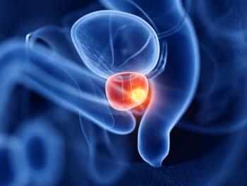
The State of Prostate MRI in 2013
Our aims in this article are to describe the various imaging sequences that comprise the multiparametric MRI exam, as well as to review current literature on the strengths/weaknesses of these sequences; to delineate strategies for standardizing interpretation and reporting of MRI results; and to expound on the role of prostate MRI in clinical practice.
Advertisement
Compared with earlier prostate MRI techniques that suffered from relatively poor sensitivity and specificity for detecting prostate cancer because of their reliance predominantly on morphology, multiparametric prostate MRI (mpMRI) in 2013 provides a wealth of functional information that has made possible vastly improved detection and characterization of prostate cancer. Our aims in this article are to describe the various imaging sequences that comprise the mpMRI exam, as well as to review current literature on the strengths/weaknesses of these sequences; to delineate strategies for standardizing interpretation and reporting of MRI results; and finally, to expound on the role that prostate MRI currently does and potentially can play in clinical practice.
Introduction
Over the last decades, prostate-specific antigen (PSA) screening has lead to earlier detection of prostate cancer (PCa), with a resultant decrease in average clinical and pathologic stage at the time of diagnosis. In addition to facilitating early diagnosis of aggressive lesions, the relatively high sensitivity of PSA screening can also result in the detection of small and low-grade prostatic malignancies that may not be life-threatening. Radical whole-gland therapy for indolent lesions exposes patients to the potential for treatment-related morbidities, which could in some cases outweigh the benefits of therapy. Concerns about potential overtreatment of indolent disease has lead to interest in gland-preserving strategies, including active surveillance and various types of focal therapy.
Clinical parameters, including digital rectal examination results, PSA values, and the results of transrectal ultrasound (TRUS)-guided biopsies, among others, can be used to stratify patients into risk categories (D’Amico criteria),[1] to predict the likelihood of disease extension outside the prostate (Partin tables),[2] and to predict recurrence after prostatectomy (Kattan nomogram).[3] Such predictive tools aid clinicians in choosing among various treatment strategies based on a patient’s underlying risks. However, sampling error intrinsic in TRUS-guided prostatic biopsy[4] has been shown to lead to the undergrading of disease in a substantial proportion of patients, which could potentially result in a suboptimal initial choice of therapeutic strategy. In one series of 1,565 patients considered candidates for active surveillance who proceeded to radical prostatectomy, Mufarrij et al reported that approximately 46% were upgraded to a Gleason score of 7 or greater at final histopathology.[5] Three-dimensional transperineal mapping biopsies (TPMB) provide a more thorough sampling of the gland, facilitating improved pretreatment staging[6]; however, TPMB can still miss clinically significant disease, typically requires general anesthesia, and may carry a higher complication rate than TRUS-guided biopsy.
Given the backdrop of increasing diagnosis of early-stage disease and the inherent challenges in choosing an optimal treatment strategy in some patients using current clinical paradigms, there is a need for improved disease detection, staging, and risk-stratification via noninvasive imaging. Among the various imaging modalities used in the management of prostate cancer patients, only MRI has both the spatial resolution and soft-tissue contrast necessary to most accurately characterize localized PCa.
The use of MRI to evaluate the prostate dates back over 3 decades.[7] Earlier iterations of prostate MRI relied predominantly on morphologic and signal changes present on T1- and T2-weighted images and suffered from relatively poor sensitivity and specificity for detecting PCa. Newer MRI sequences, including diffusion-weighted imaging (DWI) and dynamic contrast-enhanced MRI (DCE-MRI), provide additional functional information about tissues and have enabled significant improvements in the ability of MRI to detect and characterize focal prostatic lesions. MR spectroscopic imaging is also used in some centers to help differentiate between malignant and benign tissue within the prostate. Current state-of-the-art prostate MRI consists of a multiparametric approach, in which two or more functional sequences complement the morphologic information provided by T2-weighted imaging. This article will attempt to summarize the hardware and various sequences that comprise a prostate multiparametric MRI (mpMRI) exam, and where applicable, will emphasize limitations and areas for future improvement. Strategies for interpretation and reporting of MRI results will be discussed, and finally, the role of prostate MRI in clinical practice will be explored.
Hardware
The stronger main magnetic field of 3.0 Tesla MRI systems provides a higher signal-to-noise ratio (SNR) compared with 1.5 Tesla systems.[8] This increased SNR can be used to improve the spatial resolution and/or the acquisition time of the MR images. Acceptable images of the prostate can be obtained at both 1.5 and 3.0 Tesla when using appropriate coil and sequence selection.[9] However, in the authors’ opinion, use of a 3.0 Tesla system can improve the performance of any underlying mpMRI protocol and is advantageous whenever available in one’s practice.
While prostate MRI at a minimum entails use of a phased-array coil laid directly across the patient’s pelvis, in many centers, an endorectal coil (ERC) is also employed to complement the external pelvic coil. Because an ERC is located only a few millimeters from the prostate gland, it can further augment the amount of signal available for image generation. The additional signal available from an ERC is likely most advantageous at 1.5 Tesla, whereas the higher overall signal available at 3.0 Tesla makes the necessity of ERC use at this field strength less certain. Heijmink et al did report improved image quality, tumor detectability, and staging performance with the use of an ERC compared with body array coils at 3.0 Tesla[10]; however, Kim et al more recently reported equivalent performance of ERC and body array coils for prostate cancer staging.[11] It is important to note that these two studies only evaluated the diagnostic performance of T2-weighted images, and the applicability of the findings to multiparametric exams incorporating additional functional sequences is uncertain.
Although employing an ERC can provide very high-quality images, there are substantial trade-offs to be considered, including discomfort secondary to ERC placement, which can be fairly significant in some patients and can lead to poor compliance with the exam; magnetic susceptibility and motion-related artifacts that can be related to ERCs, particularly at 3.0 Tesla; and finally, workflow considerations regarding the incorporation of ERC into a prostate MRI practice-namely, increased examination time resulting from the placement and removal of the coil, as well as the personnel required to do this and the cost of the coil itself. In summary, ERCs afford both advantages and disadvantages in mpMRI; at 3.0 Tesla, the necessity for ERC use has not been clearly established, and adoption varies on a center-by-center basis.
Components of a Multiparametric Prostate MRI Exam
T2-weighted imaging
T2-weighted fast-spin echo images acquired in multiple planes represent key components of a prostate mpMRI exam. Images should be acquired with high resolution, accomplished by using a small field-of-view and a narrow image thickness of 3 to 4 mm. High-resolution T2-weighted imaging (T2WI) currently provides the best assessment of the prostate’s morphology, margins, and internal structure. The ejaculatory ducts, urethra, and verumontanum are frequently visualized on high-quality T2WI, and these internal landmarks can aid in the correct identification of zonal anatomy, and thus in lesion localization. Differentiation between the peripheral zone (PZ) and central gland (CG) is essential for accurate lesion localization, and making this distinction is typically easy on T2WI. The term “central gland” is often used in prostate MRI reports to refer to the combined central zone (CZ) and transition zone (TZ), which may be difficult to distinguish in a fraction of patients.[12] Vargas et al recently showed that the CZ can be identified as a distinct region in most patients. Appropriate recognition of the CZ is important to avoid its misinterpretation as a potential tumor; however, a very small fraction of tumors may in fact occur within the CZ, and these tend to have more aggressive features compared with CG tumors that only involve the TZ.[12] In terms of staging, the superior morphologic information provided by high-resolution T2WI facilitates assessment of seminal vesicle invasion, extracapsular extension, and invasion of the neurovascular bundles.
The normal glandular tissue in the PZ is predominantly high in signal on T2WI, whereas PCa lesions in the PZ appear as dark (hypointense) foci, possibly round or oval in shape.[13] Unfortunately, some foci of PCa may appear isointense to adjacent benign PZ on T2-weighted images, especially when small or of lower grade. Moreover, in many patients referred for prostate MRI, the PZ is not homogeneously bright, but rather a patchwork of varying signal intensities, secondary to factors such as atrophy, prior episodes of prostatitis, or post-biopsy hemorrhage.[13] Because post-biopsy changes can mimic PCa on T2WI, it is generally recommended to delay mpMRI for at least 8 to 12 weeks after biopsy,[9] and it is also important to evaluate non-contrast T1-weighted images for evidence of biopsy-related hemorrhage.[13]
Several recent publications concerning mpMRI have reported on the diagnostic performance of T2WI for PCa localization.[14-19] In a recent meta-analysis, Tan et al incorporated data from 14 studies with histopathologic correlation and reported an overall sensitivity and specificity for T2WI alone of 0.57−0.62 and 0.74−0.78, respectively.[20] Even when images are acquired using optimal technique, the overall moderate performance of T2WI alone has resulted in the need to incorporate additional functional sequences to facilitate more accurate lesion identification.
Diffusion-weighted imaging
DWI provides functional information about tissue microstructure. (For a concise review of the technical aspects of DWI and its applications in oncologic imaging, a recent review by Koh and Collins[21] is recommended.) DWI relies on the application of a diffusion-weighting gradient to water protons, which generates sensitivity of the measured signal to the ability of water protons to move freely in space. Water protons that are relatively hindered in their motion will appear as bright on DWI due to the application of this additional gradient. Changing the amplitude of the diffusion-weighting gradients varies the degree of diffusion weighting for a given set of DWI images, which is referred to as its “b-value.” At different b-values, DWI interrogates water movement over different length scales[21]; at low b-values, DWI predominantly reflects water movement over larger length scales, mainly secondary to capillary perfusion, whereas at higher b-values, DWI interrogates movement over small length scales, such as within cells or intracellular organelles. Importantly, the signal present on a DWI image represents a combination of both diffusion-related signal and signal secondary to T2 relaxation. If images with a minimum of two different b-values are acquired, an apparent diffusion coefficient (ADC) map can be generated that is intended to better represent the diffusivity of protons within tissue without the confounder of T2-related signal.[21] The ADC map generated from DWI is particularly helpful for PCa identification.
The role of DWI in the noninvasive evaluation of malignancy has grown tremendously over the past decade. The higher cellular density present in many tumors, in conjunction with more complex intracellular microstructure and likely other factors, leads to restricted diffusion of water, which is detectable on DWI images. The value of DWI for PCa imaging has been established in numerous publications in recent years.[16-20,22,23] In a recent meta-analysis pooling data from 627 patients, including both PZ and CG tumors, Wu et al reported a summary sensitivity and specificity for DWI combined with T2WI of 0.76 and 0.82, respectively, and this was shown to be superior to T2WI alone.[22] A separate meta-analysis by Tan et al, pooling data from a total of 5,892 lesions, reported a sensitivity, specificity, and area under the receiver operating characteristic (ROC) curve of 0.69, 0.89, and 0.85, respectively, for DWI alone, which was significantly better than the performance of T2WI alone (0.60, 0.76, and 0.75, respectively).[20] Interestingly, in this study, the specificity of DWI alone was superior to the combination of DWI+T2WI and was also found to be superior to the specificity of DCE imaging.
In addition, ADC values derived from DWI images have been shown to be inversely correlated with the Gleason score of lesions at biopsy or surgery.[24-27] Attempts have been made to define ADC cutoff values, both to differentiate PCa from benign tissue and to further subdivide foci of PCa into categories of different degrees of aggressiveness (see section on assessment of lesion aggressiveness below). However, the wide overlap in the ranges of ADC values for normal PZ tissue and PCa,[27-30] as well as differences in scanner hardware/software between centers, have made it difficult to define consistently reliable cutoff values. Furthermore, there is a strong dependence of the calculated ADC values on the specific b-values employed by a DWI sequence,[29] and this is also thought to contribute to the differing ranges of ADC values reported for PZ tissue and PCa in different publications.
Dynamic contrast-enhanced imaging
Angiogenesis may lead to varying combinations of increased blood flow, microvascular density, and capillary leakiness in malignant lesions compared with the organ from which a cancer arises. DCE-MRI takes advantage of these vascular differences to facilitate lesion detection, and Verma et al recently provided a useful overview of the applications of DCE-MRI in PCa.[31] Several reports have demonstrated the utility of DCE-MRI for PCa lesion identification, either alone or in combination with other sequences.[14,15,32,33] There are a wide variety of techniques for performing and interpreting DCE-MRI. In general, a T1-weighted spoiled gradient-recalled echo sequence is run prior to contrast administration, and the sequence is re-run repetitively (dynamically) after the rapid infusion of an intravenous bolus of a low-molecular-weight gadolinium chelate.
There are three methods of interpreting DCE-MRI: qualitative, semi-quantitative, and quantitative. Qualitative analysis can range from visual inspection of the images for rapidly and intensely enhancing lesions, to the plotting of kinetic curves of signal intensity vs time. The semi-quantitative approach to DCE-MRI interpretation begins with the calculation of various kinetic parameters (such as time to peak enhancement, wash-in rate, or wash-out rate) on a voxel-by-voxel basis; then, colorized parametric maps representing these parameters are overlaid on top of morphologic images, such as T2WI, to facilitate interpretation. Finally, one can perform a quantitative interpretation of DCE-MRI data, which attempts to assess microvascular changes in tumors by converting the acquired signal intensity into the true gadolinium concentration in tissues; this method employs pharmacokinetic models to determine the rate of exchange of contrast between plasma and the extravascular, extracellular space.[31] Specific quantitative vascular parameters, such as Ktrans and Kep, have been shown to be elevated in tumors, and assessment of these parameters has been shown to improve the specificity of prostate MRI compared with T2WI alone.[32]
The different approaches to DCE-MRI interpretation each have advantages and disadvantages. Although simplistic, simple visual assessment of the source images has been shown to be effective[34] and is employed at some centers; in fact, recent mpMRI guidelines provided by the European Society of Urogenital Radiology (ESUR) rely on analysis of kinetic curves for DCE-MRI interpretation.[9] The advantages of this approach include the lack of a need for special dedicated software or computer workstations. The semi-quantitative and quantitative approaches to DCE-MRI interpretation require dedicated software and/or computer hardware for interpretation, and can be time-consuming. Proponents of the quantitative approach have noted that analysis based on kinetic curves or semi-quantitative kinetic parameters depends heavily on the rate of contrast injection, whereas the quantitative approach may be more robust in its resistance to variation in injection-related factors.[31] The quantitative approach has also been proposed to be more reproducible, enabling serial measurements of perfusion parameters over time to facilitate evaluation of treatment response. As an example, Low et al recently reported promising results by using serial quantitative DCE-MRI to monitor the response of PCa to stereotactic radiation therapy.[35] Ultimately, to the best of our knowledge, no report has clearly established the superiority of one DCE-MRI interpretation strategy over the others.
MR spectroscopic imaging
MR spectroscopic imaging (MRSI) provides information about relative concentrations of various metabolites in tissues and is most commonly performed with 3-dimensional chemical shift imaging.[9,36] The use of an ERC is strongly suggested for MRSI at 3.0 Tesla and is required for MRSI at 1.5 Tesla. Spectral tracings providing assessment of relative metabolite concentrations are acquired and overlaid on the T2 morphologic images. The metabolites of most interest in the setting of PCa are citrate and choline. Choline plays a key role in cell membrane synthesis, and choline and several related metabolites are present in higher concentrations in PCa compared with benign prostate tissue.[37] Citrate, on the other hand, is present in normal PZ tissue but is reduced in PCa. In general, the ratio of choline+creatine to citrate is higher in PCa compared with benign tissue, and the degree to which this ratio is elevated is used to determine the likelihood of PCa within a given voxel. Prostate MRSI is technically challenging and time-consuming, and many centers performing mpMRI do not include MRSI in their protocols. A prospective multicenter study conducted by the American College of Radiology Imaging Network evaluated 110 patients with MRSI (at 1.5 Tesla using ERC) and T2WI, and MRSI+T2WI showed an accuracy in PCa localization equivalent to that of T2WI alone.[38] Recent guidelines for mpMRI performance and interpretation provided by the ESUR list MRSI as optional, not required, for lesion detection and staging.[9]
Strategies for Interpretation and Reporting of Prostate mpMRI Exams
The addition of DWI, DCE-MRI, and/or MRSI has been shown to significantly improve the performance of prostate MRI compared with T2WI alone, especially in the setting of PZ lesions.[14,15] However, given the large amount of functional information provided by a multiparametric exam, the optimal means of combining the findings of the various sequences into a comprehensive interpretation is not always clear. While large or high-grade cancers tend to be well visualized on all imaging sequences (
Given the complexity of prostate mpMRI, standardized interpretation and reporting schemes could help simplify and standardize the exam, thus enabling broader and more consistent adoption of the technique. Barentsz et al recently reported comprehensive guidelines for the performance, interpretation, and reporting of mpMRI based on opinion from members of an expert panel convened by the ESUR.[9] An interpretation and reporting scheme termed PI-RADS (Prostate Imaging Reporting and Data System) was detailed in this report.
For interpretation, PI-RADS provides explicit criteria for assigning a lesion a score from 1 to 5 for each sequence (T2WI, DWI, DCE, and MRSI); these scores reflect the lesion’s degree of abnormality on each particular sequence. A separate overall score from 1 to 5 is also assigned, reflecting the likelihood that the lesion reflects a clinically significant cancer. The prostate is divided into discrete anatomic regions (either 16 or 27 regions, based on models recommended by the ESUR/PI-RADS guidelines), and any identified individual lesions are assigned to these various regions. An area for future research remains how to “weight” the individual scores to create the overall score; that is, it may be the case that one sequence (or combination of sequences) is ideal for lesion localization, another best for assessment of aggressiveness, and yet another best for precisely defining lesion size and margins.
Utility of Prostate mpMRI in Clinical Practice
Prostate mpMRI can provide substantial information about focal prostatic lesions, including localization, characterization, tumor volume, lesion aggressiveness, and staging. Data from mpMRI studies can be used for biopsy guidance and can contribute substantially to the choice of an optimal therapeutic strategy.
Lesion localization/tumor volume assessment
MpMRI can contribute significantly to patient care by facilitating more accurate lesion localization, especially in anterior tumors that can be missed on TRUS-guided biopsy (
Although mpMRI has shown good diagnostic performance in the detection of large and high-grade lesions, sensitivity for small and lower-grade lesions, as well as sparse tumors (more than 50% normal PZ tissue intermixed with malignant tissue), has been shown to be lower.[15,40] Additionally, although the majority of prostate cancers occur in the PZ, approximately a third occur in the CG, and the detection of CG lesions by mpMRI is generally poor compared with PZ lesions.[15] Delongchamps et al nevertheless recently reported promising results for the detection of CG lesions using a combination of DWI+T2WI, with a sensitivity and specificity of 0.88 and 0.86, respectively.[16]
The volume of cancer within a prostate gland has been shown to have prognostic significance. The volume of the “index cancer,” ie, the largest lesion and the lesion thought to be responsible for the overall biological behavior of the cancer, has as much prognostic significance as total tumor volume.[41,42] Recently, mpMRI has been shown to have a high degree of accuracy in assessing lesion volume, compared with histopathologic findings after radical prostatectomy[43]; accurate lesion volume assessment may be of prognostic significance in patients who do not undergo radical prostatectomy and whose tumor volume cannot otherwise be accurately ascertained. Knowledge of the location and spatial relationship may also become more important for treatments such as brachytherapy, where radiation can be preferentially loaded at the tumor.
Lesion aggressiveness
For optimal clinical decision making, it is important not only to know the location of clinically significant cancer foci, but also the likelihood that such foci demonstrate an aggressive phenotype. Because higher-grade lesions tend to have higher cellular density, it follows that higher-grade lesions would demonstrate more pronounced restricted diffusion on DWI sequences. Indeed, several publications have reported an inverse correlation between Gleason score and ADC values derived from DWI.[24,25,27,44-47] Attempts to define ADC cutoff values to differentiate between low-grade lesions and intermediate- to high-grade lesions have been hampered by the overlap in ADC values for different aggressiveness categories described in different publications. Litjens et al recently reported substantial inter-patient variation in ADC values in normal PZ tissue, and showed a significantly improved ability to discriminate between low-grade and high-grade lesions when a given patient’s background ADC values in noncancerous PZ tissue are taken into account.[30] In the authors’ experience, DWI performs better for aggressiveness assessment than DCE-MRI.
Due to the sampling errors intrinsic to TRUS-guided biopsy, the clinical utility of determining lesion aggressiveness by MRI cannot be overstated. For instance, if a patient’s TRUS-guided biopsy yielded a Gleason score of 3+3=6 but the MRI findings were suggestive of higher-grade pathology, targeted repeat biopsy could be performed in order to sample the potentially higher-grade component of the lesion prior to deciding on a treatment plan.
Staging
Accurate staging of localized PCa depends on proper assessment of lesion volume and tumor laterality, and correct identification of the presence or absence of extracapsular extension. Using T2-weighted imaging only, Augustin et al reported a high accuracy of prostate MRI for staging of localized prostate cancer and an improved performance relative to the Partin tables.[48] Renard-Penna et al reported good staging accuracy using T2WI and DCE-MRI at 1.5 Tesla.[49] MpMRI-based PCa staging can even be useful after radical prostatectomy, as shown in a study by Nishida et al.[50] Pinto et al recently comprehensively reviewed the role of imaging in PCa staging, with a summary of the capabilities of mpMRI.[51]
Biopsy guidance
Both in-bore MR-guided biopsy and TRUS-guided biopsy using computer-assisted MRI-ultrasound fusion can be performed to enable precise targeting of lesions identified on a diagnostic mpMRI exam.[4] Based on the known limitations of TRUS-guided biopsy, these techniques may lead to more accurate lesion sampling, with a decreased rate of Gleason score down-grading or up-grading at radical prostatectomy.
Therapeutic planning
For localized disease, mpMRI data have been shown to be complementary to clinical staging parameters,[48] and may therefore be of assistance in choosing between gland-preserving or radical whole-gland therapies.[52] If active surveillance is being considered, mpMRI can help assess for foci of high-grade tumor or more extensive disease possibly missed at TRUS-guided biopsy; this can identify patients for whom active surveillance may not be ideal. Likewise, the absence of high-risk lesions on mpMRI has been reported to accurately identify patients without high-risk pathology for whom active surveillance could be an appropriate strategy.[53]
In choosing between focal ablative therapies vs hemi-gland or whole-gland ablation, accurate MRI assessment of lesion size, “index lesion” identification, tumor multifocality within a lobe, and tumor bilaterality will be particularly helpful.[52,54] MpMRI can be of great benefit for the implementation of focal therapy, including tumor localization, targeting, and treatment follow-up[55-57] (although the true benefit of focal therapy remains undefined, and this still requires long-term validation in larger patient cohorts). Also, mpMRI can influence the potential preservation of the neurovascular bundles during radical prostatectomy.[58]
MRI can also impact radiation therapy in a number of aspects. Although encountered less commonly in the era of PSA screening, bulky locally advanced extracapsular extension of tumor on MRI was shown in a recent study to be independently associated with poorer prognosis[59]; this observation on imaging may facilitate appropriate patient selection for radiation treatment. Also, localization of tumor within the prostate on MRI may be useful for local dose escalation strategies.[60,61] In addition, numerous studies show a role for mp-MRI in detecting local recurrence following external beam radiation therapy or brachytherapy[62-65]; this is of particular value given the nonspecificity of PSA fluctuations for early recurrence.[66,67] Finally, MRI may detect local recurrences following radical prostatectomy, which can be subsequently targeted with radiation therapy.[35,68]
Summary and Future Directions
Compared with earlier prostate MRI techniques that relied predominantly on morphology, mpMRI provides a wealth of functional information that has enabled vastly improved detection and characterization of PCa. Standardized schemes for performing, interpreting, and reporting mpMRI will be essential to ensure quality exams and increased adoption of this modality, and the guidelines set forth by the ESUR expert panel in the form of PI-RADS will be very helpful in achieving this goal.
We expect that future studies will help determine the value of each individual sequence within mpMRI, which will in turn clarify the optimal means for synthesizing the various types of information provided by mpMRI into a cohesive and clinically useful interpretation. Given the growing management shift from radical prostatectomy to focal therapy or active surveillance for patients with low/intermediate-risk prostate cancer, the capabilities of mpMRI in lesion detection, assessment of lesion aggressiveness, staging, biopsy guidance, and treatment planning should lead to an expanded role for and more widespread adoption of the modality over time.
Financial Disclosure:Dr. Taneja serves as a consultant for Eigen, Healthtronics, Bayer, GTx, and Janssen. The rest of the authors have no significant financial interest or other relationship with the manufacturers of any products or providers of any service mentioned in this article.
References:
1. D’Amico AV, Whittington R, Malkowicz SB, et al. Biochemical outcome after radical prostatectomy, external beam radiation therapy, or interstitial radiation therapy for clinically localized prostate cancer. JAMA. 1998;280:969-74.
2. Eifler JB, Feng Z, Lin BM, et al. An updated prostate cancer staging nomogram (Partin tables) based on cases from 2006 to 2011. BJU Int. 2013;111:22-9.
3. Kattan MW, Eastham JA, Stapleton AM, et al. A preoperative nomogram for disease recurrence following radical prostatectomy for prostate cancer. J Natl Cancer Inst. 1998;90:766-71.
4. Yacoub JH, Verma S, Moulton JS, et al. Imaging-guided prostate biopsy: conventional and emerging techniques. Radiographics. 2012;32:819-37.
5. Mufarrij P, Sankin A, Godoy G, et al. Pathologic outcomes of candidates for active surveillance undergoing radical prostatectomy. Urology. 2010;76:689-92.
6. Onik G, Miessau M, Bostwick DG. Three-dimensional prostate mapping biopsy has a potentially significant impact on prostate cancer management. J Clin Oncol. 2009;27:4321-6.
7. Steyn JH, Smith FW. Nuclear magnetic resonance imaging of the prostate. Br J Urol. 1982;54:726-8.
8. Chang KJ, Kamel IR, Macura KJ, et al. 3.0-T MR imaging of the abdomen: comparison with 1.5 T. Radiographics. 2008;28:1983-98.
9. Barentsz JO, Richenberg J, Clements R, et al. ESUR prostate MR guidelines 2012. Eur Radiol. 2012;22:746-57.
10. Heijmink SWTPJ, Fütterer JJ, Hambrock T, et al. Prostate cancer: body-array versus endorectal coil MR imaging at 3 T-comparison of image quality, localization, and staging performance. Radiology. 2007;244:184-95.
11. Kim BS, Kim T-H, Kwon TG, et al. Comparison of pelvic phased-array versus endorectal coil magnetic resonance imaging at 3 Tesla for local staging of prostate cancer. Yonsei Med J. 2012;53:550-6.
12. Vargas HA, Akin O, Franiel T, et al. Normal central zone of the prostate and central zone involvement by prostate cancer: clinical and MR imaging implications. Radiology. 2012;262:894-902.
13.Turkbey B, Albert PS, Kurdziel K, et al. Imaging localized prostate cancer: current approaches and new developments. Am J Roentgenol. 2009;192:1471-80.
14. Turkbey B, Pinto P, Mani H, et al. Prostate cancer: value of multiparametric MR imaging at 3 T for detection-histopathologic correlation. Radiology. 2010; 255:89-99.
15. Turkbey B, Mani H, Shah V, et al. Multiparametric 3T prostate magnetic resonance imaging to detect cancer: histopathological correlation using prostatectomy specimens processed in customized magnetic resonance imaging based molds. J Urol. 2011;186:1818-24.
16. Delongchamps NB, Beuvon F, Eiss D, et al. Multiparametric MRI is helpful to predict tumor focality, stage, and size in patients diagnosed with unilateral low-risk prostate cancer. Prostate Cancer Prostatic Dis. 2011;14:232-7.
17. Tamada T, Sone T, Higashi H, et al. Prostate cancer detection in patients with total serum prostate-specific antigen levels of 4-10 ng/mL: diagnostic efficacy of diffusion-weighted imaging, dynamic contrast-enhanced MRI, and T2-weighted imaging. Am J Roentgenol. 2011;197:664-70.
18. Isebaert S, Van den Bergh L, Haustermans K, et al. Multiparametric MRI for prostate cancer localization in correlation to whole-mount histopathology. J Magn Reson Imaging. 2012 Nov 21. [Epub ahead of print]
19. Chen M, Dang H-D, Wang J-Y, et al. Prostate cancer detection: comparison of T2-weighted imaging, diffusion-weighted imaging, proton magnetic resonance spectroscopic imaging, and the three techniques combined. Acta Radiol. 2008;49:602-10.
20. Tan CH, Wei W, Johnson V, et al. Diffusion-weighted MRI in the detection of prostate cancer: meta-analysis. Am J Roentgenol. 2012;199:822-9.
21. Koh D-M, Collins DJ. Diffusion-weighted MRI in the body: applications and challenges in oncology. Am J Roentgenol. 2007;188:1622-35.
22. Wu L-M, Xu J-R, Ye Y-Q, et al. The clinical value of diffusion-weighted imaging in combination with T2-weighted imaging in diagnosing prostate carcinoma: a systematic review and meta-analysis. Am J Roentgenol. 2012;199:103-10.
23. Delongchamps NB, Rouanne M, Flam T, et al. Multiparametric magnetic resonance imaging for the detection and localization of prostate cancer: combination of T2-weighted, dynamic contrast-enhanced and diffusion-weighted imaging. BJU Int. 2011;
107:1411-8.
24. Woodfield C, Tung G, Grand DJ, et al. Diffusion-weighted MRI of peripheral zone prostate cancer: comparison of tumor apparent diffusion coefficient with Gleason score and percentage of tumor on core biopsy. Am J Roentgenol. 2010;194:W316-W322.
25. Verma S, Rajesh A, Morales H, et al. Assessment of aggressiveness of prostate cancer: correlation of apparent diffusion coefficient with histologic grade after radical prostatectomy. Am J Roentgenol. 2011; 196:374-81.
26. Hambrock T, Somford DM, Huisman HJ, et al. Relationship between apparent diffusion coefficients at 3.0-T MR imaging and Gleason grade in peripheral zone prostate cancer. Radiology. 2011;259:453-61.
27. Vargas HA, Akin O, Franiel T, et al. Diffusion-weighted endorectal MR imaging at 3 T for prostate cancer: tumor detection and assessment of aggressiveness. Radiology. 2011;259:775-84.
28. Pang Y, Turkbey B, Bernardo M, et al. Intravoxel incoherent motion MR imaging for prostate cancer: An evaluation of perfusion fraction and diffusion coefficient derived from different b-value combinations. Magn Reson Med. 2012;69:553-62.
29. Thörmer G, Otto J, Reiss-Zimmermann M, et al. Diagnostic value of ADC in patients with prostate cancer: influence of the choice of b values. Eur Radiol. 2012;22:1820-8.
30. Litjens GJS, Hambrock T, Hulsbergen-van de Kaa C, et al. Interpatient variation in normal peripheral zone apparent diffusion coefficient: effect on the prediction of prostate cancer aggressiveness. Radiology. 2012;265:260-6.
31. Verma S, Turkbey B, Muradyan N, et al. Overview of dynamic contrast-enhanced MRI in prostate cancer diagnosis and management. Am J Roentgenol. 2012;
198:1277-88.
32. Ocak I, Bernardo M, Metzger G, et al. Dynamic contrast-enhanced MRI of prostate cancer at 3 T: a study of pharmacokinetic parameters. Am J Roentgenol. 2007;189:849.
33. Kim JK, Hong SS, Choi YJ, et al. Wash-in rate on the basis of dynamic contrast-enhanced MRI: usefulness for prostate cancer detection and localization. J Magn Reson Imaging. 2005;22:639-46.
34. Girouin N, Mège-Lechevallier F, Tonina Senes A, et al. Prostate dynamic contrast-enhanced MRI with simple visual diagnostic criteria: Is it reasonable? Eur Radiol. 2007;17:1498-509.
35. Low RN, Fuller DB, Muradyan N. Dynamic gadolinium-enhanced perfusion MRI of prostate cancer: assessment of response to hypofractionated robotic stereotactic body radiation therapy. Am J Roentgenol. 2011;197:907-15.
36. Verma S, Rajesh A, Fütterer JJ, et al. Prostate MRI and 3D MR spectroscopy: how we do it. Am J Roentgenol. 2010;194:1414-26.
37. Awwad HM, Geisel J, Obeid R. The role of choline in prostate cancer. Clin Biochem. 2012;45:1548-53.
38. Weinreb JC, Blume JD, Coakley F V, et al. Prostate cancer: sextant localization at MR imaging and MR spectroscopic imaging before prostatectomy-results of ACRIN prospective multi-institutional clinicopathologic study. Radiology. 2009;251:122-33.
39. Riches SF, Payne GS, Morgan VA, et al. MRI in the detection of prostate cancer: combined apparent diffusion coefficient, metabolite ratio, and vascular parameters. Am J Roentgenol. 2009;193:1583-91.
40. Langer DL, Van der Kwast TH, Evans AJ, et al. Intermixed normal tissue within prostate cancer: effect on MR imaging measurements of apparent diffusion coefficient and T2-sparse versus dense cancers. Radiology. 2008;249:900-8.
41. Noguchi M, Stamey T, McNeal JE, et al. Prognostic factors for multifocal prostate cancer in radical prostatectomy specimens: lack of significance of secondary cancers. J Urol. 2003;170:459-63.
42. Wise AM, Stamey T, McNeal JE, et al. Morphologic and clinical significance of multifocal prostate cancers in radical prostatectomy specimens. Urology. 2002;
60:264-9.
43. Turkbey B, Mani H, Aras O, et al. Correlation of magnetic resonance imaging tumor volume with histopathology. J Urol. 2012;188:1157-63.
44. Kobus T, Vos PC, Hambrock T, et al. Prostate cancer aggressiveness: in vivo assessment of MR spectroscopy and diffusion-weighted imaging at 3 T. Radiology. 2012;265:457-67.
45. Hambrock T, Hoeks C, Hulsbergen-van de Kaa C, et al. Prospective assessment of prostate cancer aggressiveness using 3-T diffusion-weighted magnetic resonance imaging-guided biopsies versus a systematic 10-core transrectal ultrasound prostate biopsy cohort. Eur Urol. 2012;61:177-84.
46. Turkbey B, Shah VP, Pang Y, et al. Is apparent diffusion coefficient associated with clinical risk scores for prostate cancers that are visible on 3-T MR images? Radiology. 2011;258:488-95.
47. Oto A, Yang C, Kayhan A, et al. Diffusion-weighted and dynamic contrast-enhanced MRI of prostate cancer: correlation of quantitative MR parameters with Gleason score and tumor angiogenesis. Am J Roentgenol. 2011;197:1382-90.
48. Augustin H, Fritz G, Ehammer T, et al. Accuracy of 3-Tesla magnetic resonance imaging for the staging of prostate cancer in comparison to the Partin tables. Acta Radiol. 2009;50:562-9.
49. Renard-Penna R, Rouprêt M, Comperat E, et al. Accuracy of high resolution (1.5 Tesla) pelvic phased array magnetic resonance imaging (MRI) in staging prostate cancer in candidates for radical prostatectomy: results from a prospective study. Urol Oncol. 2011 Jul 18. [Epub ahead of print]
50. Nishida K, Yuen S, Kamoi K, et al. Incremental value of T2-weighted and diffusion-weighted MRI for prediction of biochemical recurrence after radical prostatectomy in clinically localized prostate cancer. Acta Radiol. 2011;52:120-6.
51. Pinto F, Totaro A, Palermo G, et al. Imaging in prostate cancer staging: present role and future perspectives. Urol Int. 2012;88:125-36.
52. Rosenkrantz AB, Scionti SM, Mendrinos S, et al. Role of MRI in minimally invasive focal ablative therapy for prostate cancer. Am J Roentgenol. 2011; 197:W90-6.
53. Yerram NK, Volkin D, Turkbey B, et al. Low suspicion lesions on multiparametric magnetic resonance imaging predict for the absence of high-risk prostate cancer. BJU Int. 2012;110(11 Pt B):E783-8.
54. Rosenkrantz AB, Deng F-M, Kim S, et al. Prostate cancer: multiparametric MRI for index lesion localization-a multiple-reader study. Am J Roentgenol. 2012;199:830-7.
55. Ahmed HU, Hindley RG, Dickinson L, et al. Focal therapy for localised unifocal and multifocal prostate cancer: a prospective development study. Lancet Oncol. 2012;13:622-32.
56. Ahmed HU, Freeman A, Kirkham A, et al. Focal therapy for localized prostate cancer: a phase I/II trial. J Urol. 2011;185:1246-54.
57. De la Rosette J, Ahmed H, Barentsz J, et al. Focal therapy in prostate cancer-report from a consensus panel. J Endourol. 2010;24:775-80.
58. McClure TD, Margolis DJA, Reiter RE, et al. Use of MR imaging to determine preservation of the neurovascular bundles at robotic-assisted laparoscopic prostatectomy. Radiology. 2012;262:874-83.
59. Muglia VF, Westphalen AC, Wang ZJ, et al. Endorectal MRI of prostate cancer: incremental prognostic importance of gross locally advanced disease. Am J Roentgenol. 2011;197:1369-74.
60. Pinkawa M, Schoth F, Böhmer D, et al. Current standards and future directions for prostate cancer radiation therapy. Expert Rev Anticancer Ther. 2013;13:75-88.
61. Groenendaal G, Van den Berg CAT, Korporaal JG, et al. Simultaneous MRI diffusion and perfusion imaging for tumor delineation in prostate cancer patients. Radiother Oncol. 2010;95:185-90.
62. Tamada T, Sone T, Jo Y, et al. Locally recurrent prostate cancer after high-dose-rate brachytherapy: the value of diffusion-weighted imaging, dynamic contrast-enhanced MRI, and T2-weighted imaging in localizing tumors. Am J Roentgenol. 2011;197: 408-14.
63. Westphalen AC, Reed GD, Vinh PP, et al. Multiparametric 3T endorectal MRI after external beam radiation therapy for prostate cancer. J Magn Reson Imaging. 2012;36:430-7.
64. Morgan VA, Riches SF, Giles S, et al. Diffusion-weighted MRI for locally recurrent prostate cancer after external beam radiotherapy. Am J Roentgenol. 2012;198:596-602.
65. Westphalen AC, Coakley FV, Roach M, et al. Locally recurrent prostate cancer after external beam radiation therapy: diagnostic performance of 1.5-T endorectal MR imaging and MR spectroscopic imaging for detection. Radiology. 2010;256:485-92.
66. Nguyen PL, Chen M-H, Zhang Y, et al. Updated results of magnetic resonance imaging guided partial prostate brachytherapy for favorable risk prostate cancer: implications for focal therapy. J Urol. 2012;188:1151-6.
67. Jouyaux F, De Crevoisier R, Manens J-P, et al. High dose for prostate irradiation with image guided radiotherapy: contribution of intensity modulation arctherapy. Cancer Radiother. 2010;14:679-89.
68. Panebianco V, Barchetti F, Sciarra A, et al. Prostate cancer recurrence after radical prostatectomy: the role of 3-T diffusion imaging in multi-parametric magnetic resonance imaging. Eur Radiol. 2013. [Epub ahead of print]
Articles in this issue
over 13 years ago
Oligodendrogliomas: Questions Answered, Answers Questionedover 13 years ago
Accelerated Partial-Breast Irradiation: Does the Evidence Stack Up?over 13 years ago
The State of Prostate MRI in 2013: Into the Breachover 13 years ago
Multiparametric MRI: Standardizations Neededover 13 years ago
Management of Recurrent EOC: The State of the ArtAdvertisement


Advertisement
Advertisement
Trending on CancerNetwork
1
Optimizing Dosing Strategies for Bispecific Antibodies in Relapsed/Refractory Multiple Myeloma
2
Sonesitatug Vedotin Improves Overall Survival in CLDN18.2+ Gastric Cancer
3
FDA Approves Blood-Based Test for Colorectal Cancer Screening
4
Belzutifan Combo Exhibits Comparable HRQoL Vs Cabozantinib in RCC
5