
|Articles|November 1, 1999
Oncology NEWS International
- Oncology NEWS International Vol 8 No 11
- Volume 8
- Issue 11
Tumors Form New Vessels Without Angiogenesis
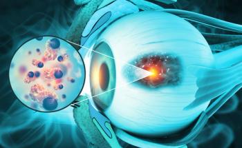
IOWA CITY, Iowa-A surprising new study shows that some melanoma cells can form themselves into vascular channels and provide a tumor’s blood supply without the need for angiogenesis (the growth of new blood vessels from existing blood vessels). The resulting channels, which are lined by melanoma cells and basement membrane (see Figure 1), function as a vascular system for the tumor without the endo-thelium-lined blood vessels produced through angiogenesis.
Advertisement
IOWA CITY, IowaA surprising new study shows that some melanoma cells can form themselves into vascular channels and provide a tumors blood supply without the need for angiogenesis (the growth of new blood vessels from existing blood vessels). The resulting channels, which are lined by melanoma cells and basement membrane (see Figure 1), function as a vascular system for the tumor without the endo-thelium-lined blood vessels produced through angiogenesis.
University of Iowa cancer biologist Mary J. C. Hendrix, PhD, and her colleagues at Iowa, Hadassah University Hospital in Jerusalem, and the National Human Genome Research Institute, discovered this phenomenon of vasculo-genic mimicry in studies of human intraocular (uveal) melanomas. Their work is reported in the September 1999 issue of the American Journal of Pathology (155:739-752, 1999).
The study suggests that aggressive melanomas have a special ability to form their own tumor supply apart from the usual mechanisms of angiogenesis, and that these vascular networks (which can be detected with angiography) may provide a noninvasive method for detecting poor-prognosis disease.
The researchers found that:
Patterned vascular channels found in aggressive primary and metastatic melanomas are different from angiogenic vessels derived from endothelium.
Highly invasive melanoma cells, but not poorly invasive ones, can make similar patterned vascular channels in vitro in the absence of endothelium.
These aggressive melanoma cells are deregulated and express genes associated with embryonic cells, including genes associated with vascular development.
The fact that these deregulated aggressive melanoma cells can build vascular channels suggests a means by which they provide tumor microcirculation and promote metastasis.
Our collaborative team addressed an important question that Dr. Robert Folberg (the pathologist on the study) had been focusing on for years: What is the biological basis for the appearance of microcirculatory networks and patterns in the tissues of patients with poor-prognosis uveal melanoma? Dr. Hendrix said in an interview. The study revealed that the aggressive tumor cells (isolated from patients with a poor prognosis) had the ability to form their own microcirculation in the absence of endothelial cells.
Combined Expertise
The combined biomechanical expertise of Dr. Andrew J. Maniotis (who developed the 3-dimensional assays to prove that aggressive tumor cells could form their own microvasculature) and my cancer biology background, along with Dr. Folbergs pathology expertise, made this study a once in a lifetime discovery, Dr. Hendrix commented. The collaboration also included Paul S. Meltzer, MD, of the National Human Genome Research Institute, who performed the genetic analysis of the tumor cells.
The investigators conducted both in vivo and in vitro studies of human uveal melanomas. Their goal was to determine why melanomas that produce fine web-like patterns of vascular channels are associated with poor prognosis. They approached this by comparing the tube-forming ability of cells from aggressive and nonaggressive melanomas when cultured with endothelial cells.
They expected to find a difference in the ability of cells from these two tumor groups to induce endothelial cells to form blood vessels, but they actually found something much more surprising: Cells from aggressive melanomas could form vascular channels even without the help of endothelial cells. Furthermore, these channels strongly resembled the microcirculatory structures observed in tissue from patients with aggressive uveal melanomas but not in tissue from patients with less aggressive disease.
Possible Use in Tumor Staging
The researchers also found similar channels in metastatic cutaneous melanoma tissue and in liver metastases from uveal melanomas. Dr. Hendrix said that they have made similar observations in other aggressive tumor models and have received independent confirmation from other laboratories that their findings have been repeated and validated.
This is particularly important because channels of this type can be visualized noninvasively in vivo using indocyanine green angiography (Figure 2, left photo), and histopathologic staining shows that these channels are periodic acid-Schiff (PAS)-positive looping patterns and networks (Figure 2, right photo). The association between the density of tumor-generated vascular channels and prognosis may mean that examination of these networks could be used as a noninvasive method for tumor staging.
There is good evidence from our report and also from previous studies by Dr. Folberg and others to suggest that imaging may provide important information regarding the metastatic potential of the tumors under examination, Dr. Hendrix said.
The basic structure of these vascular channels is an inner basement membrane covered by an outer layer of melanoma cells. Red blood cells were frequently observed within these channels. The investigators are currently characterizing the constituents of the basement membrane.
The 3D culture method developed by Dr. Maniotis was key to this project. These cultures are produced by first establishing a polymerized Matrigel or Type I collagen matrix on a coverslip, then seeding tumor cells on top of the gels and watching to see what happens. When cells from aggressive melanomas were seeded onto the gels, they readily formed vascular loops and networks that looked very much like those seen with angiography and in PAS-stained tissue samples. The researchers also found that these channels could hold injected dye and distribute it through the generated network, even in culture. Cells from less invasive melanomas did not form these networks.
Furthermore, this process was not affected by conditioned medium from invasive cells or by a variety of growth factors. It was not inhibited by conditioned medium from poorly invasive cells or by blocking antibodies. These two findings suggest that vascular channel formation is a process intrinsic to the melanoma cells themselves rather than a response to some external signal.
Cultures from aggressive melanomas also had a strong ability to contract and alter the shape of floating collagen gels. Dr. Hendrix said that this is a good test of the ability of cells to remodel and contact extracellular matricesan important mechanism in the formation of blood vessels and microvascular channels.
These behavior differences in cells from the aggressive melanomas led the investigators to ask whether they were expressing different genes than cells from poorly invasive melanomas. Hybridization to cDNA microarrays was used to analyze the expression of 5,000 genes. More than 200 genes were differentially expressed in these aggressive vs less aggressive tumors, and 15 of those have been associated with phenotypes characteristic of endothelial or vascular cells.
This suggests that the more aggressive melanoma cells have moved back toward a more pluripotent, embryonic-like genotype. The aggressive (but not the nonaggressive) melanoma tumor cells somewhat resemble embryonic cells in their ability to express myriad markers and characteristics, Dr. Hendrix said. They also express certain genes similar to those expressed by endothelial cells. However, endothelial cells produce blood vessels, and the aggressive tumor cells in our study produced vascular channels that are not lined by endothelial cells.
The researchers noted that formation of a microcirculation by cells other than endothelial cells has been reported in normal embryonic tissues but not previously in the context of tumor progression.
Treatment Implications
The investigators concluded that their findings call for reappraisal of the current assumption that endothelial-cell-mediated angiogenesis is the only mechanism underlying tumor growth and metastasis. Dr. Hendrix said, This work adds a new dimension to our thinking about the microcirculation of aggressive tumors. It provides an interesting vascular channel formation mechanism and new molecular markers for diagnostic application and therapeutic intervention.
These studies suggest that angiogenesis inhibitors alone are not going to be the magic bullet that kills all solid tumors, since the aggressive tumors in this study were able to form their own functional vascular supply system without angiogenesis or endothelial cells.
I feel that our work offers new opportunities and strategies to target aggressive tumors and may, in fact, complement the ongoing studies of Folkman and colleagues in a very important mannerby directly targeting the tumor cells themselves within an aggressive tumor, Dr. Hendrix said. We must begin testing the efficacy of antiangiogenic agents in our 3D models, in addition to developing specific agents that will target the molecular markers revealed in our study.
This study also raises new questions about drug testing. For example, in studies of angiogenesis inhibitors, will it be necessary to test patients for the presence of endothelium-lined tumor vessels as, for example, breast cancer patients are tested for HER2 before starting treatment with Herceptin (trastuzumab)?
Articles in this issue
over 26 years ago
Anti-VEGF MoAb Promising in Phase II Renal Cancer Studyover 26 years ago
Government Lawsuit Seeks Billions From Tobacco Industryover 26 years ago
NCI Plans a Large Phase III Trial of Lymphoma Vaccineover 26 years ago
NCCN Database Expanding to Include Cancer Pain Outcomesover 26 years ago
Fewer Blacks Than Whites Receive Surgery for Early Stage Lung Cancerover 26 years ago
NSABP Trial Examines Surgery’s Role in Breast Cancerover 26 years ago
Congressional Spouses Honor Three for Cancer Preventionover 26 years ago
Prophylactic Tamoxifen Debated at ECCOover 26 years ago
Survival Advantage for Simultaneous Goserelin and RTover 26 years ago
NCCR Honors Seven Members of Congress as ‘Champions’Advertisement
Related Content
Advertisement


Advertisement
Advertisement
Trending on CancerNetwork
1
Where Does LAG-3/PD-1 Inhibition Fit In PD-1–Refractory Hodgkin Lymphoma?
2
VS-7375 Exhibits Activity in KRAS G12D–Mutated Solid Tumors
3
GLP-1 Receptor Agonists Reduce Colorectal Cancer Risk in IBD Populations
4
Sacituzumab Govitecan Receives FDA Approval Across 2 TNBC Indications
5