patient is a 39-year-old premenopausal woman who presents with a new diagnosis of breast cancer to our multidisciplinary second opinion clinic.
Meenakshi Singh, MD
Advertisement
Articles by Meenakshi Singh, MD
We present a case of intracystic papillary carcinoma of the breast associated with low-grade ductal carcinoma in situ in a young woman. This is a distinct subtype of intraductal carcinoma that typically presents in postmenopausal women with a favorable prognosis.
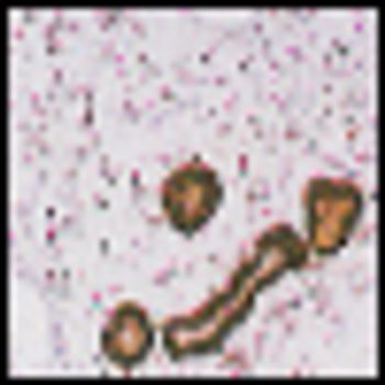
The review of the histology slides revealed predominantly decidual tissue with exaggerated placental site and a small focus of trophoblastic tissue composed of cytotrophoblast and syncytiotrophoblast with mild atypia (Figure 1). However, no necrosis or tissue invasion was identified. No villi were seen.
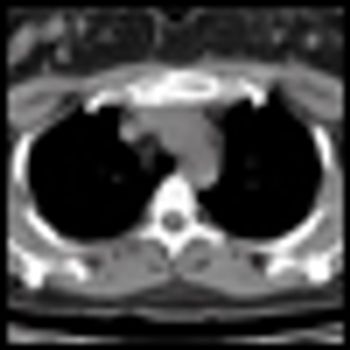
The patient presented to her primary care physician 3 months prior with an inverted left nipple and a palpable lump that was highly suggestive of neoplasm on mammogram. An ultrasound-guided core biopsy revealed an infiltrating solid-type ductal carcinoma in situ. The estimated size of the mass was approximately 1 cm. She had no symptoms suggestive of metastatic disease.
A 46-year-old multiparous (gravida 3, para 3) woman presented to her primary care provider with a palpable vulvar polypoidal mass, measuring 7 cm in greatest dimension. The mass was painless and had been growing in size over the past 2 years. Her medical history was remarkable for obesity, hypothyroidism, and an appendectomy at age 17. Her family history was significant for a sister with breast cancer, diagnosed at age 34. A core biopsy was performed.
In this installment of Second Opinion, we are presenting two cases of tumors of the female genital tract, specifically, the ovary and uterus, which contain both epithelial and mesenchymal components and therefore have unique diagnostic and therapeutic implications. The first has an unusually poor prognosis and the second is notoriously difficult to diagnose.
Advertisement
Latest Updated Articles
 Biphasic Tumors of the Female Genital Tract
August 1st 2005
 Polypoid Lesions of the Lower Female Genital Tract
April 1st 2006
- Does This Woman Have Gestational Trophoblastic Disease?
November 17th 2006

Advertisement
Advertisement
Trending on CancerNetwork
1
From Conference to Practice: Top 5 Takeaways From EHA 2026
2
ASCO 2026: Key Insights In Renal Cell Carcinoma, Merkel Cell Carcinoma, and Chronic Myeloid Leukemia
3
3 Things You Should Know About Cutaneous Squamous Cell Carcinoma
4
Emerging T-Cell Engagers and Novel Immunotargets in Multiple Myeloma
5