Articles by Anthony D. Elias, MD

98 A Phase 1/2 Trial of LY4064809, A Pan-Mutant-Selective PI3Kα Inhibitor in HR+/HER2− Advanced Breast Cancer, Updated Results From PIKALO-1
ByKomal L. Jhaveri,Dejan Juric,Antonio Giordano,Antoine Italiano,Gennaro Daniele,Jordi Rodón,Amita Patnaik, MD,Jorge Bartolomé,Anthony D. Elias, MD,Pamela N. Munster, MD,Patricia Lorusso, DO,Aixa Soyano,Maria de Miguel,Joyce O’Shaughnessy, MD,Robert Wesolowski,Giuseppe Curigliano,Tatiana Hernández,Aurélie Lombard,Shawn T. Estrem,Lin Du,Xiaofan Xu,Alberto J. Montero, MD, MBA, CPHQ 
Breast Cancer Following Radiation for Hodgkin Lymphoma: Clinical Scenarios and Risk-Reducing Strategies
ByLinda Overholser, MD, MPH,Elena Shagisultanova, MD, PhD,Rachel Rabinovitch, MD,Nicole Kounalakis, MD,Jennifer Diamond, MD,Christina A. Finlayson, MD,Christine M. Fisher, MD, MPH,Peter Kabos, MD,Anthony D. Elias, MD,Virginia F. Borges, MD, MMSc,Jose Mayordomo, MD, PhD We review available strategies for screening and risk reduction through chemoprevention or risk-reducing surgery, as well as challenges for management of breast cancer in patients with prior exposure to radiation for Hodgkin lymphoma.
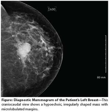
A 55-year-old perimenopausal woman presented with a palpable lump in her left breast. Diagnostic mammogram showed a 1.8-cm spiculated mass with scattered microcalcifications within the mass. Comparison with her most recent prior mammogram (about 9 months earlier) showed this to be a new mass.
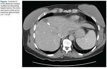
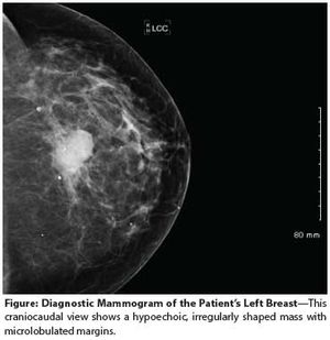
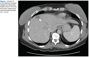
Multiple Hepatic Lesions in a Patient With a History of DCIS
ByJagar Jasem, MD, MPH,Basel Altoos, BS,Christine M. Fisher, MD, MPH,Anthony D. Elias, MD,Nicole Kounalakis, MD,Virginia F. Borges, MD, MMSc,Peter Kabos, MD An asymptomatic 45-year-old woman presented for a screening mammogram and was noted to have a soft-tissue opacity with calcifications in the left breast. Ultrasound revealed a highly suspicious mass.
Management of Young Breast Cancer Patients With de Novo Genetic Mutations
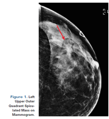
ByChristine M. Fisher, MD, MPH,Catherine E. Klein, MD,Laxmi A. Kondapalli, MD, MSCE,Anthony D. Elias, MD,Virginia F. Borges, MD, MMSc A 24-year-old woman presents to her primary care provider with a mass in her left breast. Examination confirms a 2.2-cm mass in the upper outer quadrant, with a single mobile axillary node that is firm to palpation.
The Case: A 48-year-old perimenopausal woman noted a lump in her left breast. She had had a mammogram 9 months earlier without abnormality. After ultrasound imaging confirmed a solitary mass measuring about 1.5 cm, a core needle biopsy demonstrated a poorly differentiated mammary carcinoma with chondroid features.
Diagnosis of Invasive Lobular Carcinoma in a Young Woman Presenting With Pleomorphic Lobular Carcinoma in Situ on Core Biopsy
ByNicole Kounalakis, MD,Jennifer Diamond, MD,Kyle Rusthoven, MD,Wendy Horn, MD,Sonali Jindal, MD,Josh Wisell, MD,Catherine E. Klein, MD,Anthony D. Elias, MD,Christina A. Finlayson, MD,Virginia F. Borges, MD, MMSc A 40-year-old premenopausal woman with a new diagnosis of invasive lobular carcinoma occurring in a background of lobular carcinoma in situ presents to a multidisciplinary second opinion clinic.
Early-Stage BRCA2-Linked Breast Cancer Diagnosed in the First Trimester of Pregnancy Associated With a Hypercoagulable State
ByJennifer R. Diamond, MD,Christina A. Finlayson, MD,Christiane Thienelt, MD,Peter Kabos, MD,Laura Hardesty, MD,Linda Barbour, MD, MSPH,Catherine E. Klein, MD,Rachel Rabinovitch, MD,Anthony D. Elias, MD,Virginia F. Borges, MD, MMSc This feature examines the case of a patient with newly diagnosed breast cancer in the setting of a first-trimester pregnancy presenting to our multidisciplinary breast cancer clinic.
A Young Woman With a Small ER-Positive Breast Cancer, a Micrometastatic Axillary Lymph Node, and an Intermediate Oncotype DX Recurrence Score
ByDaniel W. Bowles, MD,Rachel Rabinovitch, MD,Virginia F. Borges, MD, MMSc,Alexander Urquhart, MD,Meenakshi Singh, MD,Christina A. Finlayson, MD,Anthony D. Elias, MD patient is a 39-year-old premenopausal woman who presents with a new diagnosis of breast cancer to our multidisciplinary second opinion clinic.
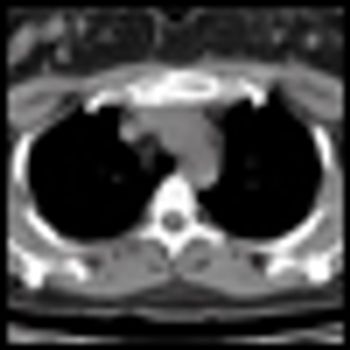
A Woman With Primary Breast Cancer and a Solitary Sternal Metastasis
ByAlexander Menter, MD,Alexander Urquhart, MD,Virginia F. Borges, MD, MMSc,Rachel Rabinovitch, MD,Meenakshi Singh, MD,William Robinson, MD, PhD,Paul Seligman, MD,Christina A. Finlayson, MD,Anthony D. Elias, MD The patient presented to her primary care physician 3 months prior with an inverted left nipple and a palpable lump that was highly suggestive of neoplasm on mammogram. An ultrasound-guided core biopsy revealed an infiltrating solid-type ductal carcinoma in situ. The estimated size of the mass was approximately 1 cm. She had no symptoms suggestive of metastatic disease.