
|Articles|February 11, 2009
Addressing Bone Loss in the Cancer Survivor
Osteoporosis, the most common late effect of cancer treatment in the US, occurs with greater frequency among cancer survivors than the general population. Survivors of breast cancer, prostate cancer, and childhood leukemia are at particularly high risk for changes in bone mineral density (BMD) / osteoporosis that can lead to fractures.[1] In breast and prostate cancer patients, bone effects are often the result of endocrine therapy–induced alterations in bone microarchitecture. They also can be caused by other types of cancer therapy, vitamin D deficiency, and other physiological changes that may or may not be related to cancer or its treatment. In childhood leukemia patients, bone effects can be caused by a variety of factors, including corticosteroid therapy, radiation therapy to the brain, and the disease itself.
Advertisement
Osteoporosis, the most common late effect of cancer treatment in the US, occurs with greater frequency among cancer survivors than the general population. Survivors of breast cancer, prostate cancer, and childhood leukemia are at particularly high risk for changes in bone mineral density (BMD) / osteoporosis that can lead to fractures.[1] In breast and prostate cancer patients, bone effects are often the result of endocrine therapy–induced alterations in bone microarchitecture. They also can be caused by other types of cancer therapy, vitamin D deficiency, and other physiological changes that may or may not be related to cancer or its treatment. In childhood leukemia patients, bone effects can be caused by a variety of factors, including corticosteroid therapy, radiation therapy to the brain, and the disease itself.
Osteoporosis can exist for many years without symptoms, and the presence of a clinically symptomatic fracture may be the first indication that bone loss has occurred. Given that the majority of cancer cases occur in people over 65 years of age, and even healthy older people are at higher risk of bone changes than younger ones (eg, owing to hormonal changes, nutritional differences, lifestyle changes including reduced mobility and lack of exercise, etc), the health care team must be vigilant in screening, monitoring, and intervention for bone effects in the cancer survivor.
Naturally, bone loss in the growing adolescent also is devastating, as this is the time of life when peak bone mass normally is achieved. Efforts to preserve bone integrity are critically important in this patient population as well. In both adult and pediatric cases, patient management can be complicated by bone metastases.
Bone health in both childhood and adult cancer survivors is receiving increased attention. The American Society of Clinical Oncology (ASCO) recommends that “cancer survivors at risk for bone and joint problems, especially survivors of breast and prostate cancers and childhood leukemia, can lower their risk by not smoking, eating foods rich in calcium, participating in regular physical activity, and limiting the amount of alcohol they drink.”[1]
Oncology nurses are well positioned to educate cancer survivors about newer agents (eg, bisphosphonates) and lifestyle changes that can help to protect against treatment- and disease-related bone loss. This article presents two case studies that highlight key risk factors for bone loss in pediatric and adult cancer survivors and provide guidance on appropriate assessment and nursing intervention.
PATIENT OVERVIEWS
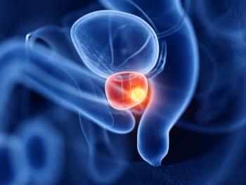
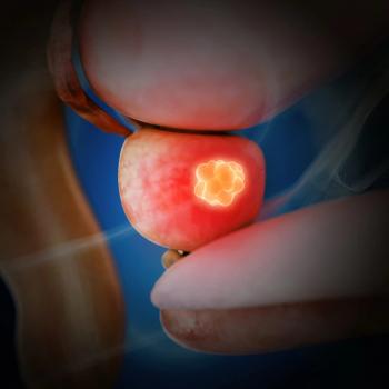
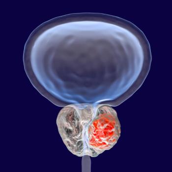
Patient 1: Stage II Prostate Cancer Survivor
JM is a 60-year-old male who was diagnosed 6 years ago with prostate cancer, clinical stage II. The primary tumor was considered to be confined within the prostate, but with both lobes involved. His prostate specific antigen (PSA) level was 18 ng/mL and his Gleason score at diagnosis was 3+5, or 8, indicating the cells were poorly differentiated and his disease course was likely to be aggressive.[2] After carefully considering his treatment options, he chose radical prostatectomy. National Comprehensive Cancer Network (NCCN) guidelines indicate he was at high risk for recurrence (based on his high Gleason score).[3]
Following surgery, JM’s disease was reclassified as Stage IV, because cancer was detected in two lymph nodes. He was given the option of active surveillance or treatment with androgen-deprivation therapy (ADT). He underwent ADT using a combination of flutamide (Eulexin) and leuprolide acetate (Lupron) for the purpose of medical castration.
JM experienced predictable side effects from his ADT regimen, including erectile dysfunction, hot flashes, and fatigue. One year following initiation of ADT, his dual energy X-ray absorptiometry (DXA) scan indicated a 2% loss in bone density in his hip and a 5% radial ulnar loss, compared with his baseline scan. This finding is consistent with the research literature, wherein BMD decreases ranging from 1.8% to 3.9% at the hip and up to 10% at the radius have been reported.[4]
Even greater losses have been documented at other sites such as the lumbar spine and femoral neck (7.1% and 6.6% respectively).[5] A review by Saad et al. of research related to BMD loss suggests bone density decreases progressively with increased duration of ADT, and fractures are independent adverse predictors of survival among prostate cancer patients.[6] The major risk factors for osteoporosis in men, aside from hypogonadism, include prior fracture after age 40, family history of osteoporotic fractures, low baseline bone density, advanced age (greater than 65 years), and apparent osteopenia on X-ray. Minor contributory risk factors include comorbidities such as chronic inflammatory disease (eg, rheumatoid arthritis, lupus), celiac disease, concomitant use of mediations such as heparin, anticonvulsants, barbiturates, etc, weight < 57 kilograms, cigarette smoking, excessive alcohol intake (three or more drinks per day), low dietary calcium intake, and high caffeine intake.[6]
Along with his ADT, JM’s risk factors for osteoporosis and future fracture included a 20 pack-year history of smoking and one fracture sustained at age 41.
Although there is no US Food and Drug Administration–approved treatment or pharmaceutical prevention for ADT-induced bone density loss, recent findings support use of bisphosphonates to attenuate bone loss. Optimal dosing has not yet been established, however. The length of follow-up is insufficient at this point to evaluate the long-term effects of bisphosphonate therapy. JM’s bone loss was treated with zoledronic acid (Zometa), one of the most commonly investigated bisphosphonates, at a dose of 4 mg intravenously every 3 months.
The scientific rationale for use of bisphosphonates is their ability to decrease bone resorption through their preferential binding to mineralized bone matrix, especially in areas of increased turnover or resorption. Their mechanism of action is multifaceted and includes inhibition of tumor cell binding to bone matrix, reduction of osteoclast development, apoptosis induction in both tumor cells and osteoclasts, reduction in IL-6 production, and inhibition of angiogensesis.[7]
One year following the initiation of bisphosphonate therapy, JM’s follow-up DXA scan has revealed no further bone loss. A 1.4% increase in bone density was documented at the hip.
NURSING MANAGEMENT
Appropriate nursing management begins with risk assessment and the implementation of preventive strategies. JM was at high risk for development of bone loss owing to his history of ADT, smoking, and fracture. He was also at high risk for development of bone metastases, another threat to his skeletal integrity. Teaching at this time included information about his risk factors and lifestyle changes that could be initiated immediately, emphasizing that osteoporosis is manageable and fractures are potentially preventable.
Specific nursing interventions for JM included assistance with smoking cessation, which included identification of local resources and assessment of his commitment to quit, as well as discussion with the physician about use of nicotine patches and potentially augmentive therapies. Serum 25-hydroxy-vitamin D levels were assessed and calcium therapy with vitamin D supplementation was begun.
He was instructed on the benefits of weight-bearing and weight-training exercise, and an initial exercise plan was developed that both he and his wife felt they could commit to together. Weight-bearing exercise, in which bones and muscles work against gravity, includes activities such as walking, jogging, Tai-Chi, etc, while muscle-strengthening exercises include weight and resistance training. JM was advised to consult his primary care provider prior to initiating any vigorous exercise program, for cardiopulmonary risk assessment.
Treatment with zoledronic acid, at a dose of 4 mg IV every 3 months, was initiated. Dosing regimens are variable, however, and more frequent dosing may be required based on the desired level of effect. Dosing and infusion time also may require adjustment based on the patient’s renal function. Monitoring serum creatinine is a critical nursing function.
JM tolerated the zoledronic acid well, although within 24 hours of both the first and second treatments he developed a fever, with oral temperatures of 100.8 °F and 101°F. When other causative factors were ruled out, it was concluded that he was experiencing a febrile response to the zoledronic acid. Such responses have been documented in the literature, as well as the observation that zoledronic acid induces increased levels of interleukin-6 (IL-6) and tumor necrosis factor alpha.
Increases in these circulating cytokines, particularly IL-6, were found to be higher among patients experiencing fever post zoledronic acid vs patients who remained afebrile.[8] JM was treated with acetaminophen 650 mg every 6 hours as needed, with successful resolution of his fever. Intravenous bisphosphonates are generally well tolerated with reports of mild to moderate flu-like symptoms following initial infusions. Symptoms are generally managed with over-the-counter remedies.[9]
Although JM experienced no further side effects of bisphosphonate therapy, one other important aspect of nursing management included assessment for and teaching about osteonecrosis of the jaw. Though this is a rare occurrence, all patients should have a complete dental examination prior to receiving bisphosphonate therapy.[10]
DISCUSSION
Treatment of prostate cancer now includes use of ADT even in the absence of bone metastases. For men receiving ADT in the adjuvant setting, the cancer survival period can be quite long, making bone health preservation a significant issue in terms of quality of life and reduction of further morbidity. Use of ADT in the setting of bone metastases increases the risk of skeletal fractures in already fragile bones.
Assessment and monitoring of serum vitamin D levels (normal serum 25 hydroxy vitamin D level is > 30 ng/mL) is essential for optimal absorption of calcium. The National Osteoporosis Foundation recommends that adults under 50 years of age require 400–800 IU of vitamin D daily, while adults age 50 and older need 800–1,000 IU of vitamin D daily. There are primarily two vitamin D supplements available: vitamin D3 (cholecalciferol) and vitamin D2 (ergocalciferol). Research indicates that they are equally efficacious. Dietary sources rich in vitamin D include fortified milk, egg yolks, saltwater fish, liver, and supplements.[11]
Although there is no FDA-approved preventive therapy for cancer treatment–induced bone loss at this time, numerous studies have demonstrated reductions in skeletal fractures and the mitigation of bone loss by early initiation of bisphosphonate therapy.[6] Bisphosphonates are now used fairly commonly for prevention of ADT-induced bone loss among men with a history of prostate cancer, or with active disease. Further research is needed (controlled double-blind studies) to determine optimal timing for initiation of therapy, optimal dosing, and which agents are preferable for the prevention vs treatment of ADT-induced bone loss.
Patient 2: Pediatric ALL Survivor
le who in 1990 was diagnosed with standard risk ALL. She was treated with chemotherapy including vincristine, L-asparaginase, 6-mercaptopurine, methotrexate, prednisone, and central nervous system (CNS) prophylaxis with intrathecal methotrexate, hydrocortisone, and cytarabine. During maintenance therapy, CC had a CNS relapse and went on to have a matched unrelated donor bone marrow transplant (BMT). Her preparative regimen included cyclophosphamide and total body irradiation (1,200 cGy) as well as a 600 cGy cranial spinal boost.
At age 15, she was noted to have inappropriate secondary sex characteristics and no menarche, and was referred to endocrinology. She was diagnosed with gonadal failure and begun on birth control pills for hormone replacement.
At a routine survivor visit, CC underwent BMD screening with a DXA scan. Results of the DXA included total body BMD T score = −1.0, lumbar spine 1–4 T score = −2.0, and hip/femur T score = −2.1. CC therefore was diagnosed with osteopenia. Upon inquiry it was noted that CC had limited dairy/calcium-rich foods in her diet and engaged in no regular exercise. Intervention for her osteopenia included calcium supplementation at a dose of 1,200 mg with vitamin D 800 IU supplements.CC also was given verbal and written information about calcium-rich foods and instructed on ways to increase physical activity. The planned follow-up for CC will include repeat DXA scan at a 1-year interval.
Nursing Management
Adequate BMD is important for any age group but is particularly important for children and adolescents. Peak bone mass is achieved around the age of 20 years and declines with further aging. One of the most important predictors of risk for developing osteoporosis during one’s lifetime is peak bone mass. Disease factors including cytokine effects and alterations in vitamin D metabolism may put cancer survivors at risk for diminished BMD.[12,13]
In addition, chemotherapy regimens including methotrexate and corticosteroids have been shown to disrupt the delicate balance between osteoblast (bone forming) and osteoclast (bone resorbing) mechanisms, which are responsible for maintaining normal BMD.[14] Finally, nutritional disruptions and decreased physical activity during therapy as well as long-term complications related to the endocrine system may contribute to diminished BMD in childhood cancer survivors.[15]
DXA, the gold standard for evaluating BMD, provides Z-scores (comparison to age-matched norms) and T-scores (comparison to young adult norms). As defined by the World Health Organization, T scores of – 1 to – 2.5 indicate osteopenia and scores of – 2.5 or lower indicate osteoporosis, where T scores < – 1 represent normal BMD.[16] DXA is used primarily to evaluate BMD of the total body, lumbar spine, and hip/femur.
It is important for nurses to also be aware of appropriate interventions for patients found to have osteopenia or osteoporosis at screening. Dietary counseling, use of calcium and vitamin D supplements, and instruction about appropriate weight-bearing exercises should be made available to patients with osteopenia (Table 3). For the more serious condition of osteoporosis, referral to endocrinology should be considered, and pharmacologic intervention, including with bisphosphonates, may be necessary.[17] Patients with complicated medical conditions, such as a history of renal calculi, may need coordination of specialty care to treat diminished BMD safely.
CONCLUSION
Studies confirm that as many as 21% to 39% of childhood cancer survivors may be at risk of diminished BMD.[15] The asymptomatic nature of this condition makes it critical that nurses utilize appropriate screening to identify patients who may have osteopenia or osteoporosis. Furthermore, knowledge of the interventions to treat diminished BMD will allow the nurse to provide appropriate care and will help to ensure that long-term skeletal complications such as fracture do not occur. A lifetime of healthy bones begins in childhood. For children at risk for diminished BMD, nurses are key advocates of a strong, healthy skeleton.
References:
1. American Society of Clinical Oncology. Cancer.Net, Late Effects. Available at: http://www.asco.org/patient/Survivorship/Late+Effects. Accessed on December 16, 2008.
2. Gleason D, Mellinger G: Prediction of prognosis for prostate adenocarcinoma by combined histological grading and clinical staging. J Urol 11:58â64, 1974.
3. National Comprehensive Cancer Network (2009). Clinical Practice Guidelines in Oncology. Prostate cancer. Available at: http://www.nccn.org/professionals/physician_gls/PDF/prostate.pdf. Accessed on December 6, 2008.
4. Eastham JA: Bone health in men receiving androgen deprivation therapy for prostate cancer. J Urol 177(1):17â24, 2007.
5. Maillefert JF, Sibilia J, Michel F, et al: Bone mineral density in men treated with synthetic gonadotropin-releasing hormone agonists for prostatic carcinoma. J Urol 161(4):1219â1222.
6. Saad F, Adachi JD, Brown JP, et al: Cancer treatment-induced bone loss in breast and prostate cancer. J Clin Oncol 26(33):5465â5476, 2008.
7. Tu SM, Lin SH: Current trials using bone-targeting agents in prostate cancer. Cancer J 14(1):35â39, 2008.
8. Dicuonzo G, Vincenzi B, Santini D, et al: Fever after zoledronic acid administration is due to increase in TNF-alpha and IL-6. J Interferon Cytokine Res 23(11):649â654, 2003. Available at: http://www.liebertonline.com/doi/abs/10.1089/107999003 322558782. Accessed on December 8, 2008.
9. Maxwell C, Swift R, Goode M, et al: Advances in supportive are of patients with cancer and bone metastases: Nursing implications of zoledronic acid. J Oncol Nurs 7(4):403â408, 2003.
10. American Dental Association (2006). Dental management of patients receiving oral bisphosphonate therapy-Expert panel recommendations . Report of the council on scientific affairs. Available at: http://haughtondentist.com/pdf/topics_osteonecrosis_recommendations.pdf. Accessed on December 7, 2008.
11. National Osteoporosis Foundation (2008). Clinicians guide to prevention and treatment of osteoporosis. Available at: http://www.nof.org/professionals/NOF_Clinicians_Guide.pdf. Accessed on December 7, 2008.
12. Hansen MA, Overgaard K, Riis BJ, et al: Role of peak bone mass and bone loss in postmenopausal osteoporosis: 12 year study. BMJ 303:961â964, 1991.
13. Arikoski P, Komulainen J, Riikonen P, et al: Alterations in bone turnover and impaired development of bone mineral density in newly diagnosed children with cancer: A 1-year prospective study. J Clin Endocrinol Metab 84(9):3174â3181, 1999.
14. van Leeuwen BL, Kamps WA, Jansen HW, et al: The effect of chemotherapy on the growing skeleton. Cancer Treat Rev 26(5):363â376, 2000.
15. Leung W, Ahn H, Rose SR, et al: A prospective cohort study of late sequelae of pediatric allogeneic hematopoietic stem cell transplantation. Medicine (Baltimore) 86(4):215â224, 2007.
16. World Health Organization: Prevention and Management of Osteoporosis: Report of a WHO Scientific Group. (WHO Technical Report Series, No. 921). Geneva, Switzerland, 2003. Available at: http://whqlibdoc.who.int/trs/WHO_TRS_921.pdf. Accessed on January 23, 2009.
17. Adler RA: Cancer treatment-induced bone loss. Curr Opin Endocrinol.Diabetes Obes 14(6):442â445, 2007.
18. Kaste SC: Bone-mineral density deficits from childhood cancer and its therapy. A review of at-risk patient cohorts and available imaging methods. Pediatr Radiol 34(5):373â378, 2004.
19. Children’s Oncology Group: Bone Health/Health link. Children’s Oncology Group/Survivorship Guidelines [online]. Version 2, March 2006. Available at: http://www.survivorshipguidelines.org/pdf/BoneHealth.pdf. Accessed on December 22, 2008.
20. Halton JM, Atkinson SA, Fraher L, et al: Mineral homeostasis and bone mass at diagnosis in children with acute lymphoblastic leukemia. J Pediatr 126(4):557â564, 1995.
21. Mayo Clinic: Exercising With Osteoporosis: Stay Active the Safe Way. Mayo Foundation for Medical Education and Research [online]. October 2008. Available at: http://www.mayoclinic.com/health/osteoporosis/HQ00643. Accessed December 22, 2008.
22. National Institutes of Health (2005). Dietary Supplement Fact Sheet: Calcium. Office of Dietary Supplements [online]. Available at: http://ods.od.nih.gov/factsheets/calcium.asp
23. Taylor R: Weight Bearing Exercises: 8 Workouts for Strong Bones. Web MD [online] (2007). Available at: http://www.webmd.com/osteoporosis/living-with-osteoporosis-7/exercise-weight-bearing. Accessed on December 22, 2008.
24. Wasilewski-Masker K, Kaste SC, Hudson MM, et al: Bone mineral density deficits in survivors of childhood cancer: Long-term follow-up guidelines and review of the literature. Pediatrics 121(3):e705âe713, 2008.
Advertisement


Advertisement
Advertisement
Trending on CancerNetwork
1
FDA Approves Blood-Based Test for Colorectal Cancer Screening
2
Steel Particulate Matter Prompts Cyclophosphamide for Injection Recall
3
Belzutifan Combo Exhibits Comparable HRQoL Vs Cabozantinib in RCC
4
Optimizing Frontline Therapeutic Strategies in EGFR-Mutated Advanced NSCLC
5