- ONCOLOGY Vol 14 No 4
- Volume 14
- Issue 4
Adenocarcinoma of the Esophagus: Risk Factors and Prevention
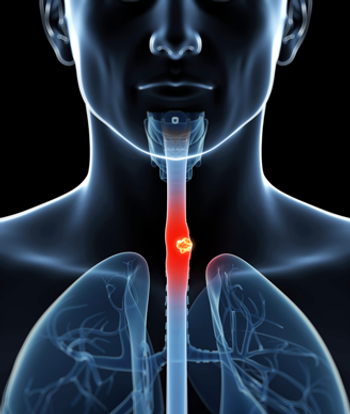
Esophageal cancer poses an interesting challenge for oncologists. Esophageal squamous cell cancer has the most varied geographical incidence of any cancer, suggesting the existence of critically important environmental and molecular epidemiologic factors. These factors remain largely unrecognized.
Esophageal cancer poses an interesting challenge for oncologists. Esophageal squamous cell cancer has the most varied geographical incidence of any cancer, suggesting the existence of critically important environmental and molecular epidemiologic factors. These factors remain largely unrecognized.
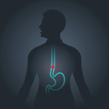
Equally puzzling is the dramatic increase in the incidence of adenocarcinomas of the esophagus and gastro-esophageal junction or cardia that has occurred in western societies during the past 3 decades.[1,2] This increase in incidence is particularly disturbing in view of the highly lethal nature of esophageal cancer. For the year 2000, the estimated number of new cases of esophageal cancer in the United States is 12,300 and the estimated number of deaths due to this cancer is 12,100.[3]
In response to these challenges, there has been a great increase in the amount of research and the number of publications on malignant and premalignant esophageal diseases. In the 2-week period following January 26, 2000, for example, 136 new English language entries using any of the key words esophageal neoplasms, Barretts esophagus, gastric cardia, and gastro-esophageal reflux were added on
Medline. Many readers of Oncology are thus likely to welcome the efforts by Forastiere et al to review this increasing mass of information.
Barretts Esophagus and Esophageal Adenocarcinoma
The main risk factor for esophageal adenocarcinoma is the presence of Barretts esophagus. This is currently defined by most investigators as the replacement of the normal squamous epithelium of the distal esophagus by a visible segment of columnar mucosa containing intestinal metaplasia on microscopic examination.
Similar to adenocarcinoma, the incidence of Barretts esophagus has been rising rapidly,[4,5] suggesting that the increase in esophageal adenocarcinoma incidence is explained, at least in part, by this increase in Barretts. However, another possible explanation is that the proportion of patients with Barretts who progress to malignancy has increased. The latter explanation is not supported by recent prospective analyses of the risk of cancer developing in patients with Barretts,[6,7] but large population-based studies are needed to properly evaluate this possibility.
It may be that the incidence of nonvisible intestinal metaplasia, termed either ultra-short segment Barretts esophagus or cardiac mucosa with intestinal metaplasia, is increasing. Indeed, recent studies have found intestinal metaplasia at the gastroesophageal junction in 9% to 36% of individuals undergoing endoscopy.[8-10] The normal-appearing gastroesophageal junction was rarely studied prior to the mid-1990s. Consequently, a rise in intestinal metaplasia at this site cannot be confirmed. It has been hypothesized that the increasing incidence of adenocarcinoma at the gastroesophageal junction or cardia is a consequence of this putative increase in the incidence of nonvisible areas of intestinal metaplasia.
As Forastiere et al note, there is considerable variability in the reported risk of developing adenocarcinoma within a segment of Barretts esophagus. In part, this reflects the fact that none of the reported prospective studies has included sufficient numbers of patients to make definitive estimates, and that the size of a study required to provide these estimates is prohibitively large.
Furthermore, because only a small proportion of Barretts mucosa is usually biopsied at endoscopy, there is considerable risk that patients with Barretts are staged incorrectly for the presence and grade of dysplasia, thus confounding estimates of the cancer risk in supposedly nondysplastic Barretts epithelium. Even when Barretts segments are carefully evaluated histologically, with a large number of biopsies taken throughout the Barretts segment, conventional examination of the biopsy specimens using histopathologic techniques may be insufficient to stratify patients according to cancer risk. This is because areas of esophageal mucosa that are similar histologically can be very different genetically.
Studies at our institution have found that molecular differences are particularly marked if either adenocarcinoma or dysplasia is present within the esophagus, even if adenocarcinoma or dysplasia is not present in the biopsy sample.[11-13] Thus, it is likely that individual patients with Barretts esophagus will eventually need to be staged for risk of disease progression by a combination of molecular and conventional pathologic methods (molecular pathology), and that the risk of malignant degeneration in populations with Barretts esophagus will need to be investigated using the tools of molecular epidemiology.
GERD and Esophageal Adenocarcinoma
In both studies that addressed this question, gastroesophageal reflux disease (GERD), as identified by symptoms of that disease, was found to be a significant risk factor for esophageal adenocarcinoma.[14,15] As Forastiere et al note, Lagergren et al found that the strength of the association with reflux symptoms was very similar in esophageal adenocarcinoma patients in whom Barretts esophagus was and was not detected.[15]
This finding has been interpreted by Lagergren et al, and by Forastiere et al in this review, to indicate that reflux has an effect on the development of esophageal adenocarcinoma that is independent of Barretts esophagus, and that gastroesophageal reflux, rather than Barretts esophagus may be the crucial factor in causing esophageal adenocarcinoma.[15] This controversial statement has been dismissed by some commentators because of the likelihood of tumor overgrowth of the Barretts segment and the risk of sampling error. Without Barretts columnar metaplasia as an intermediate step, it is difficult to explain how adenocarcinoma can develop from squamous epithelium.
Nevertheless, the findings of Lagergren et al and similar findings by others compel us to consider alternative mechanisms of tumor development. One possibility is that a renegade cell and its clone of progeny may progress to malignancy without significant clonal expansion in terms of the surface area of the esophagus involved.
Alternatively, the traditional thinking that goblet cells are required for cancer risk may be incorrect. Indeed, our observations suggest that goblet cells are progressively lost in the sequence of progression from Barretts metaplasia to dysplasia to adenocarcinoma. It may be that, if sufficient genetic alterations are present, cancer can arise in some cases from columnar metaplasia without goblet cells.
Antireflux Surgery
Forastiere et al fail to include antireflux surgery among the available treatments for GERD in their discussion of acid peptic disorders. Although in their subsequent review of cancer prevention, they note the ability of surgical fundoplication to prevent esophageal exposure to both acid and duodenal alkaline reflux, no details or references are provided. In view of the large number of studies that have confirmed the effectiveness of antireflux surgery for GERD, this omission is an oversight.
Both the two randomized trials[16,17] and two nonrandomized trials[18,19] that compared medical and surgical therapy for GERD found significant advantages for surgical treatment. Furthermore, a prospective, randomized comparison of medical and surgical therapy in patients with Barretts esophagus showed that both treatments provided good symptom control, but the medically treated patients had a higher prevalence of post-treatment persistent esophagitis (53%) and stricture (45%) than did the operated group (esophagitis, 5%; stricture, 15%). Dysplasia developed in 6 (22%) of 27 patients during medical treatment but in only 1 (3%) of 32 patients after antireflux surgery.[20] In the surgical patient who developed dysplasia, pH monitoring revealed that fundoplication had been ineffective.[20]
Similarly, an analysis of patients with Barretts esophagus in the American College of Gastroenterology registry indicated that dysplasia developed in 19.7% of patients treated medically but in only 3.4% of those treated surgically.[21] Our group reported that complete regression of visible segments of intestinal metaplasia was very unusual after antireflux surgery (as it is with medical therapy), but that complete regression of microscopic-only intestinal metaplasia at the gastroesophageal junction occurred in 73% of patients.[22]
Interestingly, in the population-based study by Lagergren et al discussed above, patients who used medications to relieve symptoms of reflux for at least 5 years before being interviewed had a greater risk of esophageal adenocarcinoma than did individuals who were matched for severity of symptoms but did not use such medications (odds ratio, 2.9; 95% confidence interval [CI], 1.9 to 4.6). Antireflux surgery, in contrast, was not associated with an increased risk of adenocarcinoma of the esophagus.[15]
Mechanisms by which acid suppressant medications may adversely affect the risk of developing esophageal adenocarcinoma are discussed elsewhere.[23] Large, randomized studies are needed to further investigate the significance of this trend toward the superiority of surgical treatment. Until the results of those trials are available, however, we believe that the published data justify regarding a properly performed antireflux operation as the gold standard therapy for reflux disease and for Barretts esophagus with intestinal metaplasia or low-grade dysplasia.
Mucosal Endoscopic Ablation
For patients in whom low-grade dysplasia fails to regress after antireflux surgery, consideration of post-fundoplication mucosal endoscopic ablation[24] may be appropriate. Forastiere et al review the use of mucosal ablation in patients who have adenocarcinoma or Barretts esophagus with high-grade dysplasia. As they note, significant complications (including the presence of subsquamous foci of cancer after treatment) can follow these mucosal ablation procedures. These procedures remain experimental, and it is important that physicians, and the patients to whom they are offered, are aware of their limitations.
Mucosal endoscopic ablation is being used in patients with early cancers that are confined to the esophageal wall, with no apparent lymph node spread. Unfortunately, it is impossible to be certain of the lymph node status of these patients. Currently available staging methods, including endoscopic ultrasound, cannot accurately detect the presence of lymph node metastases in many patients. In our experience, 41% of patients with tumors confined to the esophageal wall will have lymph node involvement.[25] The fact that some patients with locoregional disease, including lymph node metastases, are cured after surgery alone indicates that resection of involved lymph nodes in patients without distant metastases can be beneficial. This benefit is not obtained with mucosal ablation techniques.
The appropriateness of mucosal ablation for treating high-grade dysplasia can also be questioned. Previously undetected cancers are present in the resected esophagus in almost 50% of patients undergoing esophagectomy for high-grade dysplasia.[26] The 5-year survival rate for these patients after esophagectomy is 90%.[27]
Thus, esophagectomy in patients with high-grade dysplasia is not the most aggressive form of prevention, as Heath et al state; rather, it is the most prudent therapy, preventing cancer in half of patients and curing this usually incurable cancer in 90% of the others. We believe that it is more aggressive to use an experimental procedure in patients who could expect a high cure rate if treated by standard methods. If mucosal ablation techniques are to be used in patients with cancer and high-grade dysplasia, this should be done only in the context of a randomized, controlled trial.
Esophageal Adenocarcinoma in Patients With Achalasia
Although Forastiere et al correctly state that there is no known association between achalasia and esophageal adenocarcinoma, it is worth noting that
Barretts esophagus and esophageal adenocarcinomas can occur in patients with achalasia. In these patients, severe GERD has developed after iatrogenic disruption of the lower esophageal sphincter, with the impaired clearance of refluxed material by the aperistaltic esophageal body no doubt a contributing factor.[28] For this and other reasons, we advocate laparoscopic Hellers myotomy with partial fundoplication for the treatment for achalasia.
Chemoprevention
As Forastiere et al note, there is considerable interest in developing rational chemoprevention strategies to prevent the development of adenocarcinoma in patients with Barretts esophagus. One of the most promising approaches involves inhibition of cyclooxygenase 2 (COX-2) expression using COX inhibitors, such as aspirin and nonsteroidal anti-inflammatory drugs (NSAIDs), or selective COX-2 inhibitors, such as celecoxib (Celebrex) and rofecoxib (Vioxx). Cyclooxygenase-2 activity has been implicated as an important factor in tumorigenesis in general, and COX-2 is involved in a large number of processes fundamental to tumor development.
As reviewed by Forastiere et al, three epidemiologic studies have shown that use of aspirin or NSAID is associated with a significantly lower risk of either developing, or of dying from, esophageal cancer, including esophageal adenocarcinoma. Importantly, several groups have shown that expression levels of COX-2 are elevated in patients with Barretts esophagus and esophageal adenocarcinoma.[12,29,30] These results suggest that Barretts intestinal metaplasia and low-grade dysplasia lesions are suitable tissues for chemoprevention trials using COX inhibitors.[31]
Forastiere et al also discuss the use of retinoids for chemoprevention in patients with Barretts esophagus. Although a phase II trial of 13-cis-retinoic acid (isotretinoin [Accutane]) in patients with Barretts esophagus found no early evidence of regression of Barretts esophagus, this trial included only 16 patients studied for 6 weeks.[32]
The effectiveness of retinoid therapy can be influenced by the expression patterns of the retinoic acid receptors. Retinoic acid receptor (RAR) messenger RNA (mRNA) expression levels are altered in both Barretts and esophageal adenocarcinoma tissues, with significant upregulation of retinoic acid receptor-alpha (RAR-alpha) and significant downregulation of retinoic acid receptor-gamma (RAR-gamma).[13]
These findings may be helpful in designing future retinoid chemoprevention trials for patients with Barretts esophagus. One interesting chemoprevention approach for Barretts would be to combine retinoids and COX-2 inhibitors. The rationale for this (possibly synergistic[33]) approach is that COX-2 transcription can be suppressed by retinoids.[34]
References:
1. Blot WJ, McLaughlin JK: The changing epidemiology of esophageal cancer. Semin Oncol 26:2-8, 1999.
2. Lord RV, Law MG, Ward RL, et al: Rising incidence of oesophageal adenocarcinoma in men in Australia. J Gastroenterol Hepatol 13:356-362, 1998.
3. Greenlee RT, Murray T, Bolden S, et al: Cancer statistics, 2000. CA Cancer J Clin 50:7-33, 2000.
4. Prach AT, MacDonald TA, Hopwood DA, et al: Increasing incidence of Barretts oesophagus: Education, enthusiasm, or epidemiology? (letter). Lancet 350:933, 1997.
5. Watson A, Reed PI, Caygill CP, et al: Changing incidence of columnar-lined (Barretts) oesophagus (CLO) in the UK. Gastroenterology 116:A351, 1999.
6. Drewitz DJ, Sampliner RE, Garewal HS: The incidence of adenocarcinoma in Barretts esophagus: A prospective study of 170 patients followed 4.8 years. Am J Gastroenterol 92:212-215, 1997.
7. OConnor JB, Falk GW, Richter JE: The incidence of adenocarcinoma and dysplasia in Barretts esophagus: Report on the Cleveland Clinic Barretts esophagus registry. Am J Gastroenterol 94:2037-2042, 1999.
8. Spechler SJ, Zeroogian JM, Antonioli DA, et al: Prevalence of metaplasia at the gastro-oesophageal junction. Lancet 344(8936):1533-1536, 1994.
9. Oberg S, Peters JH, DeMeester TR, et al: Inflammation and specialized intestinal metaplasia of cardiac mucosa is a manifestation of gastroesophageal reflux disease. Ann Surg 226(4):522-530, 1997.
10. Nandurkar S, Talley NJ, Martin CJ, et al: Short segment Barretts oesophagus: Prevalence, diagnosis and associations. Gut 40(6):710-715, 1997.
11. Lord RV, Salonga D, Danenberg KD, et al: Telomerase reverse transcriptase expression is increased early in the Barretts metaplasia, dysplasia, adenocarcinoma sequence. J Gastrointest Surg, 4:135-142, 2000.
12. Lord RV, Danenberg K, Peters JH, et al: Increased COX-2 and iNOS expression and decreased COX-1 expression in Barretts esophagus and Barretts associated adenocarcinomas (abstract). Proc Am Assoc Cancer Res 40:A2109, 1999.
13. Tsai PI, Danenberg K, Lord RV, et al: Retinoic acid receptor expression in Barretts esophagus and Barretts associated adenocarcinomas (abstract). Proc Am Assoc Cancer Res 40:A2053, 1999.
14. Chow WH, Finkle WD, McLaughlin JK, et al: The relation of gastroesophageal reflux disease and its treatment to adenocarcinomas of the esophagus and gastric cardia. JAMA 274:474-477, 1995.
15. Lagergren J, Bergstrom R, Lindgren A, et al: Symptomatic gastroesophageal reflux as a risk factor for esophageal adenocarcinoma. N Engl J Med 340:825-831, 1999.
16. Spechler SJ: Comparison of medical and surgical therapy for complicated gastroesophageal reflux disease in veterans: The Department of Veterans Affairs Gastroesophageal Reflux Disease Study Group. N Engl J Med 326:786-792, 1992.
17. Lundell L, Dalenbäck J, Hattlebakk J, et al: Omeprazole (OME) or antireflux surgery (ARS) in the long-term management of gastroesophageal reflux disease (GERD): Results of a multicentre, randomised clinical trial. Gastroenterology 114:A207, 1998.
18. Costantini M, Zaninotto G, Anselmino M, et al: The role of a defective lower esophageal sphincter in the clinical outcome of treatment for gastroesophageal reflux disease. Arch Surg 131:655-659, 1996.
19. Isolauri J, Luostarinen M, Viljakka M, et al: Long-term comparison of antireflux surgery vs conservative therapy for reflux esophagitis. Ann Surg 225:295-299, 1997.
20. Ortiz A, Martinez de Haro LF, Parrilla P, et al: Conservative treatment vs antireflux surgery in Barretts oesophagus: Long-term results of a prospective study. Br J Surg 83:274-278, 1996.
21. McCallum RW, Plepalle S, Davenport K, et al: Role of antireflux surgery against dysplasia in Barretts esophagus. Gastroenterology 100:A121,1991.
22. DeMeester SR, Campos GM, DeMeester TM, et al: The impact of an antireflux procedure on intestinal metaplasia of the cardia. Ann Surg 228:547-556, 1998.
23. DeMeester TR, Peters JH, Bremner CG, et al: Biology of gastroesophageal reflux disease: Pathophysiology relating to medical and surgical treatment. Annu Rev Med 50:469-506, 1999.
24. Salo JA, Salminen JT, Kiviluoto TA, et al: Treatment of Barretts esophagus by endoscopic laser ablation and antireflux surgery. Ann Surg 227:40-44, 1998.
25. Nigro JJ, Hagen JA, DeMeester TR, et al: Prevalence and location of nodal metastases in distal esophageal adenocarcinoma confined to the wall: Implications for therapy. J Thorac Cardiovasc Surg 117:16-23, 1999.
26. Ferguson MK, Naunheim KS: Resection for Barretts mucosa with high-grade dysplasia: Implications for prophylactic photodynamic therapy. J Thorac Cardiovasc Surg 114:824-829, 1997.
27. Nigro JJ, Hagen JA, DeMeester TR, et al: Occult esophageal adenocarcinoma: Extent of disease and implications for effective therapy. Ann Surg 230:433-438, 1999.
28. Ellis FHJ, Gibb SP, Balogh K, et al: Esophageal achalasia and adenocarcinoma in Barretts esophagus: A report of two cases and a review of the literature. Dis Esophagus 10:55-60, 1997.
29. Wilson KT, Fu S, Ramanujam KS, Meltzer SJ: Increased expression of inducible nitric oxide synthase and cyclooxygenase-2 in Barretts esophagus and associated adenocarcinomas. Cancer Res 58:2929-2934, 1998.
30. Zimmerman KC, Sarbia M, Weber A-A, et al: Cyclooxygenase-2 expression in human esophageal carcinoma. Cancer Res 59:198-204, 1999.
31. Lord RV, Danenberg KD, Danenberg PV: Cyclooxygenase-2 in Barretts esophagus, Barretts adenocarcinomas, and esophageal SCC: Ready for clinical trials. Am J Gastroenterol 94:2313-2315, 1999.
32. Sampliner RE, Garewal HS: A phase II trial of 13-cis retinoic acid (isotretinoin) in Barretts esophagus. Gastroenterology 94:A396, 1988.
33. Spingarn A, Sacks PG, Kelley D, et al: Synergistic effects of 13-cis retinoic acid and arachidonic acid cascade inhibitors on growth of head and neck squamous cell carcinoma in vitro. Otolaryngol Head Neck Surg 118:159-164, 1998.
34. Mestre JR, Subbaramaiah K, Sacks PG, et al: Retinoids suppress epidermal growth factor-induced transcription of cyclooxygenase-2 in human oral squamous carcinoma cells. Cancer Res 57:2890-2895, 1997.
Articles in this issue
almost 26 years ago
Dr. Susan Love Launches Women’s Health Web Sitealmost 26 years ago
Thalidomide Active in Advanced Multiple Myelomaalmost 26 years ago
Bexxar Plus Fludarabine Effective First-Line Therapy for NHLalmost 26 years ago
Tamoxifen Prophylaxis Is Cost-Effective, Should Be Covered by Insurancealmost 26 years ago
Results of Lymphoma Vaccine Study Prompt Large- Scale Trialalmost 26 years ago
Physicians May Not Be Removing Enough Breast Tissue in Younger PatientsNewsletter
Stay up to date on recent advances in the multidisciplinary approach to cancer.


