- ONCOLOGY Vol 19 No 6
- Volume 19
- Issue 6
Central Nervous System Germ Cell Tumors: Controversies in Diagnosis and Treatment
The variability and complexity of central nervous system germ cell tumors have led to controversy in both diagnosis and management. If a germ cell tumor is suspected, the measurement of cerebrospinal fluid and serum alpha-fetoprotein and beta–human chorionic gonadotropin is essential. A histologic specimen is not necessary if the patient has elevated levels; however, if the tumor markers are negative, a biopsy is needed to confirm the diagnosis of a germinoma. Germinomas are extremelyradiosensitive, enabling 5-year survival rates that exceed 90%. Treatment has traditionally included focal and craniospinal axis irradiation; however, multiple ongoing studies are being conducted to examinethe efficacy of reduction or elimination of radiation therapy with the addition of chemotherapy. Nongerminomatous germ cell tumors, on the other hand, are relatively radioresistant with a poorer outcome. The combination of chemotherapy and irradiation is associated with overall survival rates of up to 60%. This article provides a review of the controversies in diagnosis and treatment of central nervous system germ cell tumors.
The variability and complexity of central nervous system germ cell tumors have led to controversy in both diagnosis and management. If a germ cell tumor is suspected, the measurement of cerebrospinal fluid and serum alpha-fetoprotein and beta–human chorionic gonadotropin is essential. A histologic specimen is not necessary if the patient has elevated levels; however, if the tumor markers are negative, a biopsy is needed to confirm the diagnosis of a germinoma. Germinomas are extremely radiosensitive, enabling 5-year survival rates that exceed 90%. Treatment has traditionally included focal and craniospinal axis irradiation; however, multiple ongoing studies are being conducted to examine the efficacy of reduction or elimination of radiation therapy with the addition of chemotherapy. Nongerminomatous germ cell tumors, on the other hand, are relatively radioresistant with a poorer outcome. The combination of chemotherapy and irradiation is associated with overall survival rates of up to 60%. This article provides a review of the controversies in diagnosis and treatment of central nervous system germ cell tumors.
Germ cell tumors (GCT) of the central nervous system (CNS) are thought to be derived from totipotent primordial germ cells, capable of both embryonic and extraembryonic differentiation. Based on the histologic components and the variable degree of differentiation, CNS GCTs are classified as germinomatous and nongerminomatous germ cell tumors (NGGCT). Germinomas comprise two-thirds of the CNS GCTs, and NGGCTs account for the remaining third.
The NGGCTs may be composed of elements of choriocarcinoma, endodermal sinus (or yolk sac) tumor, embryonal carcinoma or teratoma (mature or immature). Often, the NGGCTs are a mixture of the above elements. This variability and complexity of CNS GCTs leads to controversy in both diagnosis and management.
In addition, the rarity of CNS GCTs, comprising 1% to 2% of all primary CNS neoplasms, adds to the difficulty in determining optimal treatment. Very few prospective studies are available, and retrospective studies are limited based on the low number of patients involved, the variability in tumor size and location, histology, surgical approach, chemotherapy, and/or irradiation. In the past 2 decades, international cooperative trials have been conducted and advances have been made in treatment and prognosis.
CNS GCTs are typically midline tumors, most commonly seen in the pineal and/or suprasellar regions. Peak age at diagnosis is 10 to 12 years; however, CNS GCTs may be seen throughout childhood, adolescence, and young adulthood. The clinical presentation is dependent on the location of the tumor, whether suprasellar, pineal, or both. Common presentations include symptoms from increased intracranial pressure, visual tract involvement, and/or endocrine abnormalities.
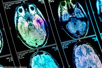
If a CNS GCT is suspected, extent of disease evaluation should include: (1) high-resolution magnetic resonance imaging of the head and spine, with and without gadolinium; (2) evaluation of the cerebrospinal fluid (CSF) for cytology by lumbar puncture or sampling of ventricular fluid at time of shunt placement; (3) CSF and serum measurement of alpha-fetoprotein (AFP) and beta-human chorionic gonadotropin (BHCG); (4) baseline endocrine and neuropsychologic evaluations; and (5) a formal visual examination.
Diagnosis
Radiologically, CNS GCTs cannot be distinguished from other CNS tumors. In the past, if patients had a pineal and/or suprasellar tumor, and suspected GCT, they were given a diagnostic trial of radiotherapy. If an early complete clinical response was seen, the patient was diagnosed with a germinoma. However, other pineal region tumors, as well as mixed NGGCT, may respond initially in the same manner and require very different treatment in order to prevent relapse. Therefore, this practice is no longer used.
The issue then arises regarding the necessity of a biopsy. A histologic specimen is unnecessary if the patient has a positive AFP or BHCG in the CSF and/or serum. Germinomas are generally negative for tumor markers, although they may secrete low levels of BHCG in the CSF (less than 100 mIU/mL). In NGGCTs, endodermal sinus tumors are associated with increased levels of AFP, while choriocarcinomas are associated with raised levels of BHCG. The secretion of these tumor markers in the CSF is pathognomonic for NGGCT, and no further histologic specimen is indicated. When low levels of BHCG are detected in the CSF, it is likely a germinoma with syncytiotrophoblastic cells, and the need for a histologic specimen is debatable.[1] In the French Society of Pediatric Oncology (SFOP) experience, four out of nine patients with secreting germinomas were treated without a histologic diagnosis, and the outcome was the same as for germinomas with a histologic diagnosis.
All other patients with suspected GCT and negative tumor markers require a histologic specimen for diagnosis and treatment. Germinomas are exquisitely sensitive to radiotherapy with excellent cure rates, whereas NGGCTs have a poorer prognosis and require more intensive chemotherapy and irradiation. Given the differing natural histories and responses to treatment of germinomas and NGGCTs, histopathology to determine an optimal treatment strategy in tumor marker-negative patients is important.[2]
It is possible, however, to make an erroneous diagnosis from a small biopsy specimen due to sampling error of a mixed GCT. Specifically, a diagnosis of a germinoma may be made from a small biopsy of a mixed GCT containing germinomatous elements (usually immature or mature teratoma). A gross total resection may provide greater tissue for histologic diagnosis, but given the location of these tumors and the resultant risk of postsurgical morbidity, a partial or total resection for tissue diagnosis is currently not recommended. Due to this risk of histologic sampling error, if any residual radiographic abnormality is present after two to four cycles of chemotherapy, the patient should undergo a "second-look" surgery.[3]
Treatment
Role of Surgery
Again, given the location of GCTs and the high postsurgical morbidity, the risks and benefits of surgery must be considered in light of the excellent response to irradiation and chemotherapy in germinoma patients. Based on the retrospective study of Sawamura et al, no further benefit was found in performing a resection of any kind--partial or complete--beyond treatment with irradiation and chemotherapy.[4]
Unlike germinomas, the role of radical resection in NGGCTs is unclear, with no definitive studies having been conducted. It is possible that radical resection may increase survival rates in NGGCT. Current studies have supported the use of delayed resective surgery, or "second-look" surgery, if residual radiographic abnormalities are seen after chemotherapy and tumor markers have normalized. In this case, the residual lesion is likely to be teratoma or necrosis/fibrosis devoid of tumor. If it is a mature teratoma, surgery may be curative. These patients are then spared any further radiation therapy by performing the "secondlook" surgery.
If immature teratoma is present, then local-field irradiation is initiated without further chemotherapy.[ 3,5,6] In patients whose tumor markers have not normalized, the pathology from "second-look" surgery was also often consistent with either fibrosis or teratoma. However, the risk of subsequent recurrence or progression of disease was significant. Therefore, "second-look" surgery was not supported in cases with any elevation of tumor markers, as the surgery did not improve outcome or allow for a change in therapy.[3]
Germinomas
Radiation Therapy. Germinomas are extremely radiosensitive. Five-year overall survival rates of over 90% are seen with radiation therapy alone. Traditionally, patients were treated with 50 Gy of radiation to the primary tumor site. Additionally, prophylactic craniospinal axis irradiation was given to patients secondary to the concern about CSF seeding, in light of previous reports of approximately 10% of patients with germinomas having CSF dissemination.[7]
Given the concern about the effects of irradiation on the pediatric population, multiple studies have been conducted to examine the efficacy of reduced volume and dosage of irradiation in the treatment of intracranial germinomas. It has been established through multiple retrospective investigations that survival outcome is not affected by a decrease in the local irradiation dose, from traditional doses of 50 to 60 Gy, to doses of approximately 40 Gy.[8-10] In February 2001, Shibamoto et al evaluated lower-dose irradiation in the treatment of germinomas based on tumor volume-based irradiation doses, and found all doses to be effective with no increased risk of local failure. This study examined 38 patients with intracranial germinomas, all treated with irradiation administered locally and to the craniospinal axis, for prophylaxis without adjuvant chemotherapy. Intracranial germinomas 4 cm or less in diameter were cured with doses of 40 to 45 Gy. A dose of 36 Gy was used for germinomas after total removal, 40 Gy for tumors less than 2.5 cm, 45 Gy for 2.5- to 4-cm tumors, and 50 Gy for tumors greater than 4 cm. All doses were effective without risk of local failure, with 10-year overall and relapse-free survival rates of 95% and 91%, respectively.[10]
In the prospective, multicenter Maligue Keimzelltmoren (MAKEI) 83/86/89 trials, intracranial germinomas were treated with reduced-dose irradiation.[8] In these trials, the craniospinal axis dose was reduced to 30 Gy with a local boost of 15 Gy. The overall 5-year survival rate was 94%, and the relapse-free survival rate was 91%, which are comparable to survival rates with higher radiation doses.
Chemotherapy and Radiation Therapy. The attempt to minimize radiation was further explored with the use of adjuvant chemotherapy. This was modeled after the successful treatment of extracranial GCTs with chemotherapy, as these tumors were found to be extremely sensitive to chemotherapy as well as radiotherapy. The chemotherapeutic agents chosen as an adjunct in CNS GCTs were the same agents that were used in the treatment of extracranial GCTs--namely, etoposide, cyclophosphamide, and the platinum-based chemotherapeutic agents.
In fact the use of adjuvant chemotherapy has allowed for even further dose reductions in radiation treatment.[ 11] Allen et al initially examined the effect of neoadjuvant carboplatin in a prospective trial, with decreased local radiation of 30 Gy and craniospinal radiation of 21 Gy, and survival rates were comparable to those obtained with higher-dose radiation alone.[12] In Matsutani's review of 153 histologically verified cases of intracranial GCTs, seven patients with germinomas were given chemotherapy (carboplatin and etoposide) prior to radiation therapy at decreased doses of 30 Gy. The survival rate was 100%, without recurrence, for over 7 years.[13]
In 1998, both Fouladi et al [14] and Sawamura et al [15] examined the use of adjuvant platinum-based chemotherapy and etoposide, with decreased local radiation doses of 30 and 24 Gy, respectively. No prophylactic craniospinal radiation was administered. The investigators found comparable rates of survival with this strategy vs higher-dose radiation therapy alone.
While a decrease in the radiation dose given locally has been supported by multiple studies, the appropriate volume of irradiation remains to be elucidated. While prophylactic craniospinal irradiation may be avoided due to the low rate of relapse in the spine [16,17], a significant difference was seen in the rate of intracranial relapse between patients treated locally at the tumor site (with or without chemotherapy), and those treated with larger irradiation fields: 41.6% vs 11%, respectively. These findings suggest that local irradiation may not be sufficient to prevent local relapse.
Several studies have evaluated the optimal volume of radiotherapy. In one such study, Sawamura et al evaluated 17 patients with germinomas treated with neoadjuvant chemotherapy followed by localized-field irradiation. [15] Patients were treated with EP (etoposide, cisplatin) or ICE (ifosfamide, cisplatin, etoposide) with subsequent involved-field irradiation at a dose of 24 Gy. According to research examining the effect of cranial irradiation on endocrinologic function, this is the maximal dose that can be administered without causing damage to the anterior pituitary gland.[18] At 2-year follow-up, overall and relapse-free survival rates of 100% and 94%, respectively, were realized. In conclusion, the use of adjuvant chemotherapy allowed for the reduction of dosage and volume of irradiation without compromising outcome.
Another study by the SFOP utilized systemic chemotherapy and local irradiation of 40 Gy without craniospinal irradiation. [17] Fifty-seven patients were treated with neoadjuvant etoposide and carboplatin alternating with etoposide and ifosfamide, followed by local tumor irradiation with 2-cm safety margins. The estimated 3-year follow-up probability was 98% overall survival and 96.4% event-free survival, which are comparable to other survival rates with larger-volume radiation therapy. However, 4 out of 57 patients experienced a relapse within the intracranial compartment (cerebellum in one patients, third ventricle in the second, pineal area in the third, and pineal area and lateral ventricle in the last patient).
The question remains: What is the optimal volume of local irradiation? It is unclear whether generous local-field irradiation encompassing the tumor site is sufficient, or inclusion of the third and lateral ventricles is necessary. Further prospective multicenter trials will be needed to evaluate treatment questions.
These concerns were again noted after studies performed by Matsutani and the Japanese Pediatric Brain Tumor Study Group. [19] Patients were treated with carboplatin and etoposide or cisplatin and etoposide, followed by a 24-Gy dose of irradiation to the tumor site. The irradiation field consisted of a limited field with less than a 1-cm margin. The overall tumor-free rate after initial treatment was 92%; however, a 12% recurrence rate within 2½ years was also noted. Seven out of the nine patients relapsed outside the irradiated area.
These findings support the previous suggestion that a larger field be employed in the future. While irradiation to the whole brain is unnecessary, the field may need to be larger than a limited local field. Aoyama et al examined 27 germinoma patients treated with etoposide and cisplatin (for pure germinoma) or etoposide, cisplatin, and ifosfamide (for disseminated germinoma or BHCG-secreting germinoma), followed by 24 Gy of involved-field local radiation therapy. [20] The clinical target volume of irradiation was the gross tumor volume as assessed by magnetic resonance imaging. Followup ranged from 18 to 102 months. Recurrence of disease was seen in 1 out of 16 patients with pure germinomas, with an actuarial relapse-free 5-year survival rate of 90%. Five out of nine patients with BHCG-secreting germinomas relapsed, with an actuarial relapse-free survival rate of 44%. Consistent with previous studies, the overall relapse rate was higher after local-field irradiation. However, the authors concluded that for the treatment of pure germinomas, limited local-field irradiation is sufficient. On the other hand, the BHCG-secreting germinomas were thought to require a higher dose of radiotherapy to attain control without relapse.
Other studies have evaluated the difference in prognosis and response to the treatment of pure germinomas vs BHCG-secreting germinomas, concluding that the latter may require a more intensive treatment protocol. [16] In more recent studies where a more intensive chemotherapeutic regimen was administered to BHCG-secreting germinomas, equal outcomes were noted. [17]
Chemotherapy Only. Some investigators have attempted to treat germinomas with chemotherapy alone, without radiation therapy. In a case review of nine patients who refused radiation treatment after chemotherapy, eight of the nine showed a complete response to chemotherapy. However, four patients experienced recurrence within 1½ years of treatment. [19]
In 1999, Shibamoto et al also reviewed five cases in which the patients were treated with chemotherapy alone by the primary physician and subsequently presented for salvage radiation therapy. [21] The patients were given standard doses of 40 to 45 Gy of local radiotherapy, with complete response and no recurrence after 90 months. A multicenter, prospective trial was then initiated to further evaluate the possibility of treatment with chemotherapy without concomitant radiotherapy. In the First International CNS Germ Cell Cooperative Trial, 45 patients with germinomas were treated with chemotherapy alone. The patients were given carboplatin, etoposide, and bleomycin, which produced a complete remission rate of 84%; however, 50% of the patients recurred. Most of the patients with pure germinomas who relapsed were then treated with salvage radiotherapy successfully. The investigators concluded that approximately 50% of the patients were spared radiotherapy, without deleterious effects. However, the early years of this multinational study showed a 10% mortality rate overall, including the germinoma and NGGCT patients, secondary to chemotherapeutic toxicity.[22]
The risks and benefits of chemotherapeutic toxicity need to be weighed against those of irradiation. In addition to immediate toxicity from chemotherapy, including myelosuppression and increased risk of infection, late effects from chemotherapy can include infertility in males, ototoxicity, and secondary neoplasm. Therefore, controversy continues to exist regarding the treatment of germinomas with chemotherapy alone. Although chemotherapy alone does not appear to produce a benefit over combined treatment, it may be considered in younger patients, or in those with pretreatment neurocognitive deficits, for whom radiotherapeutic toxicity is of greater concern. In these circumstances, any possibility of sparing or delaying radiotherapy without adverse effect may benefit the outcome and the patient's quality of life.
Nongerminomatous Germ-Cell Tumors
Radiation Therapy.Compared with germinomas, NGGCTs are relatively radioresistant and associated with a poorer outcome. Traditionally, patients with NGGCTs have been treated with at least 50 Gy of local boost and craniospinal axis irradiation, with 3-year overall survival rates of approximately 20% to 45%.[23] This wide range is attributed to variations in histology, which respond to treatment differently and carry different prognoses. In a report by Matsutani et al, patients with pure choriocarcinoma, endodermal sinus tumor, or embryonal carcinoma had a 3-year survival of 27%, patients with mixed tumors composed primarily of malignant elements had a 3-year survival of 9%, and patients with predominantly germinoma or teratoma mixed with other NGGCT elements had a 3-year survival of 70%.[13]
Many controversies complicate the role of irradiation in children with NGGCTs. The first such issue is craniospinal radiation. Most authors agree that low-dose craniospinal irradiation in patients who present beyond puberty is acceptable, especially in the setting of positive CSF cytology or overt metastatic spread. For patients with positive CSF cytology, doses of 20 to 24 Gy are recommended, and for patients with metastatic disease, Bamberg et al reported equivalent results with a reduction in dose from 36 to 30 Gy (and a concomitant reduction in the dose to the primary from 50 to 45 Gy). Patients with focal spinal disease visualized on magnetic resonance imaging have received a local boost of up to 50 Gy.[8]
The second key issue is radiation field size for the primary tumor. Local radiation volume may vary from the inclusion of the primary tumor with a 1- to 2-cm margin to primary tumor with adjacent ventricle to whole-brain radiation. Some evidence supports whole-brain radiation. In the MAKEI, the incidence of leptomeningeal disease in patients treated with local irradiation alone was 28%, as compared with 2% for those treated with whole-brain and craniospinal irradiation.[8] However, whole-brain irradiation is associated with neurocognitive deterioration, which may be minimized with a reduction in the radiation field and the use of conformal radiation and intensity-modulated radiation therapy. Dose-response data support a dose range of 45 to 50 Gy to restricted radiation fields. Total doses of less than 45 Gy have been associated with higher relapse rates of 40% to 50%, as compared with 10% to 20% in patients receiving greater than 50 Gy. Several reports have recommended lower doses of radiation when combined with chemotherapy.[23]
Combined Chemotherapy and Radiation Therapy. The addition of chemotherapy to the treatment of children with CNS NGGCT has been reported to be associated with overall survival rates of up to 60%. Robertson et al reported on 18 patients who were treated with radiation and three or four cycles of cisplatin and etoposide. Their study demonstrated a 4-year event-free survival of 67% and overall survival of 74%. All patients received more than 50 Gy of involved-field radiation, and six received additional whole-brain or craniospinal irradiation. Survival after recurrence was brief.[24]
Balmaceda et al reported on the First International Central Nervous System Germ Cell Tumor Study. Twenty-six patients had NGGCTs. Patients were initially treated with four cycles of carboplatin, etoposide, and bleomycin. Patients who achieved a complete remission received two more cycles of chemotherapy. For patients who did not achieve complete remission, radiation was given either before or after two more cycles of the same chemotherapy but with the addition of cyclophosphamide. Second-look surgery was strongly recommended if less than a complete remission was achieved. Twenty-one patients achieved a complete remission after four cycles of induction chemotherapy. However, 50% of the patients progressed or relapsed, and only 6 of those 13 were alive and without disease at the time of the report. The investigators observed a 10% rate of toxic deaths, and the overall survival rate for patients with NGGCTs was 62%.[22]
Kellie et al reported on the Second International CNS GCT Cooperative Trial. Patients were treated with combinations of cisplatin, etoposide, cyclophosphamide, bleomycin, and carboplatin. With a median follow-up of approximately 6½ years, 14 out of 20 patients were alive at between 38+ and 86+ months from diagnosis. Five patients received treatment with chemotherapy alone, and three received local irradiation after four cycles of chemotherapy in violation of the protocol. In addition, 6 of the 12 patients who progressed or relapsed were salvaged with more chemotherapy and irradiation. Only 1 of 20 patients died of toxicity, and the overall survival rate was 75%.[25]
Although the First and Second International studies showed a higher rate of recurrence with chemotherapy alone, approximately half of the patients with NGGCTs were salvaged with the addition of irradiation and more chemotherapy. This is encouraging for very young patients, in whom radiation therapy may ideally be avoided or delayed.
Teratoma
Pure CNS teratomas occur primarily in neonates with a female predominance. Complete resection is the therapy of choice. In patients with immature teratoma, chemotherapy has been reported to be useful.[26] The value of additional irradiation is unclear. Patients with incompletely resected teratoma have a 10% risk of recurrence, and those with immature teratoma, a 20% risk irrespective of previous chemotherapy.[27] However, a significant proportion of CNS germinomas or NGGCTs have components of immature or mature teratoma that may remain following eradication of the more malignant elements, requiring surgical intervention alone (for mature teratoma) or with the addition of focal irradiation (for immature teratoma).[3]
Relapse
Many investigators have shown that while more than 90% of relapses occur at the primary site of the tumor, 30% are combined with leptomeningeal spread. The outcome of relapsed patients, particularly those with NGGCTs, is bleak. Salvage therapy has included surgery, craniospinal irradiation, and a focal boost with and without high-dose chemotherapy and stem cell rescue.
In patients with germinoma who have not received radiation therapy in the past, the benefit of craniospinal and focal irradiation is clear. Merchant et al reported on eight patients who relapsed after treatment with chemotherapy alone for primary CNS germinoma. Patients were then treated with high-dose cyclophosphamide followed by craniospinal irradiation (25.2 to 36 Gy) and a boost to the site of recurrent disease (45 Gy). All patients were alive at a median follow-up of 32 months following treatment.[28]
For patients who relapse after receiving irradiation as part of primary therapy, it is recommended that they be treated with surgery, if possible, followed by myeloablative chemotherapy and stem cell rescue. Modak et al reported on 21 patients with recurrent or progressive CNS germ cell tumors who underwent treatment with thiotepa-based high-dose chemotherapy with autologous hematopoietic stem cell rescue. Half of the patients received consolidation therapy with radiation. Seven of the nine patients with germinoma were disease-free at a median time of 48 months after high-dose chemotherapy. On the other hand, 4 of the 12 patients with NGGCT were disease-free at a median of 33 months after high-dose chemotherapy.[29] Other investigators have reported similar results with high-dose chemotherapy regimens in this setting.[30]
Long-Term Effects of Therapy
Radiation Therapy
Given the improved outcomes in the treatment of pediatric brain tumors, increasing concern has been given to the effects of irradiation on the developing brain in survivors. Irradiation to the brain can adversely affect neurocognitive function and resultant quality of life.[15] In children and adults, cranial irradiation also frequently causes damage to the hypothalamic-pituitary axis, including deficiency in growth hormone, hypothyroid, and gonadal dysfunction.[31]
In addition to neuroendocrine effects, cranial irradiation increases a patient's risk for a second malignancy. In a retrospective review of 111 cases of intracranial GCTs, at a median followup of 115 months, the incidence of secondary brain neoplasm was 4.8%. This incidence is expected to increase with time, and the estimated incidence over 19 years is 16.8%.[15]
The effects of irradiation have been shown to have a more detrimental effect in younger patients as opposed to older patients. Age at diagnosis has been positively correlated with overall psychosocial and physical health summary scores in studies examining quality of life after cranial radiotherapy. [ 32,33] It is difficult to determine the effects of irradiation on patients with CNS GCTs, as they often have significant deficits prior to the initiation of radiation therapy.[33] In 2002, Merchant et al evaluated pre- and posttreatment neurocognitive function, and found no significant differences in IQ scores after patients received 25.6 Gy of craniospinal irradiation and a 50.4-Gy boost to the primary site. The patient group studied, however, was small, consisting of 12 patients with CNS GCTS aged 9 to 16 years old.[34] Further prospective studies need to be obtained with preand posttreatment analysis of neurocognitive function.
In addition to neurocognitive dysfunction, irradiation is associated with endocrine dysfunction. However patients with CNS germ cell tumors often present with neuroendocrine deficits prior to any therapy. [35] In a 1998 report on the SFOP experience, Bouffet et al observed that 29 of 57 patients presented with growth hormone deficiency or panhypopituitarism and short stature.[17] In treating these patients, the risk of short stature with craniospinal irradiation needed to be considered, regardless of age at presentation.
Chemotherapy
Chemotherapy, on the other hand, has been associated with a variety of short- and long-term toxicities. Platinum- based agents carry the risk primarily of renal tubular nephropathy and hearing loss. Etoposide is associated with an increased risk of secondary leukemia, and the pulmonary toxicity of bleomycin has been well described. In addition, cyclophosphamide has been associated with sterility and bladder fibrosis.
Conclusions
The excellent outcome of patients with pure CNS germinoma and the morbidity associated with standard radiation therapy justifies attempts to limit both the total irradiation dose and field size, with or without chemotherapy, and to limit surgery to a biopsy for diagnostic purposes. In contrast, the poor outcome of NGGCTs justifies a more aggressive approach employing chemotherapy and irradiation, and in select cases, surgical resection. Additional cooperative, international studies are needed to optimize treatment strategy relative to tumor histology and the patient.
Disclosures:
The authors have no significant financial interest or other relationship with the manufacturers of any products or providers of any service mentioned in this article.
References:
1. Shibamoto Y, Takahashi M, Sasai K: Prognosis of intracranial germinoma with syncytiotrophoblastic giant cells treated with radiation therapy. Int J Radiat Oncol Biol Phys 37:505-510, 1997.
2. Matsutani M, Takakura K, Sano K: Primary intracranial germ cell tumors: Pathology and treatment. Prog Exp Tumor Res 30:307-312, 1987.
3. Weiner HL, Finlay JL: Surgery in the management of primary intracranial germ cell tumors. Childs Nerv Syst 15:770-773, 1999.
4. Sawamura Y, de Tribolet N, Ishii N, et al: Management of primary intracranial germinomas: Diagnostic surgery or radical resection? J Neurosurg 87:262-266, 1997.
5. Sawamura Y, Kato T, Ikeda J, et al: Teratomas of the central nervous system: Treatment considerations based on 34 cases. J Neurosurg 89:728-737, 1998.
6. Branzelli MC, Patte C, Bouffet E, et al: Non-metastatic intracranial germinomas. The experience of the French Society of Pediatric Oncology. Cancer 80:1792-1797, 1997.
7. Brada M, Rajan B: Spinal seeding in cranial germinomas. Br J Cancer 61:339-340, 1990.
8. Bamberg M, Kortmann R-D, Calaminus G, et al: Radiation therapy for intracranial germinoma: Results of the German Cooperative Prospective trials MAKEI 83/86/89. J Clin Oncol 17:2585-2592, 1999.
9. Ayoyama H, Shirato H, Kakuto Y, et al: Pathologically-proven intracranial germinoma treated with radiation therapy. Radiother Oncol 47:201-205, 1998.
10. Shibamoto Y, Takahashi M, Abe M, et al: Reduction of radiation dose for intracranial germinoma: A prospective study. Br J Cancer 70:984-989, 1994.
11. Buckner JC, Peethambram PP, Smithson WA, et al: Phase II trial of primary chemotherapy followed by reduced dose radiation for CNS germ cell tumor. J Clin Oncol 17:933-940, 1999.
12. Allen JC, Darosso RC, Donahue B, et al: A phase II trial of preirradiation carboplatin in newly diagnosed germinoma of the central nervous system. Cancer 74:940-944, 1994.
13. Matsutani M, Sano K, Takakura K, et al: Primary intracranial germ cell tumors: A clinical analysis of 153 histologically verified cases. J Neurosurg 86:446-455, 1997.
14. Fouladi M, Grant R, Baruchel S, et al: Comparison of survival outcomes in patients with intracranial germinomas treated with radiation alone versus reduced-dose radiation and chemotherapy. Childs Nerv Syst 14:596-601, 1998.
15. Sawamura Y, Shirato H, Ikeda J, et al: Induction chemotherapy followed by reduced volume radiation therapy for newly diagnosed CNS germinoma. J Neurosurg 88:66-72, 1998.
16. Sawamura Y, Ikeda J, Shirato H, et al: Germ cell tumors of the central nervous system: Treatment considerations based on 111 cases and their long term clinical outcomes. Eur J Cancer 34:104-110, 1998.
17. Bouffet E, Baranzelli MC, Patte C, et al: Combined treatment modality for intracranial germinomas: Results of a multicentre SFOP experience. Br J Cancer 79:1199-1204, 1999.
18. Rappaport R, Brauner R: Growth and endocrine disorders secondary to cranial radiation. Pediatr Res 25:561-567, 1989.
19. Matsutani M, Japanese Pediatric Brian Tumor Study Group: Combined chemotherapy and radiation therapy for CNS germ cell tumors- the Japanese experience. J Neurooncol 54:311-316, 2001.
20. Aoyama H, Shirato H, Ikeda J, et al: Induction chemotherapy followed by low dose involved field radiotherapy for intracranial germ cell tumors. J Clin Oncol 20:857-865, 2002.
21. Shibamoto Y, Sasai K, Kokubo M, et al: Salvage radiation therapy for intracranial germinoma recurring after primary chemotherapy. J Neurooncol 44:181-185, 1999.
22. Balmaceda C, Heller G, Rosenblum M, et al: Chemotherapy without radiation-a novel approach for newly diagnosed CNS germ cell tumors: Results of an international cooperative trial. J Clin Oncol 14:2908-2915, 1996.
23. Borg M: Germ cell tumors of the central nervous system in children: Controversies in radiotherapy. Med Pediatr Oncol 40:367-374, 2003.
24. Robertson PL, DaRosso RC, Allen JC: Improved prognosis of intracranial non-germinoma germ cell tumors with multimodality therapy. J Neurooncol 32:71-80, 1997.
25. Kellie SJ, Boyce H, Dunkel IJ, et al: Primary chemotherapy for intracranial nongerminomatous germ cell tumors: Results of the Second International CNS Germ Cell Study Group protocol. J Clin Oncol 22:846-853, 2004.
26. Garre ML, El-Hossainy MO, Fondelli P, et al: Is chemotherapy effective therapy for intracranial immature teratoma? A case report. Cancer 77:977-982, 1996.
27. Gobel U, Schneider DT, Calaminus G, et al: Germ cell tumors in childhood and adolescence. Ann Oncol 11:263-271, 2000.
28. Merchant TE, Davis BJ, Sheldon JM, et al: Radiation therapy for relapsed CNS germinoma after primary chemotherapy. J Clin Oncol 16:204-209, 1998.
29. Modak S, Gardner S, Dunkel I, et al: Thiotepa based high dose chemotherapy with autologous stem cell rescue in patients with recurrent or progressive CNS germ cell tumors. J Clin Oncol 22;1934-1943, 2004.
30. Mahoney DH Jr, Strother D, Camitta B, et al: High dose melphalan and cyclophosphamide with autologous bone marrow rescue for recurrent/progressive malignant brain tumors in children: A pilot Pediatric Oncology Group study. J Clin Oncol 14:382-388, 1996.
31. Constine LS, Woolf PD, Cann D, et al: Hypothalamic-pituitary dysfunction after radiation for brain tumors. N Engl J Med 328:87-94, 1993.
32. Sands SA, Kellie SJ, Davidow AL, et al: Long term quality of life and neuropsychologic functioning for patients with CNS germ cell tumors: From the First International CNS Germ Cell Tumor Study. Neur Oncol 3:174-183, 2001.
33. Sutton LN, Radcliffe J, Goldwein JW, et al: Quality of life of adult survivors of germinomas treated with craniospinal irradiation. Neurosurgery 46:1292-1298, 1999.
34. Merchant TE, Sherwood SH, Mulhern RK, et al: CNS germinoma: Disease control and long term functional outcome for 12 children treated with craniospinal irradiation. Int J Radiat Oncol Biol Phys 46:1171-1176, 2000.
35. Merchant TE, Williams T, Smith JL, et al: Preirradiation endocrinopathies in pediatric brain tumor patients determined by dynamic tests of endocrine function. Int J Radiat Oncol Biol Phys 54:45-50, 2002.
Articles in this issue
almost 21 years ago
Revisiting Induction Chemotherapy for Head and Neck Canceralmost 21 years ago
Commentary (Brown/Armstrong): Pregnancy and Breast Canceralmost 21 years ago
Commentary (Theriault/Buzdar): Pregnancy and Breast Canceralmost 21 years ago
The Role of Statins in Cancer Prevention and Treatmentalmost 21 years ago
Pregnancy and Breast CancerNewsletter
Stay up to date on recent advances in the multidisciplinary approach to cancer.


