|Articles|August 1, 2002
Oncology NEWS International
- Oncology NEWS International Vol 11 No 8
- Volume 11
- Issue 8
Diagnostic Dilemma
Author(s)Henry Chun, MD, Matthew J. Mckinley, MD
A 48-year-old man is referred for evaluation. He has a history of hypercholesterolemia and obesity. Treatment with cholesterol-lowering medication was associated with elevated liver chemistries. When the elevated liver chemistries persisted
Advertisement
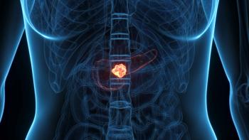
A 48-year-old man is referred for evaluation. He has a history of hypercholesterolemia and obesity. Treatment with cholesterol-lowering medication was associated with elevated liver chemistries. When the elevated liver chemistries persisted after cessation of the medication, imaging studies were obtained. Ultrasound and CT scan both revealed a heterogeneous mass in the head of the pancreas. There was dilatation of the main pancreatic duct in this area.
The patient denied abdominal pain, nausea, vomiting, and fever. He had a history of heavy alcohol use. There was no history of pancreatitis. Past medical history was positive only for hypercholesterolemia. Past surgical history was significant for cholecystectomy for gallstones 10 years earlier. Physical examination was unremarkable. Laboratory evaluation was normal except for mild elevation of alkaline phosphatase.
Endoscopic retrograde cholangiopancreatography (ERCP) was performed to evaluate the abnormal findings and to rule out chronic pancreatitis.
- The photograph demonstrates:a. A duodenal ulcer
b. A pigmented lesion in the duodenum
c. A worm
d. The major papilla with the orifice impacted with mucin
- This finding is pathognomic for what condition?
a. Malignant melanoma
b. Intraductal papillary mucinous tumor of the pancreas (IPMT)
c. Strongyloidiasis
d. Zollinger-Ellison syndrome
The correct answer to question 1 is d. The endoscopic photograph reveals the typical "fish eye" appearance of the main papilla seen with intraductal neoplasia of the pancreas and bile duct.
The correct answer to question 2 is b. Intraductal papillary mucinous tumor of the pancreas (IPMT), also known as intraductal papillary hyperplasia, mucinous ductal ectasia, mucin-producing tumor, and other names, was first reported by Ohashi and Maruyama in 1982. Since then, more than 300 cases of this rare entity have been reported worldwide.
It is characterized radiographically by cystic dilation of the main pancreatic duct or secondary branches with copious mucus secretion in the ductal lumen causing filling defects, and endoscopically by a swollen papilla extruding mucin.
The true incidence of IPMT in the population is unknown. One surgical study suggests that IPMT accounts for between 8% and 20% of pancreatic resections for malignancy. It is frequently found in men in the age range of 60 to 70 years; it is usually seen in the head of the pancreas but sometimes may involve the entire gland.
The etiology of IPMT is unclear. Environmental factors like tobacco and alcohol do not appear to play a significant role. There is no clear-cut genetic predisposition. Data from animal studies suggest nitroamines play a role in IPMT.
Common clinical manifestations include abdominal pain with features of acute and chronic pancreatitis secondary to intermittent obstruction of the main pancreatic duct, weight loss, anorexia, and steatorrhea. Diabetes, jaundice, and cholangitis can also occur. However, many patients are asymptomatic, and the tumor is found incidentally on imaging studies performed for unrelated problems.
A variety of imaging methods are available for the diagnosis of IPMT but none is superior, so a combination of methods is usually required for the diagnosis, with endoscopy and ERCP the most frequently used.
IPMT is a potentially curable disease if the lesion is resectable and has not metastasized. Total resection should be performed in main duct type (MDT)-IPMT tumors of any size originating in the main pancreatic duct, and local resection when IPMT is localized to a segment of the pancreas.
With branch duct type (BDT)-IPMT, total resection should be done when dilatation of the main duct is present even if the lesion is extremely small. However, if the lesion is less than 2.5 cm in diameter with a thin wall and normal main pancreatic duct, close monitoring with serial imaging studies can be done because these lesions are generally benign and growth is slow when it occurs.
The prognosis for patients who have undergone surgical resection for IPMT is good, with reports of 60% to 80% survival at 3 years in some studies. This is much better than survival for ductal adenocarcinoma of the pancreas. With invasive cancer, however, the 3-year disease-free survival decreases to as low as 21%. Overall, patients with BDT-IPMT do better than those with MDT-IPMT.
Selected Readings
Farrell J, Brugge W: Intraductal papillary mucinous tumor of the pancreas. Gastrointest Endosc 55:701-714, 2002.
Hurwitz L, Daniels A, Barkin J, et al: Preoperative diagnosis of intraductal papillary mucinous tumors of the pancreas by endoscopic pancreatic biopsy. Gastrointest Endosc 53:510-513, 2001.
Articles in this issue
almost 24 years ago
Medicaid Gives New Protections to Beneficiaries in Managed Care Plansalmost 24 years ago
Prostate Cancer Cell Line Vaccine Promising in Phase II Trialalmost 24 years ago
MoAb Targeting Death Receptor in Breast Canceralmost 24 years ago
Hanford I-131 Releases Did Not Increase Thyroid Cancer Riskalmost 24 years ago
Chemo/Rituximab Is Effective as First-Line CLL Therapyalmost 24 years ago
Viral Agent ONYX-015 Targets p53-Deficient Cancer Cellsalmost 24 years ago
Program Educates Teens About Testicular Cancer Self-Examalmost 24 years ago
Better Colon Cancer Imaging With PET/CT Than With PET Alonealmost 24 years ago
Delayed Lung Cancer Screening Trial to Begin Enrollment 50,000 Subjectsalmost 24 years ago
Results Similar With Weekly or Every-3-Week Irinotecan Dosing RegimensAdvertisement
Related Content
Advertisement


Advertisement
Advertisement
Trending on CancerNetwork
1
FDA Approves Gedatolisib for HR+, HER2– PIK3CA Wild-Type Advanced Breast Cancer
2
Agenus Discontinues Phase 3 BATTMAN Trial in Metastatic MSS Colorectal Cancer
3
FDA Traditionally Approves Selpercatinib in RET+ Solid Tumors
4
Subcutaneous Isatuximab Approval Expands Options in Multiple Myeloma
5