
News|Videos|January 29, 2025
Fluorescent-Based Imaging with AI Can Improve Surgery in Breast and Ovarian Cancer
Author(s)Farzad Fereidouni, PhD
The FIBI technology, created by Farzad Fereidouni, PhD, when combined with AI eliminates the need for frozen sections and other labor-intensive oncology surgery practices.
Advertisement
Farzad Fereidouni, PhD, and Anant Madabhushi, PhD, are currently developing a tool, at the Winship Cancer Institute of Emory University, that combines new artificial intelligence (AI) technologies with fluorescent imitating brightfield imaging (FIBI) microscopy technology that will image the surface of tissue while a patient with breast or ovarian cancer is on the operating table, instead of forcing surgeons to use the frozen section procedure.
Fereidouni, PhD, an associate professor at the Emory University School of Medicine, spoke with CancerNetwork® regarding the MarginCall project following their recent awarding of funds from the Advanced Research Projects Agency for Health (ARPA-H).1 Madabhushi, PhD, is a researcher of Cancer Immunology at the Winship Cancer Institute of Emory University. “We are trying to create an end-to-end solution [where] everything is automated…” Fereidouni said. The hope is that the new technology will greatly simplify the surgery process and reduce the impact that the quality of the surgeon has on the outcome.
The FIBI technology, created by Fereidouni and his team, is an approach to histology that doesn’t require the use of slides or methods like fixation, paraffinization, and sectioning, but instead captures images directly from tissue surfaces using fluorescence-based methods.2
By taking large images of the tissue, the number of photos that will be created is going to be increased compared with the current standard, and the AI is going to help sort through them. With the images, the technology should then be able to create a 3D map that will help visualize and aid the process of the surgery for the surgeon.
Transcript:
We are trying to combine 2 cutting-edge technologies here. One of them is the AI tools, which are being developed by [Dr Madabhushi], and the other one is the FIBI technology that technically addresses the problem that [Dr Madabhushi] was explaining. We are trying to image the surface of the tissue without freezing it so [that], while the patient is [undergoing surgery], we can [remove] the tissue. The process is much simpler in the way that you don’t need a very experienced [surgeon] to perform it, and it’s highly automatable. That’s the whole point of this project.
We are trying to create an end-to-end solution [where] everything is automated from the way that they resect the tissue. They can slice it by hand. They can be automatically stained and can be imaged very fast, and it’s not destructive. In [real-time], these images can be fed to AI and can be diagnosed either by AI, or [the AI] can help pathologists make the diagnosis. The way that we are going to do these images is by imaging a very large piece of tissue instead of the standard inch-by-inch blocks. For that reason, the amount of images that we are going to create is going to be way more than the regular workflow. For that reason, we need AI to guide surgeons and create a diagnosis. Those diagnoses will be used to create a 3D map the surgeon can rotate and visualize, [to] guide the surgeon to do the surgery better.
References
- Emory researchers awarded up to $17.6M from ARPA-H to innovate cancer surgery, improve outcomes. Emory Winship Cancer Institute. January 6, 2025. Accessed January 22, 2025. https://tinyurl.com/y3dpmtxr
- Fereidouni lab research projects. Emory School of Medicine. Accessed January 23, 2025. https://tinyurl.com/2s4jadwe
Advertisement
Related Content
Advertisement


Advertisement
Advertisement
Trending on CancerNetwork
1
FDA Approves Palbociclib Combo in HR+, HER2+ Metastatic Breast Cancer
2
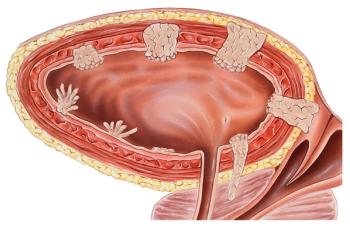
NCCN Updates Bladder Cancer Guidelines for ctDNA MRD Testing
3
Zanzalintinib/Atezolizumab Misses Survival End Point in Non-MSI-H mCRC Subgroup
4
Sigvotatug Vedotin Does Not Significantly Improve Survival in NSCLC Trial
5