- ONCOLOGY Vol 21 No 3
- Volume 21
- Issue 3
Interpreting Intratumoral Hypoxia: Can It Guide Therapy?
The role of hypoxia as a key determinant of outcome for human cancers has encouraged efforts to noninvasively detect and localize regions of poor oxygenation in tumors. In this review, we will summarize existing and developing techniques for imaging tumoral hypoxia. A brief review of the biology of tumor oxygenation and its effect on tumor cells will be provided initially. We will then describe existing methods for measurement of tissue oxygenation status. An overview of emerging molecular imaging techniques based on radiolabeled hypoxic markers such as misonidazole or hypoxia-related genes and proteins will then be given, and the usefulness of these approaches toward targeting hypoxia directly will be assessed. Finally, we will evaluate the clinical potential of oxygen- and molecular-specific techniques for imaging hypoxia, and discuss how these methods will individually and collectively advance oncology.
Drs. Graves and Giaccia have written a comprehensive review of approaches that are in use or under development for the identification of cancers containing regions of reduced oxygenation (intratumoral hypoxia). A growing body of data from preclinical and clinical studies indicates that intratumoral hypoxia is a poor prognostic sign in many solid cancers and that hypoxia can induce (via changes in gene expression) and/or select for cells with altered characteristics, including (a) maintenance of replicative potential, (b) maintenance of stem cell properties, (c) genetic instability, (d) angiogenesis, (e) metabolic reprogramming, (f) autocrine growth factor signaling, (g) invasion, (h) metastasis, and (i) resistance to radiation and chemotherapy. In short, virtually every critical aspect of cancer biology can be affected.
The transcriptional regulator hypoxia-inducible factor 1 (HIF-1) has been demonstrated to contribute to each of these processes.[1] In early-stage breast and cervical carcinomas, analysis of biopsies by HIF-1-alpha immunohistochemistry has identified patients whose biopsies showed high HIF-1-alpha levels and who had significantly increased mortality relative to patients whose biopsies did not have high HIF-1-alpha levels, as reviewed by Graves and Giaccia.
Limited Utility
Although the mechanistic data from preclinical studies and the survival data from clinical studies indicate that intratumoral hypoxia and increased HIF-1-alpha expression are associated with aggressive cancers, the ability to identify individual patients at increased risk is still limited. Cancer is a heterogeneous disease, both across patients with the same diagnosis and within individual patients over space and time. As a result of this heterogeneity, any individual diagnostic test is of limited utility in predicting prognosis or response to therapy.
In the case of hypoxia and HIF-1, two important biologic properties must be appreciated. First, all cells respond to hypoxia by inducing HIF-1 activity, but the hundreds of downstream genes whose transcription is activated or repressed by HIF-1 differ from one cell type to another. Whereas hypoxia induces expression of the
VEGF
gene (which encodes vascular endothelial growth factor) in most cells, the majority of genes are regulated by hypoxia in a cell type-specific manner. Strikingly, the expression of some genes is induced by hypoxia in one cell type and repressed in response to hypoxia in another cell type, with the response in each case mediated by HIF-1.[2] Second, among the proteins that are regulated by hypoxia are those that promote cell survival (eg, survivin) and others that promote apoptosis (eg, BNIP3). The balance between these antagonistic factors will determine the net effect of hypoxia on cell viability.
Dynamic Relationship
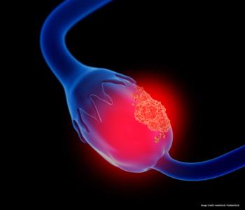
An instructive example of the dynamic relationship between proapoptotic and antiapoptotic factors is the case of ovarian cancer, in which intra-tumoral hypoxia is associated with high levels of HIF-1-alpha, apoptosis, and increased patient survival.[3] However, in a subset of biopsies with high levels of HIF-1-alpha and mutant p53 protein, the frequency of apoptotic cells was low and patient survival was significantly reduced. Thus, whereas the identification of intratumoral hypoxia may generally identify aggressive cancers that require aggressive therapy, it is likely that this measure will be most informative when integrated with other histologic (microvascular density), cytologic (rates of cell apoptosis and proliferation), and molecular (genomic sequence and gene/protein expression[4]) data, which can then be correlated with responses to various therapeutic regimens.
In addition to contributing to algorithms for individualized cancer therapy, intratumoral oxygen measurements may be useful for evaluating therapeutic responses, particularly in the case of angiogenic inhibitors such as bevacizumab (Avastin).[5] These agents may increase or, paradoxically, decrease intratumoral hypoxia, depending on when oxygen concentrations are measured during the course of therapy. Radiation therapy is another treatment modality in which oxygen measurements may be particularly useful. Because of the complex biologic consequences of hypoxia, determination of O
2
levels using techniques described by Graves and Giaccia will likely become an important addition to the oncologist's clinical database in the near future.
Gregg L. Semenza, MD, PHD
Disclosures:
The author has no significant financial interest or other relationship with the manufacturers of any products or providers of any service mentioned in this article.
References:
1. Semenza GL: Development of novel therapeutic strategies that target HIF-1. Expert Opin Ther Targets 10:267-280, 2006.
2. Kelly BD, Hackett SF, Hirota K, et al: Cell type-specific regulation of angiogenic growth factor gene expression and induction of angiogenesis in nonischemic tissue by a constitutively active form of hypoxia-inducible factor 1. Circ Res 93:1074-1081, 2003.
3. Birner P, Schindl M, Obermair A, et al: Expression of hypoxia-inducible factor 1alpha in epithelial ovarian tumors: Its impact on prognosis and on response to chemotherapy. Clin Cancer Res 7:1661-1668, 2001.
4. Chi JT, Wang Z, Nuyten DS, et al: Gene expression programs in response to hypoxia: Cell type specificity and prognostic significance in human cancers. PLoS Med 3:e47, 2006.
5. Gasparini G, Longo R, Toi M, et al: Angiogenic inhibitors: A new therapeutic strategy in oncology. Nat Clin Pract Oncol 2:562-577, 2005.
Articles in this issue
almost 19 years ago
The Future of Immunotherapy in Prostate Canceralmost 19 years ago
Oncologist-Patient Communication on Anemia Is Lacking, Study Findsalmost 19 years ago
The Complete Guide to Relieving Cancer Pain and Sufferingalmost 19 years ago
Looking Beyond Survivalalmost 19 years ago
Tumor Hypoxia and the Future of Cancer Managementalmost 19 years ago
How Do We Get More Older Patients Into Clinical Trials?almost 19 years ago
Designing Cancer Trials to Accommodate Older Patientsalmost 19 years ago
Treating Small-Cell Lung Cancer: More Consensus Than Controversyalmost 19 years ago
Prostate Cancer Immunotherapy: Promising BeginningsNewsletter
Stay up to date on recent advances in the multidisciplinary approach to cancer.


