|Articles|March 1, 2004
Oncology NEWS International
- Oncology NEWS International Vol 13 No 3
- Volume 13
- Issue 3
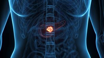
Intraoperative RT Useful in Unresectable Pancreatic Cancer
Author(s)James L. Abbruzzese, MD, FACP
This special “annual highlights” supplement to Oncology News International is acompilation of some of the major advances in the management of gastrointestinalcancers during 2003–2004, as reported in ONI. Guest editor Dr. James L. Abbruzzesecomments on the reports included herein and discusses advances in the clinicalmanagement of GI cancers, with a focus on developments in targeted therapy, newcombinations, adjuvant therapy, and what to watch for in 2004.
Advertisement
SALT LAKE CITY-Intraoperativeelectron beam radiation therapy(IOERT) may prolong the survival ofpatients who have unresectable butnonmetastatic pancreatic cancers thatare small, according to a retrospectivestudy reported at the 45th AnnualMeeting of the American Society forTherapeutic Radiology and Oncology(abstract 159). Among patients whoreceived this radiation therapy as partof their comprehensive treatment,nearly one-fifth of those with smallcancers were still alive 3 years later,but all of those with large cancers haddied."Locally advanced, nonmetastaticpancreatic cancer is a common presentation,and it is associated with profoundmorbidity and suffering forthese patients. Our therapeutic effortsare of a palliative nature," said leadauthor Christopher G. Willett, MD,clinical director of radiation oncology,Massachusetts General Hospital,and professor of radiation oncology,Harvard Medical School.Dr. Willett noted that various formsof radiation therapy have been used totreat this cancer for more than 30 years."To improve local control, intraoperativeelectron beam radiation therapy,typically 20 Gy, has been combinedwith treatment programs of externalbeam irradiation, typically on the orderof 45 to 50 Gy, and 5-FU [fluorouracil]-based chemotherapy," he said.
The investigators studied 150 patientswith unresectable, biopsy-provenductal adenocarcinoma of the pancreastreated with external beamradiation therapy, 5-FU, and IOERTbetween 1978 and 2001. Pathologistsre-reviewed the histologic findings inpatients surviving more than 3 yearsto verify the diagnosis.The patients received a wide rangeof treatments with respect to bothradiation therapy and chemotherapy,Dr. Willett noted. "As we all know,there has been a profound treatmentevolution in this disease," he said."The basic theme, however, is an effortto employ moderate- to highdoseexternal beam irradiation with5-FU and IOERT at 20 Gy."Survival BenefitThe patients had a median andmean survival of 13 and 17 months,respectively, Dr. Willett said. In Kaplan-Meier analysis, their survivalrates were 54% at 1 year, 17% at 2years, and 7% at 3 years. Eight patientswere long-term survivors, with threepatients still alive 3 to 4 years aftertreatment, and five patients still alivemore than 5 years after treatment."In an effort to assess tumor volume,we used a surrogate marker-diameter of the electron cone," Dr.Willett said. There was a significantinverse correlation between cone diameterand survival. About one-fifthof the 26 patients treated with conesthat were 5 or 6 cm in diameter werestill alive at 3 years, compared withnone of the 11 patients treated withcones that were 9 cm in diameter (17%vs 0%), he said. Patients treated withcones having a diameter of 7 or8 cm had intermediate survival.Rates of morbidity and mortalitywere reasonable, Dr. Willett noted.Overall, 0.6% of patients died duringsurgery, 20% had postoperative complications,and 15% had late complications.Whereas delayed gastric emptyingwas fairly common in theperioperative period, abdominal abscessesand fistulas were uncommon.A second laparotomy was necessary inthree patients."Those experienced with electronbeam treatment and this type of protocolfor pancreatic head carcinomasknow that the major issue is GI bleedingdue to duodenal treatment, and 16patients experienced this," he said. Asingle patient developed duodenalobstruction, and two patients had fatalgastrointestinal bleeding due to latevascular effects.The length of hospital stay for thetreatment gradually decreased over thestudy period, Dr. Willett noted. "Inour more recent time points, we weredown to about a 5- or 6-day length ofstay following a laparotomy, gastricjejunostomy, and IOERT," he said.Despite aggressive local treatment,Dr. Willett said, local control of thesecancers is likely to be very problematic."Clearly, if one is to use IOERT forthis group of patients, selection is critical."Dr. Willett noted that routinelaparoscopy (to rule out peritonealdisease) and restaging by CT after fulldosepreoperative radiation therapyand chemotherapy (to rule out progressiveand metastatic disease) havehelped identify patients who are goodcandidates for intraoperative radiationtherapy. "Our current protocol islooking at a phase I-II trial employingexternal beam irradiation with oxaliplatin[Eloxatin], gemcitabine[Gemzar], and restaging, with the appropriateuse of IOERT," he said.
Articles in this issue
over 18 years ago
Panitumumab effective in normal KRAS subsetalmost 22 years ago
XELOX Combination Is ‘Highly Active’ in Biliary System Cancersover 22 years ago
FDA Approves Erbitux for Advanced Colon Caover 22 years ago
FDA Approves Alimta/Cisplatin for Malignant Mesotheliomaover 22 years ago
Topotecan/Cisplatin Improves Cervical Cancer Survivalover 22 years ago
Multigene Assay Predicts Breast Ca Recurrenceover 22 years ago
Neoadjuvant Therapy Is Promising for Locally Advanced Gastric CancerAdvertisement
Related Content
Advertisement


Advertisement
Advertisement
Trending on CancerNetwork
1
Risvutatug Rezetecan Improves Overall Survival in Relapsed SCLC Trial
2
EMA’s Review of Daraxonrasib in PDAC Accelerated Under Phased Review Process
3
FDA OKs Pembrolizumab/EV Combo in Muscle Invasive Bladder Cancer
4
Orca-T Improves Chronic GVHD-Free Survival in Hematologic Cancers
5