|Articles|December 1, 2007
Oncology NEWS International
- Oncology NEWS International Vol 16 No 12
- Volume 16
- Issue 12
Modern multislice CT propels pancreas imaging forward
Buoyed by effective postprocessing techniques, modern multislice CT has swept away some of the modality's limitations in visualizing the complex pancreas and created new challenges for radiologists in assessing incidentally detected lesions
Advertisement
LAS VEGASBuoyed by effective postprocessing techniques, modern multislice CT has swept away some of the modality's limitations in visualizing the complex pancreas and created new challenges for radiologists in assessing incidentally detected lesions.
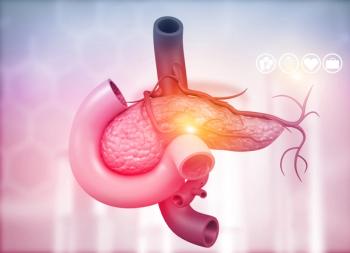
At the Stanford International Symposium on Multidetector-Row CT, radiologists demonstrated how the latest scanners are propelling CT forward in the pancreas, pushed by the steam of sophisticated multiplanar reformations and minimum intensity projections (MinIPs), which are now considered crucial (see Figure 1).
Islet cell tumors
At the Stanford symposium, R. Brooke Jeffrey, MD, discussed the use of multislice CT to distinguish islet cell tumors from ductal adenocarcinoma.
"Islet cell tumors represent only a small percentage of pancreatic neoplasms, but they are important because they have a much better prognosis than the more common ductal adenocarcinoma," said Dr. Jeffrey, professor of radiology, Stanford University.
Unlike ductal adenocarcinoma, islet cell tumor cells are usually hypervascular and rarely cystic. Features typical of ductal adenocarcinoma, such as local invasion, vascular encasement, and pancreatic duct obstruction, are far less common in islet cell tumors. Calcifications are present in up to 15% of islet cell tumors, whereas they are extremely rare in ductal adenocarcinoma cases.
For smaller tumors and multiple tumors, intraoperative ultrasound should be combined with CT for maximum sensitivity. As lesions get larger, they tend to be much more heterogeneous and are able to obstruct veins but not encase the arteries. It's also important to bear in mind that not all hypervascular lesions are necessarily islet cell tumorsthey may actually be renal cell carcinoma metastases.
"For poorly understood reasons, the pancreas is the sanctuary organ for renal cell metastases," Dr. Jeffrey said.
Incidental cystic lesions
Greater use of multislice CT in abdominal imaging has resulted in greater detection of incidental cystic lesions in the pancreas. One large study showed that cystic lesions in the pancreas are very common incidental findings on abdominal MR scans, present in 20% of cases (Radiology 223:547-553, 2002). Most of these lesions had a simple appearance, said Michael Macari, MD, another speaker at the Stanford symposium. Most of these lesions are benign.
"This is an epidemic. We don't see them as frequently on CT, due to lower contrast resolution, but we do see them often in daily practice. The radiologist and clinician need to decide what to do with these findings," said Dr. Macari, section chief of abdominal imaging at New York University School of Medicine.
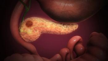
Cystic lesions can be benign or malignant, and there is often overlap in the imaging appearance of the various types, making confident diagnosis difficult. Most cystic lesions (85%) are pseudocysts, lesions that are filled with fluid and debris, and these are typically associated with a prior history of pancreatitis. Occasionally, pseudocysts have septations (see Figure 2). The other main types of cystic lesions are mucinous cystic tumor, serous cystadenoma, solid pseudopapillary tumor, and intraductal papillary mucinous tumor
Aside from prior history of pancreatitis, examiners must consider patient age, sex, andmost importantthe size of the lesion. Tiny lesions are often seen on multislice CT scans, but most are clinically irrelevant.
In general, lesions smaller than 2.5 cm with no malignant characteristics such as nodularity or thick irregular enhancing septations can be safely followed. Sometimes incidental findings of cystic lesions result in surgeries that harm the patient. The Whipple operation, for example, is associated with complications such as bowel obstruction. The procedure was once associated with 25% mortality rates, raising concern about lesions that could possibly never have become clinically significant. "Mortality is exceedingly rare, but morbidity is still quite high. We try to put the brakes on surgery if there are benign characteristics," Dr. Macari said.
Since most cystic lesions are incidentally detected, it may be necessary to perform another CT scan with protocols optimized for imaging the pancreas, such as dual-phase acquisition and thin-section multislice CT. Multiplanar reformats enable a more confident assessment of cystic lesions. MinIPs are useful in the assessment of ductal communication.
In cases of difficult diagnosis, it may prove helpful to perform cyst aspiration or endoscopic ultrasound, Dr. Macari said. Occasionally, for example, the serous cystadenoma will be unilobular and difficult to differentiate from peripheral mucinous cystic tumors. Cyst aspiration could clarify the true nature of this finding. Occasionally, surgery is still necessary to make a confident diagnosis.
Articles in this issue
over 18 years ago
Third-line single-agent cetuximab ups overall survivalover 18 years ago
Sorafenib gets ok for liver cancerover 18 years ago
Celgene to acquire Pharmionover 18 years ago
Novacea halts ASCENT-2 trialover 18 years ago
RT/temozolomide raises possibility of cure in glioblastomaover 18 years ago
Brachytherapy as effective in younger as in older menover 18 years ago
MMA not harming patientsover 18 years ago
Kinase inhibitor may prevent RT-induced lung injuryover 18 years ago
FDA approves lower starting dose for dasatinib for CMLover 18 years ago
Encouraging Avastin results in glioblastoma multiformeAdvertisement
Related Content
Advertisement


Advertisement
Advertisement
Trending on CancerNetwork
1
From Conference to Practice: Top 5 Takeaways From EHA 2026
2
Emerging T-Cell Engagers and Novel Immunotargets in Multiple Myeloma
3
Evaluating Bladder-Sparing Strategies and Key GU Data From ASCO 2026
4
Givastomig Earns FDA Fast Track Designation in HER2-Negative Gastric Cancer
5