- ONCOLOGY Vol 14 No 10
- Volume 14
- Issue 10
Multidisciplinary Management of Pediatric Soft-Tissue Sarcoma
Soft-tissue sarcomas comprise approximately 7% of all pediatric malignancies. Surgery, chemotherapy, and radiation therapy have significantly improved survival.
ABSTRACT: The management of pediatric soft-tissue sarcomas has improved drastically through the use of multimodal therapy. These tumors include rhabdomyosarcomas and nonrhabdomyosarcomas. Both are staged using physical, radiographic, and histologic evaluation, and both have intricate staging and grouping systems that correlate closely with prognosis. However, approaches to therapy for the two tumor types remain somewhat different. Rhabdomyosarcomas are treated primarily with chemotherapy. Surgical intervention is limited to initial biopsy, wide local excision when clear margins are feasible, and resection of residual disease. Radiation therapy is reserved for patients with persistent or recurrent disease and may be delivered by external beam or brachytherapy. Nonrhabdomyosarcomas are best treated primarily by surgical resection, although radiation and chemotherapy are now being used with some success. Another major difference concerns evaluation of lymphatics. Nonrhabdomyosarcomas in children frequently behave similarly to adult sarcomas, and less commonly involve regional lymph nodes, whereas pediatric patients with rhabdomyosarcomas often have nodal involvement necessitating surgical evaluation of regional lymph nodes as part of the staging protocol. Multimodal therapy has led to improved survival as well as better functional and cosmetic results. With further clinical trials and improved techniques such as brachytherapy and lymphatic mapping with sentinel node biopsy, we expect to continue to optimize therapy for pediatric patients with soft-tissue sarcomas. [ONCOLOGY 14(10):1471-1481, 2000]
Introduction
The multimodal management of pediatric soft-tissue sarcomas with the judicious use of surgery, chemotherapy, and radiation therapy has significantly improved survival, while decreasing morbidity, in these pediatric malignancies. Soft-tissue sarcomas comprise approximately 7% of all pediatric malignancies.[1]
Sarcomas are malignant tumors of mesenchymal cell origin. They are named according to the normal tissue they resemble-for example, rhabdomyosarcoma (skeletal muscle), leiomyosarcoma (smooth muscle), fibrosarcoma and malignant fibrous histiocytoma (connective tissue), neurofibrosarcoma or malignant peripheral nerve sheath tumor (neurofibromas, as seen in patients with neurofibromatosis), liposarcoma (adipose), synovial sarcoma (synovium), peripheral nerve sheath tumors (peripheral nerve), and angiosarcoma (blood and/or lymphatic vessels). Other sarcomas include rare entities such as alveolar soft-part sarcoma, extraosseous Ewing’s sarcoma, peripheral neuroectodermal tumors, epithelioid sarcoma, and hemangiopericytoma.
Although multimodal adjuvant therapy is standard treatment, the surgeon plays an important role in tissue diagnosis and staging. Surgical staging involves incisional or excisional biopsy, lymph node sampling, and/or sentinel lymph node biopsy with the use of lymphatic mapping. Primary resection is only indicated when it can be performed safely and is not mutilating. Otherwise, chemotherapy is administered as primary therapy, with delayed resection performed after local response has been assessed.
Radiation therapy is reserved for patients with recurrent or persistent disease and is most frequently given by external beam. Brachytherapy administered via the use of implants placed at the time of tumor resection is being used with increasing frequency and is particularly useful for unresectable microscopic disease. This technique allows aggressive treatment of the tumor bed while sparing the patient the morbidity of bone and soft-tissue exposure to radiation.
Rhabdomyosarcoma
Rhabdomyosarcoma comprises 50% of soft-tissue sarcomas in children and may occur anywhere in the body, even where skeletal muscle is generally not present.[2] The most common sites are the head and neck and genitourinary system. In the Intergroup Rhabdomyosarcoma Study (IRS) IV, the distribution of primary sites was 29% genitourinary, 28% head and neck, 15% extremities, 12% pelvis, 7% trunk and thorax, 6.5% orbit, and 1% gastrointestinal (Figure 1).
Tumor Biology
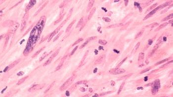
Rhabdomyosarcoma is one of the small, blue round-cell tumors of childhood. It arises from embryonic mesenchyme, which has the potential to develop into skeletal muscle. Skeletal muscle cross striations on light microscopy help to distinguish these tumors.[3] Histology is confirmed through immunohistochemical techniques, electron microscopy, and molecular genetic evaluation, which can also be used to differentiate the subtypes of rhabdomyosarcoma. There are five subtypes; however, most fall into one of two major subtypes-embryonal and alveolar. Other subtypes include botryoid, spindle-cell, and pleomorphic rhabdomyosarcomas.
The alveolar subtype is distinguished on light microscopy by the presence of small, round cells with septations that resemble a pulmonary alveolus. In IRS I and II, the tumor was only labeled as alveolar if it appeared 50% alveolar by light microscopy. Currently, a tumor is considered alveolar if any alveolar component is visualized.[4] The alveolar variant has been found to have a translocation between the long arm of chromosome 2 and chromosome 13. This has been shown to involve the PAX3 gene locus, thereby disturbing transcription during neuromuscular development.[5,6]
Alveolar rhabdomyosarcoma is most frequently seen in the extremities, trunk, or perineum. It is probably because the tumor occurs in these unfavorable sites that alveolar histology is associated with a poor prognosis. A recent review did not distinguish the alveolar subtype as a significant prognostic indicator in extremity rhabdomyosarcomas.[7]
Embryonal histology is the most common subtype and usually arises in the head and neck, genitourinary tract, and orbit. This subtype is known to have a loss of heterozygosity at the 11p15 locus, the region responsible for the IGFII gene.[8,9] The DNA content in these tumors ranges from diploid to hyperdiploid; diploid tumors are associated with a poorer prognosis.[10]
The botryoid variant, named for its resemblance to a “cluster of grapes,” is most commonly seen in infants and arises in cavitary structures such as the vagina and bladder. Another variant of embryonal histology is spindle-cell rhabdomyosarcoma, which is most commonly found in the paratesticular area, although it has also been found to arise in other regions, such as the head and neck.[11]
Rhabdomyosarcoma is associated with a p53 mutation and the Li-Fraumeni syndrome,[12] a familial cancer syndrome that exhibits autosomal dominant inheritance of a germline mutation of the p53 gene. This syndrome is characterized by a high incidence of soft-tissue or bony sarcomas, leukemia, brain or adrenal neoplasms, and maternal premenopausal breast cancer.[13]
Evaluation and Staging
The clinical presentation of rhabdomyosarcoma varies, depending on tumor site. Most of these tumors present as painless masses. However, those in the head and neck may involve the central nervous system from intracranial extension of the tumor or infiltration of the cranial nerves, meninges, or brain stem. The differential diagnosis should consider benign masses such as lipoma, neurofibroma, or even hematoma. However, in a child with a persistent mass, it must include soft-tissue sarcoma.
The evaluation of a patient with a suspected rhabdomyosarcoma should seek to define local extent of tumor (ie, resectability), lymph node involvement, and distant metastasis. The patient should initially undergo a thorough physical examination with palpation of the lymph node basins, followed by radiographic evaluation. In most cases, a computed tomography (CT) scan or magnetic resonance imaging (MRI) of the primary tumor will be necessary to assess tumor size and its extension into adjacent structures. These studies will also assist in narrowing the differential diagnosis.
A bone scan and CT scan of the chest are also recommended to rule out metastatic disease to the skeletal structures or pulmonary parenchyma. The diagnosis should then be confirmed with a tissue specimen, which may be attained via fine-needle, core, or incisional biopsy. Once the histologic diagnosis has been made, the tumor should be staged. Rhabdomyosarcoma is currently staged using the Lawrence/Gehan staging system coupled with postresection classifications. The staging system and operative grouping are shown in Table 1.
Treatment
Multimodality therapy has led to continued improvement in survival as well as cosmetic and functional outcomes in pediatric rhabdomyosarcomas. The treatment plan includes surgical staging and chemotherapy followed, in most patients, by definitive or second-look surgery.
• Surgery-The first step, prior to performing surgery, is to obtain a tissue diagnosis, with sufficient tissue samples to allow histologic evaluation. The primary surgical approach may be wide local excision, or incisional or excisional biopsy. Wide local excision should be undertaken only when the preoperative evaluation has revealed a resectable tumor and it is thought feasible to obtain negative margins. Biopsies must be performed in such a way that the scar will not interfere with subsequent wide local excision, particularly when dealing with tumors of the extremities (Figure 2). The initial procedure may include vascular access, bone marrow aspiration, brachytherapy catheter placement, and lymph node mapping with sentinel node biopsy or lymph node sampling.
The wide array of surgical options underscores the importance of adequate preoperative staging. Careful determination of surgical margins is crucial, and resection should continue until clear margins are obtained.
We recommend reexcision for all patients with microscopic positive margins as well as for those referred to our institution with unclear or positive margins or those with pathology unavailable for review by our pathologist. It has been demonstrated that reexcision of extremity and trunk sarcomas in node-negative patients with microscopic residual disease results in improved survival.[14]
We currently perform lymphatic mapping with excision of the sentinel node(s) in all our patients with extremity rhabdomyosarcoma. This is best accomplished at the time of the initial procedure, since prior biopsy, wide local excision, or partial excision may disrupt the lymphatics and, thereby, the specificity of the pathologic examination. Although experience with lymph node mapping in rhabdomyosarcoma is limited, we feel that it will allow more accurate staging of pediatric patients with extremity rhabdomyosarcoma.
Lymph node mapping is performed by injecting the tumor with technetium-labeled sulfur colloid followed by isosulfan blue dye (Lymphazurin). A radioisotope detector is used to localize the sentinel node, and an incision is made, overlying the localized area, to remove all blue nodes with high radioisotope counts (Figure 3, Figure 4, and Figure 5). Formal lymph node dissection is not recommended because it has not been shown to affect survival significantly, even in patients with histologically positive nodes.[15]
Wide local excision is generally avoided in head and neck tumors, vaginal/uterine tumors, and bladder tumors. This is particularly true in head and neck tumors when aggressive excision will result in a significant cosmetic or functional defect. Orbital exenteration for orbital rhabdomyosarcoma is reserved for recurrence.
Vaginal or uterine rhabdomyosarcoma in pediatric patients usually responds to conservative therapy, and treatment should comprise biopsy followed by chemotherapy alone or combined with radiation and followed by second-look surgery. Persistent disease may require partial vaginectomy.[16]
Whenever possible, the bladder should be conserved. Anterior pelvic exenteration is no longer performed, having been replaced by primary chemotherapy and, occasionally for persistent disease, radiation therapy. Conservative therapy, although not as successful in bladder rhabdomyosarcomas as for vaginal primaries, has led to a survival rate of 89% and the retention of functional bladders in 60% of patients at 4 years from diagnosis.[17]
• Chemotherapy-The chemotherapeutic regimen for rhabdomyosarcoma was previously based on histology as well as stage. In IRS IV, however, treatment was not altered according to histology.[4] Since then, topotecan (Hycamtin) has emerged as a new agent that may be helpful in improving results in patients with alveolar rhabdomyosarcoma and undifferentiated sarcoma.[18] Current recommendations stem from the ongoing IRS V.
In IRS V, patients are divided into low-risk, intermediate-risk, and high-risk categories based on the likelihood of disease recurrence (Table 2). This risk is determined by (1) the site of the primary tumor (the orbit and nonparameningeal head and neck sites as well as genitourinary nonbladder/nonprostate sites are considered favorable; all others are unfavorable); (2) the size of the primary tumor (tumor size £ 5 cm is favorable, > 5 cm is unfavorable); (3) the presence (unfavorable) or absence (favorable) of regional lymph node involvement and distant metastases; and (4) the histologic subtype (embryonal, botryoid, and spindle-cell rhabdomyosarcomas are favorable, whereas alveolar rhabdomyosarcoma and undifferentiated sarcoma are unfavorable).
Treatment for low-risk patients consists of VAC (weekly vincristine for nine doses plus actinomycin D [Cosmegen], with or without cyclophosphamide [Cytoxan, Neosar]) and granuloycte colony-stimulating factor (G-CSF, Neupogen) for four doses every 12 weeks beginning at weeks 0, 12, 24, and 36. Radiation therapy is added for patients with residual localized disease.
Intermediate-risk patients receive radiation therapy and VAC or VAC plus topotecan, according to randomized assignment, for nearly 1 year. High-risk patients begin treatment with irinotecan (Camptosar) followed by VAC and radiation therapy. Those who respond resume irinotecan therapy, again for about 1 year. Table 2 shows the treatment options for each risk category. The diseases and therapies are sufficiently complex that knowledgeable and experienced pediatric oncologists, surgeons, and radiation therapists should direct each patient’s treatments.
• Radiation Therapy-Radiation therapy is utilized selectively to enhance local tumor control. Brachytherapy has been valuable in cases of positive or questionable surgical margins. Burmeister et al recently reported good local control with acceptable wound complications in a small series of 12 patients.[19] Brachytherapy catheters, placed at the time of excision, allow directed local control. The catheters are afterloaded by a radiation oncologist approximately 1 to 2 weeks postoperatively (Figure 6, Figure 7, and Figure 8).
Summary
The prognosis of pediatric patients diagnosed with rhabdomyosarcoma has continued to improve through the use of multimodal therapy. Patients with locally controlled disease can expect a favorable prognosis, whereas those with metastatic or locally advanced disease remain the subjects of intense research as clinical protocols attempt to find a therapy that will improve outcome.
The advent of the Lawrence/Gehan staging system, coupled with the new grouping system, allows accurate standardized staging that can be used to predict prognosis. Recent emphasis has been placed on therapy that provides local control with less functional or cosmetic impairment. These therapies are now being applied to groups with predicted favorable outcome to ensure that tumor control is maintained while toxicity and disability decrease.
Nonrhabdomyosarcoma Soft-Tissue Sarcoma
As a group, nonrhabdomyosarcoma soft-tissue sarcomas comprise almost 50% of pediatric soft-tissue sarcomas. These tumors can occur anywhere in the body but are most commonly seen in the extremities. These uncommon malignancies include malignant fibrous histiocytoma, malignant peripheral nerve sheath tumors, extraosseous Ewing’s sarcoma, alveolar soft-part sarcoma, clear-cell sarcoma or melanoma of the soft parts, synovial sarcoma, leiomyosarcoma, liposarcoma, fibrosarcoma, angiosarcoma, epithelioid sarcoma, and hemangiopericytoma.
Tumor Biology
Each of the nonrhabdomyosarcoma soft-tissue sarcomas arises from mesenchymal cells. The tumors are distinguished from one another on the basis of light microscopy, immunohistology, and electron microscopy. The most common types in the pediatric population are synovial sarcoma, malignant peripheral nerve sheath tumor, and fibrosarcoma. These tumors may also be associated with the Li-Fraumeni syndrome, implicating a p53 mutation.[4] Nonrhabdomyosarcomas have also been reported to occur with other syndromes, including neurofibromatosis, Down syndrome, and spina bifida.[20] Histologic tumor grade remains the foremost factor in determining patient prognosis, and for this reason, a formal grading system has been developed as shown in Table 3.[20-22]
According to this system, grade 1 tumors are limited to myxoid, well-differentiated liposarcomas, dermatofibrosarcoma protuberans, extraskeletal chondrosarcoma, well-differentiated leiomyosarcoma, malignant hemangiopericytoma, peripheral nerve sheath tumors, and infantile fibrosarcoma. Grade 2 tumors are less than 15% necrotic at histologic examination, have a mitotic index of 5 to 10 per high-power field, and show no marked nuclear atypia or cellularity.
Grade 3 tumors have marked nuclear atypia or cellularity, greater than 15% necrosis, and/or more than 10 mitoses per high-power field. They include pleomorphic liposarcomas, mesenchymal chondrosarcomas, extraskeletal osteosarcoma, triton tumors, alveolar soft-part sarcomas, extraosseous Ewing’s sarcoma, and epithelioid sarcomas. Fibrosarcoma and malignant hemangiopericytomas in children less than 4 years old are excluded from the grade 3 classification.
Evaluation and Staging
As with rhabdomyosarcomas, the clinical presentation of nonrhabdomyosarcomas varies based on the region of the body. Most will manifest as painless masses. The differential diagnosis is also the same as that described for rhabdmyosarcoma. Radiographic examination should be performed to determine the extent of tumor, whether there has been invasion into adjacent structures, and whether distant metastases are present. MRI is the preferred imaging modality for tumors of the extremity and head and neck. CT scans are useful for abdominal tumors and for evaluation of lung parenchyma for metastatic disease (Figure 9).
Following radiographic evaluation, a tissue diagnosis is obtained via either needle biopsy, incisional or excisional biopsy, or wide local excision based on tumor resectability. If there is clinical evidence of nodal involvement, lymphatic staging may be performed, although nodal involvement is less frequently seen with nonrhabdomyosarcoma than with rhabdomyosarcoma.
Treatment
Multimodal therapy has also been shown to be the most effective approach to treatment for nonrhabdomyosarcoma.
• Surgery-Surgical evaluation begins at the time of tissue diagnosis and surgical staging. Complete resection with negative margins remains the most important prognostic indicator, and surgical intervention must be planned with this goal in mind.[4] Because many children present with locally advanced disease, the most common initial surgery performed is incisional or excisional biopsy in order to obtain a tissue diagnosis. Wide local excision is undertaken as an initial surgery only if complete excision is feasible.
Following any indicated adjuvant therapy, children who have undergone initial biopsy return to the operating room for second-look surgery and complete excision of the remaining tumor (Figure 10 and Figure 11). Positive or unclear margins should be treated with further resection; however, if this is not possible due to adjacent structures, or the prospect of adverse cosmetic or functional outcome, brachytherapy catheters may be implanted to allow aggressive localized radiotherapy. This method may also improve local control in limb-sparing surgery.
Malignant fibrous histiocytoma is an uncommon subtype of nonrhabdomyosarcoma; 44 pediatric patients have been diagnosed and treated for this tumor at the M. D. Anderson Cancer Center over a 15-year period. A review by Corpron et al revealed an excellent prognosis for children with resectable tumors of this type.[23] Treatment for malignant fibrous histiocytoma, as for other nonrhabdomyosarcomas, consists primarily of surgery with the goal of complete resection; chemotherapy and radiation are reserved for patients with residual, metastatic, or recurrent disease. Of note, in 24 patients with microscopic residual disease, survival was not affected, but the rate of recurrence increased.[24]
Surgical resection should also be considered for metastatic disease. According to the findings of several small studies, aggressive metastasectomy for pulmonary disease appears to increase survival.[25] However, this technique needs to be evaluated further and should be reserved for metastatic disease that is localized (Figure 12).
• Radiotherapy-Nonrhabdomyosarcoma was once believed to be radioresistant. However, radiation is now widely used in the adult population with good success.[22] Radiation therapy should be administered to patients with advanced local, residual, or recurrent disease. It may be delivered either by external beam (as is often the case for locally advanced or recurrent disease) or by brachytherapy (which should be considered for patients in whom residual disease is either confirmed or strongly suspected at the time of surgical resection). Brachytherapy treats the tumor bed aggressively, while sparing the surrounding normal tissue and bone.
• Chemotherapy-Chemotherapy has not been well studied in pediatric patients with nonrhabdomyosarcoma, because of the multiplicity of histologic types, differences in biological behavior, and relative rarity of the disease in this patient population. Therefore, based on experience in adults, most children are treated with doxorubicin and ifosfamide (Ifex). Recent studies have shown that children with grade 1 and 2 tumors do not benefit from chemotherapy and that chemotherapy should be reserved for those with either grade 3 tumors, incompletely resected local disease, or metastases at diagnosis.[24]
Summary
Pediatric nonrhabdomyosarcoma soft-tissue sarcomas are a relatively rare group of malignancies. Children with these tumors are beginning to benefit from multimodal therapy. However, the roles of radiation therapy and chemotherapy are not as clearly defined as in rhabdomyosarcoma. Ongoing research will continue to optimize therapy in these patients.
References:
1. Robinson LL: General principles of the epidemiology of childhood cancer, in Pizzo PA, Poplack DG (eds): Principles and Practice of Pediatric Oncology, 2nd ed. Philadelphia, JB Lippincott, 1993.
2. Maurer HM, Beltangady M, Gehan EA, et al: The Intergroup Rhabdomyosarcoma Study I: A final report. Cancer 61:209-220, 1988.
3. James J, Andrassy RJ: Rhabdomyosarcoma in children. Contemp Surg 30:58-65, 1987.
4. Wexler LH, Helman LJ: Pediatric soft-tissue sarcomas. CA Cancer J Clin 44:211-247, 1994.
5. Turc-Carel C, Lizard-Nacol S, Justrabo E, et al: Consistent chromosomal translocation in alveolar rhabdomyosarcoma. Cancer Genet Cytogenet 19:361-362, 1986.
6. Shapiro DN, Sublett JE, Li B, et al: Fusion of PAX3 to a member of the forkhead family of transcription factors in human alveolar rhabdomyosarcoma. Cancer Res 53:5108-5112, 1993.
7. Neville HL, Andrassy RJ, Lobe T, et al: Extremity rhabdomyosarcoma: An analysis of preoperative staging, prognostic factors, and outcome from the Intergroup Rhabdomyosarcoma Study IV. J Pediatr Surg 35:317-321, 2000.
8. Scrable HJ, Witte DP, Lampkin BC, et al: Chromosomal localization of human rhabdomyosarcoma locus by mitotic recombination mapping. Nature 329:645-747, 1987.
9. Ogawa O, Eccles MR, Szeto J, et al: Relaxation of insulin-like growth factor II gene imprinting implicated in Wilms’ tumour. Nature 362:749-751, 1993.
10. Pappo AS, Crist WM, Kuttesch J, et al: Tumor-cell DNA content predicts outcome in children and adolescents with clinical group III embyronal rhabdomyosarcoma: The Intergroup Rhabdomyosarcoma Study Committee of the Children’s Cancer Group and the Pediatric Oncology Group. J Clin Oncol 11:1901-1905, 1993.
11. Leuschner I, Newton WA Jr, Schmidt D, et al: Spindle-cell variants of embryonal rhabdomyosarcoma in the paratesticular region: A report of the Intergroup Rhabdomyosarcoma Study. Am J Surg Pathol 17:221-230, 1993.
12. Felix CA, Kappel CC, Mitsudomi T, et al: Frequency and diversity of p53 mutations in childhood rhabdomyosarcoma. Cancer Res 52:2243-2247, 1992.
13. Li FP, Fraumeni JF, Mulvihill JJ, et al: A cancer family syndrome in 24 kindreds. Cancer Res 48:5358-5362, 1988.
14. Hays DM, Lawrence W Jr, Wharam M, et al: Primary re-excision for patients with microscopic residual tumor following initial excision of sarcomas of trunk and extremity sites. J Pediatr Surg 24:5-10, 1989.
15. Andrassy RJ, Corpron CA, Hays D, et al: Extremity sarcoma: An analysis of prognostic factors from the Intergroup Rhabdomyosarcoma Study III. J Pediatr Surg 31:191-196, 1996.
16. Andrassy RJ, Hays R, Raney B, et al: Conservative surgical management of vaginal and vulvar pediatric rhabdomyosarcoma: A report from the Intergroup Rhabdomyosarcoma Study III. J Pediatr Surg 30:1034-1037, 1995.
17. Hays DM: Bladder/prostate rhabdomyosarcoma: Results of the multi-institutional trials of the Intergroup Rhabdomyosarcoma Study. Semin Surg Oncol 9:520-523, 1993.
18. Vietti T, Crist W, Ruby E, et al: Topotecan window in patients with rhabdomyosarcoma: An IRSG Study (abstract). Proc Am Soc Clin Oncol 16:510a, 1997.
19. Burmeister BH, Dickinson I, Bryant G, et al: Intra-operative implant brachytherapy in the management of soft-tissue sarcomas. Aust NZ J Surg 67:5-8, 1997.
20. Dillon PW: Nonrhabdomyosarcoma soft-tissue sarcoma in children. Semin Pediatr Surg 6:24-28, 1997.
21. Rao BN: Nonrhabdomyosarcoma in children: Prognostic factors influencing survival. Semin Surg Oncol 53:530-541, 1993.
22. Dinges S, Budach V, Budach B, et al: Local recurrences of soft tissue sarcomas in adults: A retrospective analysis of prognostic factors in 102 cases after surgery and radiation therapy. Eur J Cancer 30:746-751, 1994.
23. Corpron CA, Black CT, Raney RB, et al: Malignant fibrous histiocytoma in children. J Pediatr Surg 31:1080-1083, 1996.
24. Maurer HM, Cantor A, Salzberg A, et al: Adjuvant chemotherapy vs observation for localized nonrhabdomyosarcoma soft-tissue sarcoma in children (abstract). Proc Am Soc Clin Oncol 11:363, 1992.
25. Ueda T, Uchida A, Kodama K, et al: Aggressive pulmonary metastasectomy for soft-tissue sarcomas. Cancer 72:1919-1925, 1993.
Articles in this issue
over 25 years ago
New Awards Spotlight Courage of Cancer Survivorsover 25 years ago
Children’s Art Project at M. D. Anderson Cancer Centerover 25 years ago
Settling on an Increased NCI Budgetover 25 years ago
Computer Billing Standardover 25 years ago
Ligand Receives FDA Marketing Clearance for Bexarotene Gelover 25 years ago
Mayo Clinic Study Shows Patients Uncertain About Cancer Risk Termsover 25 years ago
Follow-up Care for Cancer: Making the Benefits Equal the CostNewsletter
Stay up to date on recent advances in the multidisciplinary approach to cancer.


