|Articles|July 1, 1997
- ONCOLOGY Vol 11 No 7
- Volume 11
- Issue 7
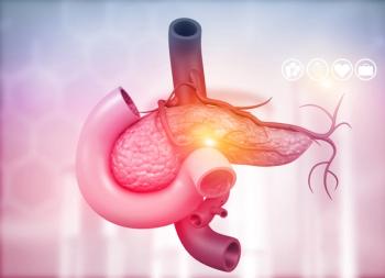
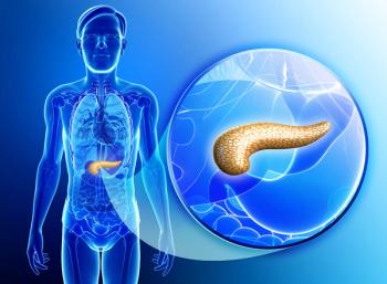
Pancreatic Cancer Surgical Practice Guidelines
The Society of Surgical Oncology surgical practice guidelines focus on the signs and symptoms of primary cancer, timely evaluation of the symptomatic patient, appropriate preoperative extent of disease evaluation, and role of the surgeon in
Advertisement
The Society of Surgical Oncology surgical practice guidelines focuson the signs and symptoms of primary cancer, timely evaluation of the symptomaticpatient, appropriate preoperative extent of disease evaluation, and roleof the surgeon in diagnosis and treatment. Separate sections on adjuvanttherapy, follow-up programs, or management of recurrent cancer have beenintentionally omitted. Where appropriate, perioperative adjuvant combined-modalitytherapy is discussed under surgical management. Each guideline is presentedin minimal outline form as a delineation of therapeutic options.
Since the development of treatment protocols was not the specific aimof the Society, the extensive development cycle necessary to produce evidence-basedpractice guidelines did not apply. We used the broad clinical experienceresiding in the membership of the Society, under the direction of AlfredM. Cohen, MD, Chief, Colorectal Service, Memorial Sloan-Kettering CancerCenter, to produce guidelines that were not likely to result in significantcontroversy.
Following each guideline is a brief narrative highlighting and expandingon selected sections of the guideline document, with a few relevant references.The current staging system for the site and approximate 5-year survivaldata are also included.
The Society does not suggest that these guidelines replace good medicaljudgment. That always comes first. We do believe that the family physician,as well as the health maintenance organization director, will appreciatethe provision of these guidelines as a reference for better patient care.
Symptoms and Signs
- Adenocarcinoma involving the pancreatic head or uncinate process oftencauses obstruction of the intrapancreatic portion of the common bile duct,resulting in jaundice.
- Tumors in the pancreatic body and tail do not obstruct the bile duct,and therefore, patients are rarely diagnosed prior to the development oflocally advanced or metastatic disease.
- In the absence of jaundice, patients often present with complaintsof vague upper abdominal or back pain, weight loss due to anorexia, decreasedenergy level, and nonspecific upper gastrointestinal or dyspeptic symptoms.
- An occasional patient may present with a change in bowel habits secondaryto pancreatic exocrine insufficiency due to tumor obstruction of the pancreaticduct; bowel movements may be loose, malodorous, and consistent with steatorrhea.
- New-onset hyperglycemia
- Nausea and vomiting secondary to duodenal obstruction
Evaluation of the Symptomatic Patient
Work-up
- History
- Physical examination
- Liver function tests and ultrasound (to confirm extrahepatic biliaryobstruction in jaundiced patients)
- Extent of disease (both locoregional and distant) is assessed by contrast-enhancedhelical CT scan.
- Distant metastatic disease:
- Confirm cytologic diagnosis.
- Fine needle aspiration (FNA) cytology--CT-guided, endoscopic ultrasound(EUS)-guided, fluoroscopy-guided paracentesis (if ascites is present)
- No distant metastatic disease:
- Mass in pancreas considered unresectable-- EUS-guided or CT-guidedFNA
- No mass in pancreas--endoscopic retrograde cholangiopancreatography(ERCP) with biopsy demonstrating a malignant stricture
Appropriate timeliness of surgical referral
- Evaluation should begin as soon as symptoms are reported.
Preoperative Evaluation for Extent of Disease
- History and physical examination
- In the absence of hyperbilirubinemia, routine laboratory studies areof little value in arriving at the diagnosis of a pancreatic neoplasm.Most patients with adenocarcinoma will evidence a mild degree of hyperglycemia,yet this finding is often subtle and difficult to discriminate from verymild adult-onset diabetes mellitus.
- In the absence of extrahepatic biliary obstruction, elevated liverfunction tests should raise the suspicion of metastatic disease (as istrue with other solid tumors).
- Intraoperative biopsy has a false-negative rate of at least 30% andhas been associated with such complications as pancreatic fistula and pancreatitis.Since a negative biopsy is unlikely to alter the therapeutic strategy,intraoperative biopsy of the pancreas should generally be avoided.
- Many surgeons, however, are reluctant to proceed with pancreaticoduodenectomyin the absence of cytologic or histologic confirmation of disease. Forthose surgeons, EUS-guided or CT-guided FNA is a reasonable alternative.In patients eligible for clinical trials examining the value of preoperativeradiation, chemotherapy, or both, preoperative EUS-guided FNA is the preferredmethod of cytologic confirmation of disease.
- Local criteria for resectability can be determined with a high degreeof accuracy by preoperative imaging modalities.
- The use of contrast-enhanced helical CT obtained at 1.5- or 3.0-mmsection thickness and 5-mm scan interval gives precise information regardingthe relationship of the tumor to the superior mesen- teric artery (SMA),hepatic artery, and celiac axis. The relationship of the tumor to the superiormesenteric-portal venous confluence is less reliably assessed by CT scan.However, the absence of a normal fat plane between the tumor and this venousstructure should suggest the potential for tumor involvement of the lateralor posterior wall of the superior mesenteric or portal vein.
- Celiac and SMA angiography with venous phase imaging in selected patients(ie, patients considered for reoperative pancreaticoduodenectomy)
- Absolute criteria for unresectability include:
- The presence of distant metastatic disease
- Encasement of the celiac axis or SMA
- Occlusion of the superior mesenteric-portal venous confluence
Role of the Surgeon in Initial Management The majority of patients with pancreatic cancer present with locallyadvanced or metastatic disease. The surgeon is often the entry point forthe patient into the realm of therapeutic options.
- Following an initial evaluation consisting of a physical examination,chest x-ray, and contrast-enhanced CT, it is possible to determine theextent of disease and the potential for surgical resection.
- Biliary decompression, when necessary, in patients with locally advanced,unresectable primary tumors and/or metastatic disease, should be performedendoscopically.
- Transhepatic decompression is a second-line alternative.
- Laparoscopically assisted cholecystojejunostomy in carefully selectedpatients
- In general, biliary enteric bypass via a standard laparotomy shouldbe reserved for patients who experience repeated episodes of stent occlusionor cholangitis. The majority of operative biliary enteric bypass proceduresshould be performed in patients who are brought to the operating room forplanned pancreatectomy and are found to be unresectable due to unsuspectedmetastatic disease or locally advanced primary tumors.
Surgical considerations
- For tumors to the right of the mesenteric vessels--pancreaticoduodenectomy(standard, pylorus-preserving, or extended pancreatectomy to include vascularresection and reconstruction in highly selected patients)
- For tumors to the left of the mesenteric vessels--distal pancreatectomyif resectable
- Palliative open or laparoscopic bypass surgery in selected patientsnot amenable to nonoperative biliary decompression
- Endoscopic, laparoscopic, and open laparotomy for biliary decompressionor gastric bypass are all useful in selected patients. The role of eachprocedure in the individual patient depends on many variables, and definitivealgorithms are lacking.
These guidelines are copyrighted by the Society of Surgical Oncology(SSO). All rights reserved. These guidelines may not be reproduced in anyform without the express written permission of SSO. Requests for reprintsshould be sent to: James R. Slawny, Executive Director, Society of SurgicalOncology, 85 W Algonquin Road, Arlington Heights, IL 60005.
Pancreatic cancer remains the fourth leading cause of cancer-relateddeaths in adults in the United States. Its etiology is unknown, and thereis currently no effective method of early diagnosis. The development ofmolecular techniques to diagnose pancreatic cancer at a time when the tumoris localized to the pancreas would allow a greater number of patients toreceive potentially curative therapy. In addition, effective treatmentof subclinical, micrometastatic disease (which exists in the liver of mostpatients at the time of removal of the primary pancreatic tumor) woulddramatically increase long-term survival rates following pancreaticoduodenectomy.These are areas of active laboratory investigation and early preclinicalstudy.
TNM staging based on radiographic and operative findings (Table1) does not directly correlate with treatment recommendations and thereforeis not commonly used. Patients are best classified as having potentiallyresectable (T1-2, selected T3, NX, M0), locally advanced (T3, NX, M0),or metastatic disease (T1-3, NX, M1). Only recently has a pathologic stagingsystem for resected specimens been suggested.[1]
Recent advances in the clinical care of patients with pancreatic cancerhave focused on: (1) decreasing treatment-related toxicity by utilizingaccurate preoperative imaging, and (2) improving the quality and lengthof patient survival through the use of innovative multimodality therapydirected at known patterns of disease recurrence.[2,3]
Preoperative Assessment
Surgical resection, as part of a multimodality approach for patientswith resectable pancreatic cancer, represents the only potentially curativetreatment strategy. However, only 16% to 30% of patients who undergo operationwith curative intent have their tumors successfully removed; the remainingpatients are found to have unsuspected liver or peritoneal metastases orlocal tumor extension to the mesenteric vessels.[4] Only patients whosetumors are completely resected enjoy a survival benefit. Therefore, themajority of patients who undergo surgical exploration for presumed cancerof the pancreatic head derive no survival benefit, yet the laparotomy resultsin a perioperative morbidity of 20% to 30%, a mean hospital stay of 14to 20 days (with the recovery period lasting an additional 15 to 20 days),and a median survival of only 6 months. Furthermore, in patients whosetumors are resected with positive margins, survival is no better than inpatients with locally advanced, unresectable disease treated by palliativechemotherapy and irradiation.[2,5]
The need for accurate preoperative assessment is evidenced by the largenumber of positive margin resections recently reported[6] and the highincidence of local tumor recurrence following pancreaticoduodenectomy.[2,4]In addition, in patients with unresectable disease, endoscopic, percutaneous,and laparoscopic methods of biliary decompression make laparotomy for palliationunnecessary.
Contrast-enhanced helical CT can accurately assess the relationshipof the tumor to the celiac axis and superior mesenteric artery (SMA). Potentiallyresectable primary tumors in the pancreatic head or uncinate process aredefined preoperatively (on CT) by: (1) the absence of extrapancreatic disease;(2) no evidence of tumor extension to the celiac axis or SMA; and (3) apatent superior mesenteric vein (SMV) and portal vein.[2] Patients withlocally advanced disease are not subjected to the potential morbidity andprolonged recovery period associated with a laparotomy, but instead, canreceive biliary decompression, if needed, by means other than a laparotomy.
Multimodality Treatment
Aggressive multimodality treatment is reserved for patients with potentiallyresectable primary tumors defined by the CT criteria stated previously.
Current treatment programs build on past experience, largely in unresectabletumors, using preoperative or postoperative chemotherapy and radiationtherapy combined with surgery.[7,8] The Gastrointestinal Tumor Study Group(GITSG) first demonstrated a survival advantage for pancreatic cancer patientstreated with adjuvant chemoradiation (40 Gy of external-beam irradiationplus concomitant fluorouracil [5-FU]) following pancreaticoduodenectomy,compared with patients treated with surgical resection alone (median survival,20 vs 11 months).[9]
This treatment advantage was confirmed in a group of 30 additional patientstreated with postoperative chemoradiation.[10] Delivery of adjuvant therapy,however, was hindered by the prolonged recovery time of most patients followingpancreaticoduodenectomy. Many patients required more than 10 weeks to achievea performance status necessary for treatment with chemoradiation.
The inability of some patients to tolerate postoperative therapy, combinedwith the growing popularity of neoadjuvant therapy in patients with othersolid tumors, caused investigators to consider giving radiation and chemotherapyprior to surgery in patients with potentially resectable pancreatic cancer.The use of preoperative chemoradiation is supported by the following considerations[2]:(1) Radiation therapy is more effective in well-oxygenated cells that havenot been exposed to the devascularization of surgery. (2) Radiation therapybefore surgical exploration may prevent the implantation and disseminationof tumor cells at the time of laparotomy, thereby decreasing subsequentperitoneal failure. (3) Patients with disseminated disease evident on restagingstudies after chemoradiation are not subjected to laparotomy.[3]
The following management principles can be applied to most patientswith presumed adenocarcinoma of the pancreas:
- Endoscopic retrograde cholangiopancreatography (ERCP) is performedwhen CT demonstrates obstruction of the intrapancreatic portion of theextrahepatic bile duct without a low-density mass in the pancreatic head.
- When CT demonstrates a low-density tumor mass in the pancreatic head,resectability is assessed preoperatively (as defined above). Patients withCT evidence of arterial encasement, venous occlusion, or extrapancreaticdisease are not considered for surgical resection. Biliary decompression,if necessary, is usually performed endoscopically.
- The decision to proceed with pancreaticoduodenectomy is made preoperativelybased on high-quality imaging studies. There are few indications for intraoperativebiopsy of the pancreas. For those surgeons who will not proceed with pancreaticoduodenectomyin the absence of tissue confirmation of malignancy, data suggest thatpreoperative CT-guided FNA provides a safe and accurate alternative.[11]Endoscopic ultrasound-guided FNA is currently replacing CT-guided FNA atmost centers. Experience with reoperative pancreatectomy supports the opinionthat there is no place for "exploratory" laparotomy in patientswith biopsy-proven or suspected pancreatic cancer; rather, the operativeplan should be carefully outlined preoperatively.[12]
- A definitive algorithm for palliative decompression of unresectablepatients is lacking. Stent placement and laparoscopic or open operativedecompression are all useful bypass strategies.
References:
1. Staley CA, Cleary KR, Abbruzzese JL, et al: Need for standardizedpathologic evaluation of pancreaticoduodenectomy specimens: Pancreaticoduodenectomyspecimens. Pancreas 12:373-380, 1996.
2. Evans DB, Abbruzzese JL, Rich TA: Cancer of the pancreas, in DeVitaVT Jr, Hellman S, Rosenberg SA (eds): Cancer: Principles and Practice ofOncology, 5th ed, pp 1054-1087. Philadelphia, Lippincott, 1997.
3. Spitz FR, Abbruzzese JL, Lee JE, et al: Preoperative and postoperatiavechemoradiation strategies in patients treated with pancreaticoduodenectomyfor adenocarcinoma of the pancreas. J Clin Oncol 15:928-937, 1997.
4. Fuhrman GM, Charnsagavej C, Abbruz-zese JL, et al: Thin-section contrastenhanced computed tomography accurately predicts resectability of malignantpancreatic neoplasms. Am J Surg 167:104-113, 1994.
5. Nitecki SS, Sarr MG, Colby TV, et al: Long-term survival after resectionfor ductal adenocarcinoma of the pancreas: Is it really improving? AnnSurg 221:59-66, 1995.
6. Willet CG, Lewandrowski K, Warshaw AL, et al: Resection margins incarcinoma of the head of the pancreas: Implications for radiation therapy.Ann Surg 217:144-148, 1993.
7. Evans DB, Staley CA, Lee JE, et al: Adenocarcinoma of the pancreas:Recent controversies, current management, and future therapies. GI Cancer1:149-161, 1996.
8. Staley CA, Lee JE, Cleary KA, et al: Preoperative chemoradiation,pancreaticoduodenectomy, and intraoperative radiation therapy for adenocarcinomaof the pancreatic head. Am J Surg 171:118-125, 1996.
9. Kalser MH, Ellenberg SS: Pancreatic cancer: Adjuvant combined radiationand chemotherapy following curative resection. Arch Surg 120:899-903, 1985.
10. Gastrointestinal Tumor Study Group: Further evidence of effectiveadjuvant combined radiation and chemotherapy following curative resectionof pancreatic cancer. Cancer 59:2006-2110, 1987.
11. Leach SD, Rose JA, Lowy AM, et al: Significance of peritoneal cytologyfindings in patients with potentially resectable adenocarcinoma of thepancreatic head. Surgery 118:472-478, 1995.
12. Robinson EK, Lee JE, Pisters PWT, et al: Reoperatiave pancreaticoduodenectomyfor periampullary carcinoma. Am J Surg 172:432-438, 1996.
Articles in this issue
almost 24 years ago
Chromosomal Abnormalities Predict Poor Outcome in Multiple Myelomaalmost 29 years ago
Tumor Board Conference from the University of Pittsburghalmost 29 years ago
Role of Diet in Cancer Hard to Study, Expert Saysalmost 29 years ago
Results of Newer Radiation Treatments for Prostate Canceralmost 29 years ago
New Evidence That NSAIDs Reduce Breast Cancer Riskalmost 29 years ago
Book Review: Tamoxifen: A Guide for Clinicians and Patientsalmost 29 years ago
Esophageal Cancer Surgical Practice GuidelinesAdvertisement
Related Content
Advertisement


Advertisement
Advertisement
Trending on CancerNetwork
1
From Conference to Practice: Top 5 Takeaways From EHA 2026
2
ASCO 2026: Key Insights In Renal Cell Carcinoma, Merkel Cell Carcinoma, and Chronic Myeloid Leukemia
3
3 Things You Should Know About Cutaneous Squamous Cell Carcinoma
4
Emerging T-Cell Engagers and Novel Immunotargets in Multiple Myeloma
5