|Articles|May 1, 1999
- ONCOLOGY Vol 13 No 5
- Volume 13
- Issue 5
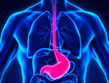
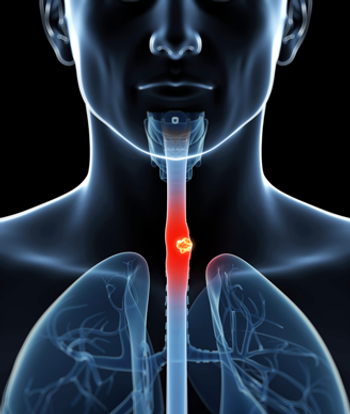
Photodynamic Therapy for Barrett’s Esophagus
For patients with high-grade dysplasia associated with Barrett’s esophagus or adenocarcinoma, photodynamic therapy
Advertisement
For patients with high-grade dysplasia associated with Barretts esophagus or adenocarcinoma, photodynamic therapy (PDT) with porfimer sodium (Photofrin) may be a useful, minimally invasive option.
While esophagectomy remains the gold standard and should be considered for every patient with high-grade dysplasia, PDT should be considered for the substantial number of patients for whom esophagectomy is contraindicated, said Scott W. Taber, MD, assistant professor, department of surgery, University of Louisville School of Medicine at a satellite symposium of the annual meeting of the Society for Thoracic Surgeons.
Dr. Taber cited several other alternatives to PDT, including photoablation with the neodymium/yttrium-aluminum-garnet (Nd:YAG) laser. The drawback of the latter, he said, is that because treatment is limited to an area of only about 2 cm, multiple endoscopies and treatments are needed. In addition, the laser entails risk of perforations, explosions, and fire.
Photodynamic therapy is approved for the palliation of dysphagia and is currently being evaluated in phase III multicenter clinical trials in the United States and Europe for high-grade dysplasia in Barretts esophagus. It can be used alone or in combination with radiation, especially external-beam radiation or brachytherapy. However, PDT is generally avoided in combination with induction chemotherapy until patients white blood cell counts have recovered, said Dr. Taber.
Optimal Management Approach Uncertain
There remain inherent uncertainties in the approach to managing Barretts esophagus because of difficulties in determining the exact anatomic location of the gastroesophageal junction in the gastric fold (where the specialized columnar epithelium in the tubular esophagus is susceptible to histologic changes). In addition, there is disagreement over the proper biopsy criteria, although most physicians biopsy every 2 cm throughout the Barretts area (in each of four quadrants with a jumbo biopsy forceps) plus in any location that looks suspicious.
What is agreed upon, Dr. Taber emphasized, is that the prevalence of adenocarcinoma has been on the rise. While the ratio of squamous cell cancer of the esophagus to adenocarcinoma of the esophagus used to be 90/10, the ratio has reversed to 80/20adenocarcinoma to squamous cell cancer. He noted further that some attribute the change to masking of gastroesophageal reflux disease (GERD) as a result of patients self medicating with over-the counter histamine-2 blockers, which promote development of Barretts esophagus. The prevalence of Barretts esophagus in the general population may be as high as 7 per 1,000, and among those with GERD, it is 8 per 1,000.
While we know that there is progression from Barretts esophagus to invasive cancer [adenocarcinoma], we really dont know how often high-grade dysplasia progresses to it, said Dr. Taber. Progression, he added, takes place within a time frame of 48 to 60 months. For Barrett s esophagus that is not high grade, treatment has classically been through acid suppression (histamine-2 blockers or proton pump inhibitors), physical management of GERD through weight loss, abstinence from caffeine, tobacco, and alcohol, other dietary restrictions, elevating the head of the bed 2 to 3 inches, and antireflux surgery (Nissen fundoplication).
Earlier Study Shows Benefit of PDT
An earlier study in 55 such patients showed that, at 5-year follow-up, 30% converted back to normal squamous mucosa after PDT (Gastrointest Endosc Clin North Am 7(2):207-216, 1997). Furthermore, a substantial additional proportion of the population improved from high-grade dysplasia to mild or moderate dysplasia. Stricture rates were high, but Dr. Taber attributed that to a high light dose (300 J). Areas where treatment overlapped may be getting much higher doses, he said, suggesting that for the thin lesions of Barretts esophagus, 200 J should be sufficient. Centering balloons currently in development may also prevent areas from being skipped or from receiving doubled dosimetry.
In the current phase III trial of 100 patients using the centering balloon and 200 J of light energy, preliminary assessment has shown lower stricture rates with fewer skipped areas. The average patient, Dr. Taber said, is treated with 200 J of light for about 500 seconds (8 minutes, 20 seconds) per treatment segment. Average treatment sessions for this patient population last about 30 to 40 minutes.
Although phototoxicity occurs in more than 20% of patients, careful attention to patient education can lower the risk.
Photobleaching (which inactivates porfimer sodium, the photoactive agent in the skin) can be promoted by asking patients to go into direct sunlight for 1 minute daily starting between the first and second week after injection. Dr. Taber recommended extending the exposure time in the third and fourth weeks. At 30 days, patients should go outside and test an area of skin.
Unanswered Questions
The question of real interest is whether or not we can start treating people in the mild to moderate dysplasia phase of Barretts esophagus. Will PDT convert or alter their dysplasia pattern? Could we then just treat GERD, the original cause of their Barretts esophagus? asked Dr. Taber.
He noted further that because important questions about the PDT success rate remain unresolved, it is essential that patients be under the rigorous screening protocol of the phase III trial. Physicians wishing information about enrollment can call Herma Neyndorff of QLT PhotoTherapeutics, Inc., at 604-872-7881.
PDT is a useful minimally invasive means for local tumor control in the esophageal lumen. It provides substantial ablation of dysplastic mucosa, said Dr.Tabor.
Articles in this issue
about 27 years ago
Thalidomide Shows Promising Results in Patients With Multiple Myelomaabout 27 years ago
Bill Would Allow Medicaid to Pay for Cervical/Breast Cancer Treatmentabout 27 years ago
Photodynamic Therapy for Palliation of Locally Advanced Lung Cancerabout 27 years ago
Zeneca Launches Education Program Aimed at Reducing Breast Cancer Riskabout 27 years ago
Reinventing Bone Marrow Transplantationabout 27 years ago
Age Should Not Determine Treatment of SCLC Patientsabout 27 years ago
Single Chest X-Ray Leads to Improved Lung Cancer Prognosisabout 27 years ago
No Link Found Between PCB Exposure and Cancer Mortalityabout 27 years ago
Arsenic Induces Remissions in Patients With Acute Promyelocytic LeukemiaAdvertisement
Related Content
Advertisement


Advertisement
Advertisement
Trending on CancerNetwork
1
Risvutatug Rezetecan Improves Overall Survival in Relapsed SCLC Trial
2
Dostarlimab Yields Sustained Complete Responses in dMMR/MSI-H Rectal Cancer
3
FDA Accepts NDA for Mezigdomide Combo in R/R Multiple Myeloma
4
Clinical Scenario - Triple-Negative Breast Cancer
5