|Poll|March 21, 2013
Young Man With a History of Headaches and Blurred Vision
Advertisement
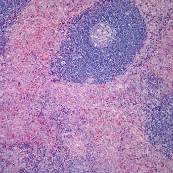
A healthy 24-year-old male presented with a history of several months of poorly localized headaches and blurred vision. Evaluation by an ophthalmologist detected the presence of bitemporal hemianopsia. MRI of the brain demonstrated a multi-lobulated mass with both cystic and solid components causing significant superior displacement of the optic chiasm. The patient subsequently underwent a subtotal resection.
The T1-weighted post-contrast image (top left), and the T2-weighted images (top right, bottom left and right) are shown here.
Based on the radiographic appearance of the mass, what is the most likely diagnosis?
Advertisement
Related Content
Advertisement


Advertisement
Advertisement
Trending on CancerNetwork
1
FDA OKs Regulatory T-Cell Immunotherapy in Hematologic Malignancies
2
Purple Reign Vs Thriller: A Duel of The Top Frontline EGFR+ NSCLC Regimens
3
ASCO Breakthrough 2026 Wrap-Up: Precision Oncology, AI, and Biomarkers Define the Future of Cancer Care
4
MARIPOSA Trial: EGFR-Mutant Lung Cancer Frontline Strategies
5