
|Articles|March 10, 2010
Cancer Management Chapter 12: Liver, gallbladder, and biliary tract cancers
Worldwide, hepatocellular carcinoma is the fifth most common malignancy and the third most common cause of cancer mortality. Most patients with hepatocellular carcinoma suffer from cirrhosis primarily caused by alcoholism or chronic infection with hepatitis B virus (HBV) or hepatitis C virus (HCV); decades may pass between infection with viral hepatitis and development of this cancer. The approximately equal annual incidence and mortality of 1 million reported around the world stands as evidence of its lethality.
Advertisement
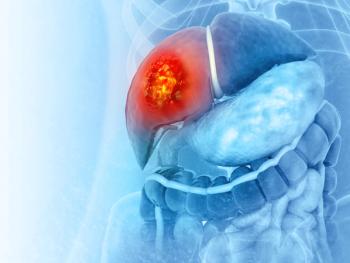
HEPATOCELLULAR CANCER
Worldwide, hepatocellular carcinoma is the fifth most common malignancy and the third most common cause of cancer mortality. Most patients with hepatocellular carcinoma suffer from cirrhosis primarily caused by alcoholism or chronic infection with hepatitis B virus (HBV) or hepatitis C virus (HCV); decades may pass between infection with viral hepatitis and development of this cancer. The approximately equal annual incidence and mortality of 1 million reported around the world stands as evidence of its lethality.
Epidemiology
Gender
Hepatocellular carcinoma is the most common tumor in males worldwide, with a male-to-female ratio of 5:1 in Asia and 2:1 in the United States.
Geography
Eighty percent of new hepatocellular cancer cases occur in developing countries, but the incidence of this cancer is also rising in developed countries. Modeling of the spread of HCV infection suggests that this number may continue to increase dramatically. In the United States, incidence of hepatocellular carcinoma is 5 per 100,000, whereas in the Far East and sub-Saharan Africa, this neoplasm occurs at an incidence of 150 per 100,000 population and comprises almost 50% of all diagnosed tumors.
Age
The incidence of hepatocellular cancer increases with age. The mean age at diagnosis is 53 years in Asia and 67 years in the United States.
Race
From 2003 to 2005, the incidence of hepatocellular tumors in the United States was higher among Asian immigrants (12 per 100,000) and blacks (7 per 100,000) than among whites (4 per 100,000). However, a study analyzing SEER data has shown that the incidence of hepatocellular carcinoma is rising in both white and black populations in the United States.
Survival
In patients who meet stringent criteria and undergo transplant, the 5-year survival is approximately 65% to 70%; the 5-year recurrence rate is less than 20%. The 5-year survival rate following liver resection (governed by a different set of operative criteria) is about equal to that following transplant. This is in spite of a majority of patients experiencing metastases or a recurrence in the remaining liver. Fewer than 20% of resected patients who have tumor recurrence are able to undergo transplant. Seventy-five percent of patients have unresectable disease that is diagnosed at an advanced stage. Median survival in these patients can range from months to years depending upon the stage of disease, the biologic aggressiveness of tumor, vascular invasion, and “background” liver function, among other important factors.
Etiology and risk factors
Hepatitis B
The annual incidence of hepatocellular carcinoma in HBV carriers is 0.5% and in patients with known cirrhosis is 2.5%. In one study, the relative risk of hepatocellular carcinoma in HBV carriers was 100 in Asian patients, who likely acquired the virus at birth. The epidemiology of HBV infection is different in the western world, where it is acquired later in life, and risk in whites appears to be related to inflammatory activity and cirrhosis.
In endemic areas of hepatitis B, approximately 90% of all patients with hepatocell-ular carcinoma are positive for hepatitis B surface antigen (HBsAg). The presence of the hepatitis B “e” antigen has been found to increase risk ninefold. An important reason to provide antiretroviral agents to patients with chronic HBV infection is the relationship between the risk of hepatocellular carcinoma development and absolute serum levels of HBV DNA. The most compelling epidemiologic evidence of a causal relationship between HBV infection and hepatocellular carcinoma is the observation of a significant decline in the incidence of childhood hepatocellular carcinoma after the introduction of a national immunization program in Taiwan. The hepatitis B “x” gene, which can interact with p53 , has been a focus of study on the pathogenesis of hepatocellular carcinoma.
Hepatitis C
The risk of hepatocellular carcinoma in patients with chronic HCV infection and established cirrhosis is 2% to 8% per year. The molecular mechanisms of HCV infection and carcinogenesis are poorly understood, yet the disease is believed to occur in the context of chronic inflammation, which leads to fibrosis and cirrhosis. Unlike patients with HBV infection, patients with hepatocellular carcinoma infected with HCV usually have cirrhotic livers at diagnosis; this finding suggests an extended period of infection (or hepatic damage) before malignancy develops.
Alcohol
Patients with alcoholic cirrhosis are at risk for hepatocellular carcinoma, and the addition of HCV infection increases that risk dramatically. About 60 to 80 g of alcohol (equivalent to four to six beers) must be consumed daily over 5 to 10 years for a male to develop cirrhosis (odds ratio, 4). The interaction between alcohol consumption and HCV infection approximately doubles the odds ratio.
Other possible etiologies
These include aflatoxin, hemochromatosis, hepatic venous obstruction, thorotrast (a contrast agent no longer used for radiologic procedures), androgens, estrogens, and α1-antitrypsin deficiency. Emerging risk factors for hepatocellular carcinoma are obesity and nonalcoholic fatty liver disease. In a recently completed, prospective, cohort study of patients with biopsy-proven nonalcoholic steatohepatitis, older age and advanced fibrosis were risk factors for hepatocellular carcinoma.
Prevention
A large 5-year randomized study performed in China has confirmed that treatment of chronic HBV infection with the antiviral lamivudine (Epivir) not only decreases the risk of progression to cirrhosis of the liver but also decreases the rate of progression to hepatocellular cancer. Treated patients had a 3.9% risk of hepatocellular cancer over 5 years, compared with 7.4% for those in the placebo group (hazard ratio: 0.49; P = .047). It remains controversial whether antiviral therapy with ribavirin (Copegus, Rebetol) and interferon decreases the risk of hepatocellular carcinoma in cirrhotic patients with HCV infection who achieve a sustained virologic response. If there is any benefit to therapy in such cases, it appears to be small.
Signs and symptoms
Nonspecific symptoms
Patients usually present with abdominal pain and other vague symptoms, including malaise, fever, chills, anorexia, weight loss, and jaundice.
Physical findings
An abdominal mass is noted on physical examination in one-third of patients. Less common findings include splenomegaly, ascites, abdominal tenderness, muscle wasting, and spider nevi. Up to 10% of patients may present with an acute abdomen due to a ruptured tumor.
Screening and diagnosis
Presently, no organization recommends routine screening of average-risk, asymptomatic adults for liver, gallbladder, and biliary tract cancers.
α-Fetoprotein
This serum marker is produced by 70% of hepatocellular carcinomas. The normal range for this serum marker is 0 to 20 ng/mL, and a level > 200 ng/mL is essentially diagnostic for hepatocellular cancer in the absence of chronic, active hepatitis B infection. In the presence of active hepatitis B infection, the diagnostic cutoff is considered to be at least 1,000 ng/mL. In the setting of HCV infection, the cutoff for diagnosis of hepatocellular carcinoma has not been well studied. In hepatitis C, values > 200 ng/mL appear to be highly predictive of hepatocellular carcinoma. False-positive results may be due to acute or chronic hepatitis, germ-cell tumors, or pregnancy. In its guidelines for diagnosis of hepatic masses suspicious for hepatocellular carcinomas, the National Comprehensive Cancer Network does not recommend biopsy for patients with an α-fetoprotein level > 400 ng/mL, except in the presence of HBsAg positivity. In that situation, a cutoff value of 4,000 ng/mL is recommended for biopsy.
Hepatitis B and C
Given the association between hepatitis B and C and hepatocellular cancer at the time of diagnosis of a hepatic mass, blood should be sent for hepatitis B and C antigen and antibody determinations.
Imaging
The initial diagnostic test in the symptomatic patient may be ultrasonography, as it is noninvasive and can detect lesions as small as 1 cm. Ultrasound findings should be followed up with more specific imaging.
Triple-phase, high-resolution CT and contrast-enhanced magnetic resonance imaging (MRI) are the primary imaging modalities used to diagnose and stage hepatocellular carcinoma. Typically, hepatocellular carcinoma is a hypervascular tumor with washout in the portal venous phase. Recent reports have documented a high number of false-positive results with CT angioportography (CTAP) and CT hepatic angiography (CTHA). CT scan predicts resectability in only 40% to 50% of cases and does not accurately determine the functional extent of cirrhosis. Major difficulties arise when the liver parenchyma is not homogeneous and the lesions are smaller than 1 cm. Use of positron emission tomography (PET) scanning is not defined in this disease, and the uptake of 18F-fluorodeoxyglucose by hepatocellular tumors is variable.
In a study following patients with chronic liver disease for development of hepatocellular carcinoma, 18,000 Chinese patients with chronic HBV infection were screened with ?-fetoprotein determinations and liver ultrasound every 6 months or no screening for 5 years. The hepatocellular carcinoma mortality was 37% lower in the screened group than in the controls, and this outcome can be attributed to early-stage tumor detection and resection (
Zhang BH et al: J Cancer Res Clin Oncol 137:417–422, 2004).
Laparoscopy
This procedure is useful for the evaluation of small tumors, the extent of cirrhosis, peritoneal seeding, and the volume of noninvolved liver and, therefore, may be used prior to open laparotomy for resection. Laparoscopic or intraoperative ultrasonography should be used to confirm preoperative imaging tests. The laparoscopic results may change surgical management in up to one-third of selected patients.
High-risk patients
Individuals at high risk should be screened for hepatocellular carcinoma using ultrasonography and serum α-fetoprotein levels. Screening increases the proportion of cancers that are resectable. However, a study comparing 6-month and 12-month survival intervals in a cohort of HCV-infected patients with hemophilia showed no substantial benefit to more frequent screening.
PATHOLOGY
Three morphologic patterns of hepatocellular carcinoma have been described: nodular, diffuse, and massive. Diffuse and massive types account for > 90% of cases. The nodular type usually has multiple lesions in both lobes.
Histologic arrangements
Several histologic arrangements have been identified: trabecular, compact, pseudoglandular or acinar, clear cell, and a fibrolamellar variant, which is associated with a relatively favorable prognosis and a younger age at diagnosis. The fibrolamellar variant is more commonly resectable and is not usually associated with infection and cirrhosis.
Staging and prognosis
The staging system for hepatocellular cancer is based on the number and size of lesions and the presence or absence of vascular invasion (
Of the roughly 20% of patients who can undergo resection, factors associated with improved survival include curative resection, small tumor size, well differentiated tumors, and normal performance status. Cirrhosis, nodal metastases, and an elevated prothrombin time are indicative of a poor prognosis, as are male sex, age > 50 years, poor performance status, duration of symptoms < 3 months, tumor rupture, aneuploidy, high DNA synthesis rate, hypocalcemia, vascular invasion, and a high serum α-fetoprotein level.
Side effects of RFA may include pleural effusions and peritoneal bleeding. In 1,000 patients treated with RFA for hepatocellular carcinoma, neoplastic seeding from percutaneous RFA occurred at a rate of 3.2% per patient or 1.8% per treatment. The patients underwent 1,845 RFA treatments for 3,837 nodules; 20% had nodules > 3 cm in size. The observation period was 5 to 64 months, and poor differentiation was the only risk factor for neoplastic seeding in multivariate analysis. Surrogate markers for poor differentiation were larger tumor size and elevated tumor marker levels. Other investigators have reported occurrences of “seeding” in as many as 12% and as few as 0.9% of patients undergoing RFA for hepatocellular carcinoma, with subcapsular tumor location being a risk factor (
Imamura J et al: Am J Gastroenterol 103:3057–3062, 2008).
Treatment
Surgery
Surgery is the form of treatment that offers the greatest potential for cure even though only a small minority of patients will actually be cured. Unfortunately, many patients whose disease is thought to be resectable are clinically understaged preoperatively.
Only stage I or II tumors have a significant likelihood of being resectable for cure. Resectability is limited by the functioning liver tissue at the completion of a negative margin operation. Therefore, even a large tumor may still be potentially resectable for cure. Moreover, contiguous involvement of large vessels (including the portal vein and inferior vena cava) or bile ducts does not automatically mitigate against a resection. Resection is contraindicated in patients with metastatic disease to non-portal nodes and in extrahepatic locations. The use of the Child-Pugh score and volumetric evaluation aids in assessment of resectability. For cirrhotic patients in whom less than 30%–35% of the liver remains at the completion of resection, operative treatment is contraindicated. Likewise, this is true for noncirrhotic patients where less than 25%–30% remains.
Bilobar disease may be addressed with formal resection, tumor ablation techniques (eg, cryoablation, radiofrequency ablation [RFA], and ethanol injection ablation), or a combination of the modalities.
Contraindications to resection
These include imminent clinical hepatic failure (jaundice in the absence of biliary obstruction), hypoalbuminemia, ascites, renal insufficiency, hypoglycemia, prolongation of the prothrombin and partial thromboplastin times, main portal vein involvement, extrahepatic metastatic disease, or other comorbid diseases that would preclude surgery of any kind.
Noncirrhotic versus cirrhotic patients
Resection should be performed in all noncirrhotic patients, when feasible. Resection of hepatocellular carcinoma in th
e presence of cirrhosis is more controversial due to its increased morbidity in this setting. Cirrhosis has been a major deterrent to resection in western nations. Resectability rates vary from 0% to 43% for cirrhotic patients, whereas up to 60% of patients without cirrhosis undergo resection. Use of the modified Child-Pugh classification of liver reserve (
When resection is performed in the presence of cirrhosis, Child-Pugh class A patients fare better than do Child-Pugh class B or C patients. Survival rates at 5 years following resection range from 4% to 36%, with noncirrhotic patients living longer than cirrhotic patients.
Methodologies for delivering transarterial chemoembolization (TACE) are numerous. A recent innovation in chemotherapy delivery during TACE is the use of drug-eluting beads (DEBs). These perforated beads allow for slow, localized release of chemotherapy after embolization. In a study by Varela et al, 27 patients with Child-Pugh class A liver disease and unresectable hepatocellular cancer underwent DEB-TACE. Pharmacokinetic samples were analyzed for doxorubicin, demonstrating lower systemic drug exposure than expected with conventional TACE. Two patients had abscesses, but the radiographic response rate was a promising 75%. Studies of this technology are now being planned and conducted at multiple sites (
Varela M et al: J Hepatol 46:474–481, 2007).
Transplantation
Owing to the risk of hepatic failure following resection in cirrhotic patients, transplantation has become an option for patients with hepatocellular cancer and cirrhosis. In a study of 181 patients with hepatocellular carcinoma, Starzl and Iwatsuki found similar overall 5-year survival rates in patients treated with transplantation versus resection (36% vs 33%). Survival rates were similar in the two groups when tumors were compared for TNM stage. However, survival was significantly improved in patients with concomitant cirrhosis if they were treated with transplantation. Tumor recurrence rates for stages II and III tumors were significantly lower after transplantation than after resection, but no differences were seen for stage IV tumors. Numerous additional studies have examined the relative value of transplantation, ablation, and resection. There is much debate over their interpretation.
Patients with cirrhosis and single tumors < 5 cm or multiple tumors (up to 3 with none > 3 cm) can be considered for transplantation. Larger tumors may be treated with resection when feasible. The University of California San Francisco has prepared “expanded criteria” for transplant eligibility. The use of transplantation is significantly limited by the scarcity and lack of immediate availability of donor organs. Recently, changes in the organ allocation system have decreased waiting times for patients with documented hepatocellular cancer.
Therapies for nonresectable hepatocellular carcinoma
Given the high risk of recurrence after resection, the multifocal nature of hepatocellular carcinoma, and its association with chronic liver disease, therapies for nonresectable disease can play an important role in management. A number of prognostic factors have been identified for patients with unresectable hepatocellular carcinoma. These factors, taken alone, can have a great effect on survival rates, making cross-treatment comparisons more difficult because considerable selection bias may be present in any nonrandomized trial. Additionally, comparisons are difficult, as there are few randomized trials among the large number of non-resectional, liver-directed therapies currently available. It is worth noting that a number of these treatments do not have overlapping toxicities and that research into combinations is potentially worthwhile.
Ablation
Ablation techniques include RFA, cryotherapy, and injection of chemicals (eg, ethanol) directly into the tumor. RFA can be performed via laparotomy or percutaneously via CT or ultrasound guidance. It is best suited for lesions < 3 cm in size. The overall recurrence rate after ablation is > 70%. Some recurrence represents undertreatment of lesions, microscopic satellites that were not included in the ablation site, poor imaging due to technical issues, inexperience of the ablator, and selection of large tumors. Ablation is safe and effective for patients who cannot undergo resection or who need a bridge to transplant.
Intratumoral ethanol injection The direct injection of 95% ethanol into a neoplastic lesion causes cellular dehydration and coagulation necrosis. This procedure has been largely replaced by RFA, which in several clinical trials has shown superior efficacy to ethanol injection.
Cryotherapy and RFA
Cryotherapy and RFA techniques are suitable for treating localized disease. Cryotherapy has been used intraoperatively to ablate small, solitary tumors outside a planned resection (ie, in patients with bilobar disease). Cryotherapy must be performed using laparotomy, which limits its use in the palliative setting. RFA creates necrosis through the use of heated electrodes at the site of the tumor and has efficacy superior to that of ethanol injections for lesions > 2 cm in size.
Hepatic Transcatheter Embolization/TACE
Normal hepatocytes receive most of their blood supply from the portal vein, whereas tumors create new blood vessels from branches of the hepatic arterial system. This target is exploited by embolization of the hepatic artery with any number of substances, resulting in radiographic responses in about 50% of patients and evidence of tumor liquefaction in over two-thirds of patients. At 2 years, the overall survival benefit from this procedure ranges from 20% to 60%. Embolization is accomplished by advancing a catheter within the tumor-feeding branch of the hepatic artery. Materials injected have included polyvinyl alcohol, iodized oil (Lipiodol), collagen, and autologous blood clot. If chemotherapy is used, it usually is suspended in iodized oil, which is retained in the tumor. Common chemotherapeutic agents used have included cisplatin and doxorubicin, which may produce systemic side effects. Postembolization syndrome, with fever, abdominal pain, and, occasionally, ileus, occurs in 50% of patients when a large volume of liver is treated. Complications include infection and abscess. Since few complete responses are achieved, treatments may be repeated on the same tumor for better results. Due to risk of liver failure, chemoembolization may be contraindicated in patients with portal vein thrombosis and for most individuals with Child-Pugh B status. Patients with Child-Pugh C liver disease cannot receive chemoembolization due to risk of liver failure.
A phase I trial of stereotactic radiation therapy used 6 fractions to deliver a dose of 24 Gy to 54 Gy, depending on the volume of normal liver irradiated. In the 31 patients with unresectable hepatocellular carcinoma, the median survival was about 12 months without any instances of radiation-induced liver disease (
Tse RV et al: J Clin Oncol 26:657–664, 2008).
The effect of TACE on survival remains controversial, with randomized studies returning mixed results. A randomized, controlled trial in Spain was stopped early due to a survival benefit. Meta-analysis of studies indicates a positive impact on overall survival.
Radiation therapy
Adjuvant treatment There is no evidence, however, that adjuvant radiation therapy can improve local or regional control after adequate resection or ablation.
Unresectable disease Whole-liver radiation therapy can provide palliation in patients with unresectable tumors but is limited to a total dose of ≤ 30 Gy due to the risk of radiation-induced liver disease. Whole-liver irradiation has been combined with chemotherapy and transcatheter arterial chemoembolization, with objective response rates of approximately 40% to 50% and median survival rates of about 18 months. Patients with tumor regrowth after chemoembolization may respond to radiotherapy.
Radiation therapy has also been delivered using yttrium-90 microspheres infused via the hepatic artery to carefully selected patients; response rates as high as 89% have been reported. This approach caused liver toxicity in 42% of patients in one series. One study reported that 56% of treated T3 tumors were downstaged to T2, which allowed subsequent liver-directed therapy or use as a bridge to transplant.
Three-dimensional conformal radiation therapy treatment planning can allow patients with nondiffuse disease to be safely irradiated to doses well above the whole-liver tolerance dose, with doses up to 90 Gy given safely to selected patients, and a response rate of 40%, and a median survival of 15 months.
Multiple institutions have reported response rates as high as 90% with acceptable toxicity when conformal radiation therapy was combined with transcatheter arterial chemoembolization. Good response and local control rates have also been reported for proton, carbon ion, and stereotactic radiotherapy.
Sorafenib is an oral multikinase inhibitor with antiangiogenic and antiproliferative effects. The SHARP trial compared sorafenib (Nexavar: 400 mg PO bid) versus placebo in 600 patients with Child-Pugh class A liver disease. About one-third of patients had undergone embolization, and the disease progressed. In all, 70% of patients had portal vein thrombosis and/or metastatic lesions. In spite of a low radiographic response rate (2% of tumors responded by radiographic criteria), median survival for the 300 patients given sorafenib was significantly better than that for the placebo group (10.7 vs 7.9 months; hazard ratio [HR], 0.69). The drug was well tolerated, with fatigue, diarrhea, rash, and hand-foot syndrome as manageable side effects. This is the first well-powered trial to convincingly demonstrate a survival benefit with the use of a systemic agent for this disease. Sorafenib now is the standard of care for hepatocellular carcinoma not amenable to locoregional therapy and the first FDA-approved agent for this indication. Studies using the drug with embolization are ongoing (
Llovet JM et al: N Engl J Med 359:378–390, 2008).
In the Asia-Pacific Trial, 226 patients with Child-Pugh class A liver disease were randomized to sorafenib and placebo. These patients primarily had HBV infection; compared with SHARP study patients, they had more local therapy before their entry into the trial and, as a whole, more advanced disease. Median survival was 6.5 months compared with 4.2 months for sorafenib and placebo, respectively (HR, 0.68) (
Cheng AL et al: Lancet Oncol 10:25–34, 2009).
More data are needed to establish the role of sorafenib in treating Child-Pugh class B liver disease
Chemotherapy
Systemically administered chemotherapy The low response rates to chemotherapy are related to the role of the hepatocyte in detoxification. Hepatocytes also have high multidrug resistance expression. Furthermore, many patients with hepatocellular carcinoma have cirrhosis or hepatic dysfunction, which complicates the administration of chemotherapeutic agents that undergo hepatic metabolism. Agents with partial response rates near or above 10% include doxorubicin, fluorouracil (5-FU), and cisplatin.
Biologic therapy Inhibiting angiogenesis in hepatocellular carcinoma is based on several factors. Hepatocellular carcinoma is a vascular tumor; increased levels of vascular endothelial growth factor are found in hepatomas compared with normal hepatic tissue, and increased endothelial growth factor levels before resection of tumor or TACE are associated with early relapse, aggressive behavior, and poor prognosis. Other targets include EGFR (which is frequently expressed in hepatoma cells), the mitogen-activated protein kinase (MAPK) pathway, and the phosphatidylinositol 3-kinase/Akt/mammalian target of rapamycin (P13K/Akt/mTOR) pathway. Conventional markers of radiographic response are poorly related to tumor cell kill in hepatocellular carcinoma. More meaningful markers of therapeutic benefit may be time to progression, progression-free survival, and overall survival.
BILIARY TRACT CANCERS
Gallbladder carcinoma is diagnosed approximately 5,000 times a year in the United States, making it the most common biliary tract tumor and the fifth most common gastrointestinal tract cancer. Approximately 4,500 cases of bile duct tumors occur each year in the United States.
Epidemiology
Gallbladder cancer
Gender
Women are more commonly afflicted with gallbladder cancer than are men, with a female-to-male ratio of 1.7:1.
Age
The median age at presentation of gallbladder cancer is 73 years.
Sunitinib (Sutent), an oral inhibitor of multiple receptor tyrosine kinases, was tested in patients with hepatocellular carcinoma in a phase II trial. The median overall survival was almost 10 months. However, sunitinib was harder to tolerate than is sorafenib, and its use was related to more serious treatment-related adverse events (eg, grade 3/4 cytopenias, transaminitis, vasculitis). It also required monitoring of cardiac function (
Zhu AX et al: J Clin Oncol 26[15S]:4521, 2008).
Race
An incidence five to six times that of the general population is seen in southwestern Native Americans, Hispanics, and Alaskans.
Bile duct cancer
Gender
Bile duct tumors are found in an equal number of men and women.
Age
Extrahepatic bile duct tumors occur primarily in older individuals; the median age at diagnosis is 70 years.
Etiology and risk factors
Gallbladder cancer
The risk of developing gallbladder cancer is higher in patients with cholelithiasis, calcified gallbladders, and in typhoid carriers. Gallstones are present in 70% or more of patients with gallbladder cancer and presumably cause chronic inflammation. The overall incidence of gallbladder cancer in individuals with cholelithiasis is 1% to 3% and in patients with so-called porcelain gallbladders, caused by chronic cholecystitis, 10% to > 50%. In patients having gallbladder polyps measuring > 1 cm, the risk of cancer is high.
Erlotinib (Tarceva), an oral agent that inhibits the epidermal growth factor receptor (EGFR, HER1) -related tyrosine kinase enzyme, was evaluated for efficacy in hepatocellular carcinoma. A number of phase II trials examining agents targeting EGFR have been tested in patients with hepatocellular carcinoma and appear to have only modest activity. In two phase II studies with erlotinib, “disease control” was greater than 50% (response rate, >10%), and median survival was 10 to 13 months. The combination of bevacizumab (Avastin) and erlotinib in a phase II study of 40 patients with Child-Pugh A or B liver disease that was not amenable to surgery or regional therapy resulted in a progression-free survival of 9 months and a median survival of 15.6 months, which compares favorably with sorafenib. Use of this regimen in the cirrhotic patient may be associated with bleeding and thrombosis (
Thomas MB et al: J Clin Oncol 27:843–850, 2009).
Bile Duct Cancer
Primary sclerosing cholangitis (PSC)
Thirty percent of cholangiocarcinomas are diagnosed in patients with PSC with or without ulcerative colitis. The annual incidence of cholangiocarcinoma in patients with PSC is estimated at 1.5% per year; their lifetime risk of developing this malignancy is 10% to 15%. These patients have a highly abnormal biliary system, making diagnosis of cholangiocarcinoma difficult.
Ulcerative colitis
Patients with ulcerative colitis have an incidence of bile duct cancer that is 9 to 21 times higher than that of the general population. This risk does not decline after total colectomy for ulcerative colitis.
Other risk factors
Congenital anomalies of the pancreaticobiliary tree, parasitic infections, biliary papillomatosis, and Lynch syndrome are also associated with bile duct tumors. No association of bile duct cancer with calculi, infection, or chronic obstruction has been found.
Signs and symptoms
Gallbladder cancer
Early and late disease
In the early stages, gallbladder cancer is usually asymptomatic. Later, symptoms similar to those of benign gallbladder disease arise; they include right upper quadrant pain, nausea, vomiting, fatty food intolerance, anorexia, jaundice, and weight loss. This nonspecificity of symptoms delays presentation for medical attention and contributes to the low curability of gallbladder cancer.
Physical findings
These may include tenderness, an abdominal mass, hepatomegaly, jaundice, fever, and ascites.
Bile duct cancer
Jaundice
This is the most frequent symptom found in patients with high bile duct tumors; it is present in up to 98% of such patients.
Nonspecific signs and -symptoms
Patients who do not present with jaundice have vague complaints, including abdominal pain, weight loss, pruritus, fever, and an abdominal mass.
Diagnosis
Gallbladder cancer
Gallbladder carcinomas are often diagnosed at an advanced stage, such that by the time symptoms have developed, most tumors are unresectable.
Laboratory values
Findings in patients with gallbladder carcinoma are nonspecific but may include anemia, leukocytosis, and an elevated bilirubin level.
Ultrasonography
This diagnostic study is useful for defining a thickened gallbladder wall and may show tumor extension into the liver. It is valuable in measuring the size of a polyp.
CT and MRI
CT is more helpful than ultrasonography in assessing adenopathy and spread of disease into the liver, porta hepatis, or adjacent structures. MRI may be used to evaluate intrahepatic spread.
A retrospective review of 73 consecutively treated patients with an R0 resection of gallbladder cancer found that adjuvant chemoradiotherapy was a significant predictor of overall survival after adjusting for prognostic factors. This suggested that a benefit to postoperative therapy may exist, although it remains to be proven in phase III studies (
Gold DG et al: Int J Radiat Oncol Biol Phys 75:150–155, 2009).
Two analyses using the SEER database have concluded that adjuvant radiation therapy may have a survival benefit for node-positive gallbladder cancer. One of these two studies also found a benefit for tumors that were T2+ (
Mojica P et al: J Surg Oncol 96:8–13, 2007 and Wang SJ et al: J Clin Oncol 26:2112–2117, 2008).
Cholangiography
Endoscopic retrograde cholangiopancreatography (ERCP), transhepatic cholangiography (THC), or magnetic resonance cholangiography (MRC) may be useful in the presence of jaundice to determine the location of biliary obstruction and involvement of the liver.
Bile duct cancer
Cholangiocarcinoma may present earlier than gallbladder cancer by virtue of the development of biliary obstruction with jaundice, which may be painless. Tissue confirmation of suspected bile duct cancer can be difficult. The goals of the diagnostic evaluation include the determination of the level and extent of obstruction, the extent of local invasion of disease, and the identification of metastases.
Many patients with cholangiocarcinoma are thought to have metastatic adenocarcinoma of an unknown primary, although occasionally the metastatic lesion may produce biliary dilatation without the primary lesion itself being radiographically visualized. Recently, microarray-based technology for genetic analysis has become available to help characterize tumors that are difficult to identify.
Ultrasonography
It is generally accepted that ultrasonography should be the first imaging procedure in the evaluation of the jaundiced patient.
CT
This diagnostic is a complementary test to ultrasonography, but both tests are accurate for staging in only 50% of patients and for determining resectability in < 45% of patients.
Cholangiography
This diagnostic technique is essential to determine the location and nature of the obstruction. Percutaneous THC is used for proximal lesions, and ERCP is used for distal lesions. Magnetic resonance cholangiopancreatography (MRCP) may replace invasive studies in the near future. Histologic confirmation of tumor can be made in 45% to 85% of patients with the use of exfoliative or brush cytology during cholangiography.
Pathology
Gallbladder cancer
Histologic types
Over 85% of gallbladder neoplasms are adenocarcinomas, and the remaining 15% are squamous cell or mixed tumors.
Route of spread
The initial route of spread of gallbladder cancer is locoregional rather than distant. For patients undergoing resection for presumed high-risk gallbladder masses or preoperatively defined disease limited to the gallbladder, 25% of patients will have lymphatic involvement, and 70% will have direct extension of disease into the liver defined at operation.
Bile duct cancer
Adenocarcinoma
Morphologically, more than 90% of bile duct tumors are adenocarcinomas. Three macroscopic appearances have been identified. The papillary and nodular types occur more frequently in the distal bile duct, whereas the sclerosing type is found in the proximal bile duct. Patients with papillary lesions have the best prognosis. Immunohistochemical staining may be positive for cytokeratin 7 and 20.
Other histologic types
Unusual malignant diseases of the biliary tract include adenosquamous carcinoma, leiomyosarcoma, and mucoepidermoid carcinoma.
Route of spread
Most bile duct tumors grow slowly, spreading frequently by local extension and rarely by the hematogenous route. Nodal metastases are found in up to one-third of patients.
The role of adjuvant radiation therapy for biliary disease has not been studied in a randomized trial; however, there is some evidence that a role may be present. A review of 3,187 cases of gallbladder cancer recorded in the SEER survey from the National Cancer Institute found that, despite a higher stage, patients treated with adjuvant radiation therapy demonstrated a longer median survival than did those treated with resection only (14 vs 8 months;
P
< .0001). On subgroup analysis, this benefit was most evident for patients with involved regional lymph nodes or liver invasion (
Mojica P et al: J Surg Oncol 96:8–13, 2007).
A Cox proportional hazards survival analysis was applied to 75 consecutive patients with hilar cholangiocarcinoma treated surgically with curative intent. It found that the most powerful predictors of outcome (in descending order) were resection type, adjuvant radiation therapy, regional lymph node involvement, and preoperative serum bilirubin (
Cheng Q et al: Eur J Surg Oncol 33:202–207, 2007).
Staging and prognosis
Gallbladder cancer
Gallbladder cancer is staged primarily at the time of surgery, and staging is determined by lymphatic involvement and extension of disease into adjacent structures (
Stage
Survival of gallbladder carcinoma is directly related to disease stage. The 5-year survival rate is 83% for tumors that are confined to the gallbladder mucosa; this rate decreases to 33% if the tumor extends through the gallbladder. For patients who have involvement of the lymph nodes or metastatic disease, 5-year survival rates range from 0% to 15%.
Type of therapy
Median survival is also improved in patients who have undergone curative resection, as compared with those who have had palliative procedures or no surgery (17 months vs 6 and 3 months, respectively).
Bile duct cancer
Over 70% of patients with cholangiocarcinoma present with local extension, lymph node involvement, or distant spread of disease. The American Joint Committee on Cancer staging system for extrahepatic tumors is shown in
Stage
Survival for these patients is poor and is directly related to disease stage. Median survival is 12 to 20 months for patients with disease limited to the bile ducts and ≤ 8 months when the disease has spread.
Tumor location
Survival is also related to tumor location, with patients with distal lesions doing better than those with mid or proximal tumors.
Success of therapy
Curative resection and negative margins result in improved survival.
Treatment
In the absence of polyps identified ultrasonographically and confirmed by CT during the work-up of suspected cholelithiasis, relatively few patients with gallbladder cancer are diagnosed prior to surgery. Only 1% to 2% of cholecystectomy specimens are found to contain malignancy.
Surgery for gallbladder cancer
Surgical management of gallbladder carcinoma is based on the local extension of the tumor. Surgery may be curative, but < 30% of patients are potentially resectable at time of diagnosis.
Early-stage disease
Tumors that invade the mucosa, those that do not penetrate the muscularis, and those that penetrate full thickness but do not abut the liver or muscularis require cholecystectomy alone. Laparoscopic cholecystectomy may be adequate for T1 tumors. If there is direct extension of disease to or through the serosa, the resection should include the gallbladder bed (segments IVb and V) and a porta hepatis lymphadenectomy. Disease that involves the gallbladder node is particularly curable and should be resected. Nodal disease beyond the pericholedochal (porta hepatis) nodes defines the surgically incurable patient. The usual pattern of spread is locoregional, although lymphangitic metastasis can be observed for T3 and T4 disease. Based on retrospective data, some surgeons will perform radical surgery in T3 and T4 disease with intent to cure.
A phase II trial of sorafenib in 46 patients with advanced cholangiocarcinoma showed disease control in 32% of patients; 54% treated for at least 12 weeks showed stable disease or partial response (
Dealis C et al: J Clin Oncol 26[15S]:4590, 2008).
Another phase II study of cetuximab (Erbitux) plus gemcitabine and oxaliplatin in patients with advanced or metastatic cholangiocarcinoma demonstrated an overall response rate of 58%. Six patients underwent curative resection after a major response (32%). One patient had a complete response, and six had stable disease (
Greunberger B et al: J Clin Oncol 269[15S], 2008).
Surgery for bile duct cancer
The rate of resectability is 15% to 20% for high bile duct tumors and up to 70% for distal lesions. Guidelines for surgery include the absence of retropancreatic and paraceliac nodal metastasis, noncontiguous liver metastasis, major vascular invasion or extrahepatic invasion of adjacent organs. Some surgeons will attempt en bloc resection with vascular reconstruction.
Assessing resectability
High-resolution CT or MRI with biliary reconstruction may be supplemented with hepatic arteriography, portal venography, or duplex imaging preoperatively to assess resectability.
A phase III trial evaluated overall survival in 410 patients with inoperable biliary tract cancer. Patients were randomized to gemcitabine and cisplatin versus gemcitabine alone. Metastatic disease was present in 75% of patients and locally advanced disease in 25% of patients. Median overall survival was 11.7 months in the combined treatment arm, compared to 8.2 months with gemcitabine. Gemcitabine and cisplatin also had an improved progression-free survival, 8.5 versus 6.5 months, and similar toxicity compared to the single agents (
Valle JW et al: J Clin Oncol 27[15S]:abstract 4503, 2009).
Preoperative treatments
Preoperative stenting for patients with jaundice caused by obstruction is controversial. Many surgeons find that placement of a biliary stent may confound imaging and impede surgery. This must be weighed against the benefit of relieving an obstruction and improving surgical outcome. Three randomized trials have shown no benefit to preoperative decompression of the biliary tree in patients with obstructive jaundice. Some authors advocate the preoperative placement of biliary stents to facilitate dissection of the hilus. This procedure should be performed close to the planned resection to reduce the risk of cholangitis and maintain the duct at its maximally dilated size.
Proximal tumors
Local excision is often possible for proximal lesions. Hepatic resection is indicated for high bile duct tumors. Resection is not indicated in situations in which a clear surgical margin cannot be obtained.
Mid-ductal and distal tumors
Mid-ductal lesions can often be removed by resection of the bile duct with associated portal lymphadenectomy. Distal or mid-ductal lesions that cannot be locally excised should be removed by pancreaticoduodenectomy.
Reconstruction techniques
Biliary-enteric continuity is usually reconstructed with a Roux-en-Y anastomosis to the hilum for high lesions and in a standard drainage pattern following pancreaticoduodenectomy.
Liver transplantation
This procedure has been attempted for unresectable tumors, but early recurrence and poor survival have prevented the widespread application of this approach. Protocols for transplantation for patients with localized, unresectable disease who respond to neoadjuvant chemoradiation are ongoing.
Surgical bypass
For patients found to have unresectable disease at surgical exploration, operative biliary bypass may be performed using a variety of techniques. Bypass results in excellent palliation and obviates the need for further intervention.
Adjuvant radiochemotherapy for gallbladder cancer
Because of the rarity of biliary cancers, there is little prospective data to guide our practices. In the United States, gallbladder cancer commonly is treated with 5-FU and radiation after resection. A randomized trial performed in Japan showed that treatment with 5-FU and mitomycin produced a survival benefit in patients with resected gallbladder cancers. These data came from a planned subset of a larger trial in which 112 patients with gallbladder cancer were randomized. At 5 years, 26% of chemotherapy-treated patients were alive, compared with 14% of those treated with surgery and observation alone. Based on these data, consideration of chemotherapy for these patients is warranted, but definitive conclusions would require a larger randomized trial.
For advanced, unresectable gallbladder cancer, palliation of obstructive jaundice and pain should be the goal. Combined-modality chemoradiation may benefit patients with advanced disease. Regarding chemotherapy, many regimens using oxaliplatin (Eloxatin), gemcitabine (Gemzar), as well as capecitabine (Xeloda) alone, or in combination with cisplatin have been tried. Modest response rates are the norm, and the indolence or aggressiveness of the tumor primarily influences survival.
Adjuvant chemoradiotherapy for cholangiocarcinoma
Delivery of adjuvant chemoradiotherapy is common for patients who have had margin-negative or microscopically positive margin resections, with observational series providing supporting evidence of improved outcomes. Chemoradiotherapy with infusional 5-FU or capecitabine is also given in the setting of positive regional lymph nodes.
Management of unresectable, locally advanced disease
The majority of patients present with unresectable disease, and palliative care is the goal of their management. Most of these patients experience a rapid decline in health. A few patients have disease that proves relatively indolent, and survival can be measured in years rather than months.
There is considerable experience using brachytherapy alone or combined with external-beam radiation therapy for unresectable bile duct tumors. Median survival ranges from 10 to 24 months, and 5-year survival rates are approximately 10% with these approaches. Cholangitis, however, may occur more frequently in people treated with brachytherapy. Photodynamic therapy (PDT) in cholangiocarcinoma is an interesting option that may provide a significant survival benefit in addition to treating biliary stenosis and cholestasis and improving quality of life. A randomized trial of stenting alone versus with PDT found a significantly longer median survival and improved quality of life in the PDT group. Although it is unlikely that all of the tumor was treated with PDT, this study did show the importance of local control and its association with quality of life. As previously discussed, another palliative option is use of yttrium-90 microspheres.
Chemotherapy
Biliary tract malignancies are uncommon cancers, and the numbers of clinical trials and of patients in those trials are limited. Generally speaking, responses to chemotherapy are infrequent and brief. However, newer drugs and drug combinations are better tolerated and stand to improve on past results.
5-FU
This drug has historically been the most active single agent, with single-agent response rates in the 10% to 20% range.
Capecitabine
This prodrug of 5-FU (see chapter 13) produced responses in 4 of 8 gallbladder cancers but in only 1 of 18 cholangiocarcinomas in a phase II study presented by Hassan et al from the M. D. Anderson Cancer Center.
Gemcitabine
Multiple studies have documented gemcitabine as an active agent, particularly in gallbladder cancer. Cisplatin and gemcitabine may be a synergistic doublet, with reported response rates for gallbladder cancer in the range of 30% to 50%.
Combination chemotherapy
Combined 5-FU and gemcitabine have modest activity in biliary malignancies. A group in Toronto published results of combination therapy with capecitabine and gemcitabine. The mix of cholangiocarcinoma and gallbladder cancer in the 45 patients studied was nearly 50:50. The overall response rate was 31%, and the median survival was a promising 14 months. This regimen is a reasonable alternative to gemcitabine and cisplatin for biliary tract neoplasms.
Hepatic arterial chemotherapy
There is limited experience with hepatic arterial chemotherapy for biliary tract neoplasms, but there are case reports of responses to floxuridine in the literature.
Treatment recommendations
In the absence of a clinical trial, patients should be offered gemcitabine and/or 5-FU (or capecitabine), with or without leucovorin. Other agents, such as doxorubicin or cisplatin, may be added, but, as noted, there is no unequivocal evidence that combination chemotherapy produces any substantial benefits in improving quality of life or survival.
References:
SUGGESTED READING
ON HEPATOCELLULAR CARCINOMA
Ghoori S, Schiavo M, Russo A, et al: First-line treatment for hepatocellular carcinoma: resection or transplantation? Transplant Proc 39:2271â2273, 2007.
Cheng BQ, Jia CQ, Liu CT, et al: Chemoembolization combined with radiofrequency ablation for patients with hepatocellular carcinoma larger than 3 cm: A randomized controlled trial. JAMA 299:1669â1677, 2008.
Donato F: Alcohol and hepatocellular carcinoma: the effect of lifetime intake and hepatitis virus in men and women. Am J Epidemiol 255:323â331, 2002.
Gupta S, Bent S, Kohlwes J: Test characteristics of alpha-fetoprotein for detecting hepatocellular carcinoma in patients with hepatitis C: A systematic review and critical analysis. Ann Intern Med 139:46â50, 2003.
Llovet J, Bruix J: Systematic review of randomized trials for unresectable hepatocellular carcinoma: Chemoembolization improves survival. Hepatology 37:429â442, 2003.
Mathurin P, Raynard B, Dharancy S, et al: Meta-analysis: Evaluation of adjuvant therapy after curative liver resection for hepatocellular carcinoma. Aliment Pharmacol Ther 17:1247â1261, 2003.
Mok TS, Yeo W, Yu S, et al: An intensive surveillance program detected a high incidence of hepatocellular carcinoma among hepatitis B virus carriers with abnormal alpha-fetoprotein levels or abdominal ultrasonography results. J Clin Oncol 23:8041â8047, 2005.
Tse RV, Hawkins M, Lockwood G, et al: Phase I study of individualized stereotactic body radiotherapy for hepatocellular carcinoma and intrahepatic cholangiocarcinoma. J Clin Oncol 26:657â664, 2008.
Yao FY, Bass NM, Ascher NL, et al: Liver transplantation for hepatocellular carcinoma: Lessons from the first year under the Model of End-Stage Liver Disease (MELD) organ allocation policy. Liver Transpl 10:621â630, 2004.
Yeo W, Mok TS, Zee B, et al: A randomized phase III study of doxorubicin versus cisplatin/interferon alpha-2b/doxorubicin/fluorouracil (PIAF) combination chemotherapy for unresectable heaptocellular carcinoma. J Natl Cancer Inst 97:1532â1538, 2005.
ON GALLBLADDER TUMORS
Doval DC, Sekhon JS, Gupta SK, et al: A phase II study of gemcitabine and cisplatin in chemotherapy-naive, unresectable gall bladder cancer. Br J Cancer 90:1516â1520, 2004.
Gold DG, Miller RC, Haddock MG, et al: Adjuvant therapy for gallbladder carcinoma: The Mayo Clinic experience. Int J Radiat Oncol Biol Phys 75:150â155, 2009.
Mojica P, Smith D, Ellenhorn J: Adjuvant radiation therapy is associated with improved survival for gallbladder carcinoma with regional metastatic disease. J Surg Oncol 96:8â13, 2007.
Patt YZ, Hassan MM, Aguayo A, et al: Oral capecitabine for the treatment of hepatocellular carcinoma, cholangiocarcinoma, and gallbladder carcinoma. Cancer 101:578â586, 2004.
Takada T, Amano H, Yasuda H, et al: Is postoperative adjuvant chemotherapy useful for gallbladder carcinoma? A phase III multicenter prospective randomized controlled trial in patients with resected pancreaticobiliary carcinoma. Cancer 95:1685â1695, 2002.
Wang SJ, Fuller CD, Kim JS, et al: Prediction model for estimating the survival benefit of adjuvant radiotherapy for gallbladder cancer. J Clin Oncol 26:2112â2117, 2008.
on Bile duct TUMORS
Alberts SR, Al-Khatib H, Mahoney MR, et al: Gemcitabine, 5-fluorouracil, and leucovorin in advanced biliary tract and gallbladder carcinoma: A North Central Cancer Treatment Group phase II trial. Cancer 103:111â118, 2005.
Cheng Q, Luo X, Zhang B, et al: Predictive factors for prognosis of hilar cholangiocarcinoma: Postresection radiotherapy improves survival. Eur J Surg Oncol 33:202â207, 2007.
Knox JJ, Hedley D, Oza A, et al: Combining gemcitabine and capecitabine in patients with advanced biliary cancer: A phase II trial. J Clin Oncol 23:2332â2338, 2005.
Rea DJ, Heimbach JK, Rosen CB, et al: Liver transplantation with neoadjuvant chemoradiation is more effective than resection for hilar cholangiocarcinoma. Ann Surg 242:451â461, 2005.
Abbreviations in this chapter
AJCC = American Joint Committee on Cancer; NCI = National Cancer Institute; NCCN = National Comprehensive Cancer Network; SEER = Surveillance, Epidemiology, and End Results; SHARP = Sorafenib Hepatocellular Carcinoma Assessment Randomized Protocol
Advertisement
Related Content
Advertisement


Advertisement
Advertisement
Trending on CancerNetwork
1
FDA Approves Gedatolisib for HR+, HER2– PIK3CA Wild-Type Advanced Breast Cancer
2
FDA Clears Manufacturing Concern Behind Rivoceranib/Camrelizumab CRL in HCC
3
Sac-TMT Combo Meets PFS End Point in PD-L1–Negative NSCLC Trial
4
Gedatolisib Approval in HR+/HER2- Breast Cancer May Be “Practice Changing”
5