|Articles|May 1, 1999
- ONCOLOGY Vol 13 No 5
- Volume 13
- Issue 5
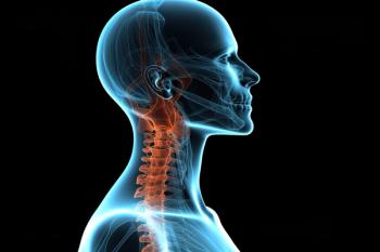
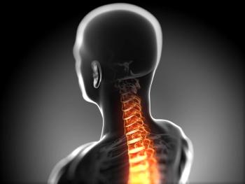
Commentary (Lanzieri): Current Imaging Techniques for Head and Neck Tumors
Author(s)Charles F. Lanzieri, MD
Head and neck imaging, in its current form, really began with the development of the computed tomographic (CT) scanner in the late 1970s and early ’80s. Originally, only CT scans of the brain were performed because of constraints on gantry size.
Advertisement
Head and neck imaging, in its current form, really began with the development of the computed tomographic (CT) scanner in the late 1970s and early ’80s. Originally, only CT scans of the brain were performed because of constraints on gantry size. Since neurosurgeons are accustomed to viewing the patient in the operating room from the top of the head, CT images were displayed from the point of view of the surgeon, with the patient’s right on the viewer’s right.
When gantry sizes increased to the point where the rest of the body could fit, radiologists began to display images in the “traditional” anatomic position, ie, with the patient’s left displayed on the viewer’s right. This created much confusion for imaging physicians and their clinical colleagues, as well as for imaging technicians. With the advent of neck imaging, the two approaches converged, and, eventually, a common format was adopted, in which the patient’s left was displayed on the viewer’s right for all cross-sectional images. This was the first major contribution of radiologists interested in head and neck imaging.
Advances in Imaging Revolutionize Patient Evaluation and Treatment
In the early 1980s, a flurry of discoveries concerning head and neck imaging were published. Led by respected and logical minds including Peter Som at Mount Sinai School of Medicine, Tom Bergeron at New York University, Toni Hasso at Loma Linda, and many others, these discoveries revolutionized the evaluation and management of patients with head and neck pathology.
Current cancer therapy for head and neck cancer derives largely from what we have learned about head and neck imaging over the past 20 years. In the accompanying article, Jane Weissman, who is a prodigy in this field, and her colleague Raucheline Akindele succinctly summarize where head and neck imaging is today and where it is about to go. One cannot improve on their review. One can only comment that the story is far from over.
There is no doubt that the efforts of investigators in head and neck imaging have improved patient care, leveraged imaging into more cost-effective patient management, and improved the quality of life of untold millions of people. These results were achieved by expediting head and neck evaluation, doing it less invasively, and providing more valuable information than was ever thought possible.
The development of this field of medicine promises to continue into the foreseeable future. The greatest impact of head and neck radiology on patient care may have yet been felt.
Recent Developments
The past few months have witnessed the development of magnetic resonance (MR)–guided biopsy of lesions that, heretofore, could only be approached surgically. Some lesions are too dangerous to biopsy under CT guidance, but recent breakthroughs in open magnetic resonance imaging (MRI) have allowed many more patients to take undergo minimally invasive surgery in the head and neck performed by radiologists.
In the not too distant future, MR spectroscopy may allow for “virtual biopsy” through tissue characterization. This promising technique may obviate the need for biopsy altogether in some instances.
Tumor control utilizing radiofrequency for thermal ablation is still experimental but promises to provide a new therapeutic approach to head and neck cancer. This modality, performed under the guidance of a high-field MR scanner, may be able to arrest tumor growth without morbidity. The use of low-field imaging in the operating room also holds huge potential for head and neck cancer patients, allowing for image-guided surgery and more complete resections.
Finally, intra-arterial chemotherapy has recently been used in patients with head and neck tumors. This technique allows more aggressive chemotherapy to be delivered directly to head and neck tumors with far less morbidity.
Summary
What began with standardizing the projection of axial images visualization and localization of head and neck tumors just 2 decades ago has led to new classification schemes for tumors based on imaging characteristics. Spectroscopy promises to provide more definitive tissue characterization, and nascent imaging techniques promise to vastly improve patient care in the next decade. Dr Weissman is to be congratulated on her fine work and should be invited to update oncology’s readers again as these techniques mature.
Articles in this issue
about 27 years ago
Thalidomide Shows Promising Results in Patients With Multiple Myelomaabout 27 years ago
Bill Would Allow Medicaid to Pay for Cervical/Breast Cancer Treatmentabout 27 years ago
Photodynamic Therapy for Palliation of Locally Advanced Lung Cancerabout 27 years ago
Zeneca Launches Education Program Aimed at Reducing Breast Cancer Riskabout 27 years ago
Reinventing Bone Marrow Transplantationabout 27 years ago
Age Should Not Determine Treatment of SCLC Patientsabout 27 years ago
Single Chest X-Ray Leads to Improved Lung Cancer Prognosisabout 27 years ago
No Link Found Between PCB Exposure and Cancer Mortalityabout 27 years ago
Arsenic Induces Remissions in Patients With Acute Promyelocytic LeukemiaAdvertisement
Related Content
Advertisement


Advertisement
Advertisement
Trending on CancerNetwork
1
Dostarlimab Yields Sustained Complete Responses in dMMR/MSI-H Rectal Cancer
2
FDA Accepts NDA for Mezigdomide Combo in R/R Multiple Myeloma
3
ADCs in the Frontline: Evolving Approaches in Metastatic TNBC
4
Risvutatug Rezetecan Improves Overall Survival in Relapsed SCLC Trial
5