- Oncology Vol 30 No 6
- Volume 30
- Issue 6
Contemporary Management of Small Renal Masses
Despite improved understanding of the molecular features of renal tumors, increasing expertise in surgical management of localized renal cancers, and multiple effective systemic therapies for metastatic cancer, mortality from renal cell carcinoma remains largely unchanged.
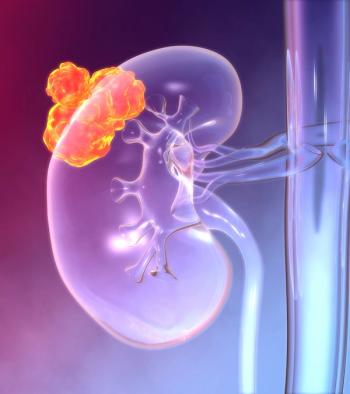
Despite improved understanding of the molecular features of renal tumors, increasing expertise in surgical management of localized renal cancers, and multiple effective systemic therapies for metastatic cancer, mortality from renal cell carcinoma (RCC) remains largely unchanged. Small renal masses (SRMs) are those that are smaller than 4 cm, clinically localized, and enhance on contrasted imaging. An improved ability to detect these tumors has led to an increased incidence of disease, as well as more complexity in managing patients for this increasingly common clinical scenario. In this issue of ONCOLOGY, Leone and colleagues describe several contemporary issues in the management of SRMs.[1] Their review of current literature and practice patterns suggests that defining one optimal management strategy remains challenging, especially given the environments that patients and providers encounter when dealing with SRMs. Contemporary management of SRMs should account for both the individual features of the patient and the tumor in question, and should attempt to provide data so that future decisions can be even more evidence-based.
The primary issue surrounding management of SRMs is the histopathologic heterogeneity of renal cortical tumors. For tumors that are 4 cm or smaller, over 20% will have a benign histopathologic diagnosis and less than 20% will harbor particularly aggressive features with concern for early metastasis.[2,3] Unfortunately, despite advances in imaging technology for computed tomography (CT), magnetic resonance imaging (MRI), and contrast-enhanced ultrasound, differentiation between benign and malignant lesions is imperfect. Molecular imaging holds great promise, but may be better at differentiating marker-positive vs marker-negative tumors. For example, if differentiation of clear cell RCC from other tumors is the endpoint in question, carbonic anhydrase IX imaging may serve this purpose well. However, if a more comprehensive description of the potential aggressiveness of a tumor is desired, it is difficult to imagine that biopsy can be avoided.
The next question at hand is whether biopsy should be performed prior to treatment selection for a given SRM. The popularity of renal mass biopsy is growing, and appropriately so, since it can assist some patients in avoiding invasive treatment. However, there are no standard protocols for renal mass biopsy, and local practice patterns and research interests generally determine its use. Leone et al correctly indicate that use of biopsy is a patient-by-patient determination that should be based on whether the result will change patient management. We would add that biopsy of SRMs should also be evaluated in the context of overall risk vs benefit compared with alternative strategies. Although select centers of excellence may be able to perform such biopsies with “major bleeding < 1%” in patients,[1] it is not yet determined whether the actual impact of biopsy will become universally applied in diverse clinical practices. What proportion of patients will spend a night under observation following renal mass biopsy, which is comparable to length of stay for a minimally invasive kidney-sparing treatment? How often will hematoma delay kidney-sparing surgery and lead to increased anxiety in these patients? Recent literature has indicated that cost-benefit analysis favors a biopsy-first approach, but the significant publication biases in reported studies (vs series that do not go to print) are a major limitation in these studies. At present, renal biopsy appears excellent for determination of histologic subtype, but has limited accuracy when evaluating tumor grade or presence of aggressive features such as sarcomatoid differentiation. When the benefits outweigh the risks, and the results are likely to impact management, biopsy is the right choice.
The third area of interest regarding SRMs is the diverse clinical practices of individual providers. How will doctor A vs doctor B interpret, communicate, and act upon results indicating 'clear cell RCC, grade 1', or 'oncocytic neoplasm, favor malignant tumor', for example? How will provider X vs provider Y manage a 75-year-old patient with multiple comorbidities and an exophytic, 2.5-cm, minimally enhancing renal mass? Clinicians factor each patient’s clinical characteristics, each tumor’s radiographic features, and the relative merits of multiple therapeutic approaches into each scenario, and do so without coming to identical conclusions. Previous studies from several institutions have confirmed that SRMs grow relatively slowly and with low rates of metastasis (1%–2%). For this reason, active surveillance is a reasonable management strategy in carefully selected patients with advanced age, multiple comorbidities, poor surgical risk, and/or limited life expectancy. Leone et al discuss the role of serial radiographic imaging for those on surveillance, and the need to identify those patients with linear growth rates higher than 0.5 cm/year in order to recommend appropriate treatment.[1,4] Significant changes are even better appreciated by direct review of serial studies and volumetric measurements, particularly when different modalities (such as CT and ultrasound) have been performed.
Studying the practice patterns at the institution and individual-provider level will reveal the high variability that currently exists.[5-7] But in order to learn from this experience, we would suggest that the field must move beyond research and into the realm of quality improvement. The chances that well-designed prospective clinical trials for patients with SRMs will inform all of the present dilemmas and those to come are fleeting. Participation in quality improvement collaborations and other initiatives in which data can be collected, information exchanged, and action taken has the best chance of effecting change for the management of SRMs.[8,9] Perhaps with such efforts, we will better apply selection criteria, standardized treatment protocols, and consistent follow-up criteria to patients with SRMs. As specific protocols become available, future studies should include patient-centered outcomes, such as health-related quality of life, and value-based outcomes (taking into account quality, appropriateness, and cost). In order to improve the value of care of patients with SRMs, the approach to diagnosis, management, and follow-up will likely continue to be individualized, with attention to preservation of renal function without compromising oncologic outcomes.
Financial Disclosure:The authors have no significant financial interest in or other relationship with the manufacturer of any product or provider of any service mentioned in this article.
References:
1. Leone AR, Diorio GJ, Spiess PE, Gilbert SM. Contemporary issues surrounding small renal masses: evaluation, diagnostic biopsy, nephron sparing, and novel treatment modalities. Oncology (Williston Park). 2016;30:507-14.
2. Frank I, Blute ML, Cheville JC, et al. Solid renal tumors: an analysis of pathological features related to tumor size. J Urol. 2003;170:2217-20.
3. Lane BR, Babineau D, Kattan MW, et al. A preoperative prognostic nomogram for solid enhancing renal tumors 7 cm or less amenable to partial nephrectomy. J Urol. 2007;178:429-34.
4. Smaldone MC, Kutikov A, Egleston BL, et al. Small renal masses progressing to metastases under active surveillance: a systematic review and pooled analysis. Cancer. 2012;118:997-1006.
5. Banerjee M, Filson C, Xia R, Miller DC. Logic regression for provider effects on kidney cancer treatment delivery. Comput Math Methods Med. 2014;2014:316935.
6. Weight CJ, Crispen PL, Breau RH, et al. Practice-setting and surgeon characteristics heavily influence the decision to perform partial nephrectomy among American Urologic Association surgeons. BJU Int. 2013;111:731-8.
7. Lane BR, Golan S, Eggener S, et al. Differential use of partial nephrectomy for intermediate and high complexity tumors may explain variability in reported utilization rates. J Urol. 2013;189:2047-53.
8. Ghani KR, Miller DC. Variation in prostate cancer care. JAMA. 2015;313:2066-7.
9. Auffenberg GB, Ghani KR, Ye Z, et al. Comparing publicly reported surgical outcomes with quality measures from a statewide improvement collaborative. JAMA Surg. 2016 Mar 16. [Epub ahead of print]
Articles in this issue
over 9 years ago
Diabetes Management in Cancer PatientsNewsletter
Stay up to date on recent advances in the multidisciplinary approach to cancer.


