|Articles|March 2, 2022
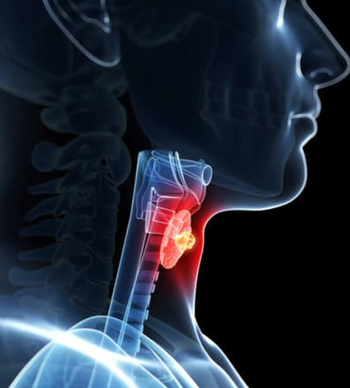
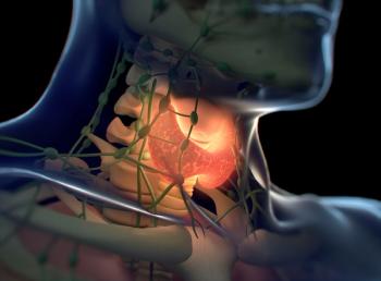
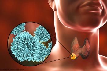
CT and Ultrasonography Have Similar Diagnostic Accuracy for Papillary Thyroid Cancer Cervical Lymph Node Metastases
Author(s)Ariana Pelosci
A system review and meta-analysis found that computed tomography and ultrasonography had comparable diagnostic accuracy in identifying cervical lymph node metastases in patients with papillary thyroid cancer.
Advertisement
CT and ultrasonography achieved similar accuracy in identifying cervical lymph node metastases (CLNM) for patients with papillary thyroid cancer, with investigators citing a need for further studies to confirm the role of CT in staging, according to a systemic review and meta-analysis published in JAMA Otolaryngology-Head & Neck Surgery.
Investigators did not observe a significant difference in sensitivity between ultrasonography and CT at 73% (95% CI, 64%-80%) and 77% (95% CI, 67%-85%), respectively (P = .11) when detecting lateral compartment CLNM. Additionally, no difference in specificity was identified, with a specificity of 89% (95% CI, 80%-94%) for ultrasonography and 88% (95% CI, 79%-94%) for CT (P = .79). Those with central compartment metastasis experienced a higher sensitivity with CT at 39% (95% CI, 27%-52%) compared with 28% (95% CI, 21%-36%) for ultrasonography (P = .004); however, specificity was higher in ultrasonography at 95% (95% CI, 92%-98%) compared with 87% (95% CI, 77%-93%) for CT (P <.001).
In this systemic review, 47 studies were included totaling 31,942 observations of patients with thyroid cancer. Forty-four studies concentrated on ultrasonography and 15 used CT; an additional 2 studies used MRI. The risk of bias was low in 21 studies, and 26 had a high risk.
A total of 17 studies using ultrasonography were included with 3162 observations of patients with thyroid cancer, of whom 1244 had lateral CLNM. Pooled estimated mean sensitivity for ultrasonography in detecting lateral compartment CLNM was 82% (95% CI, 74%-88%), 84% (95% CI, 74%-91%) for specificity, and 0.90 (95% CI, 0.87-0.92) for the area under the curve. For CT, investigators included 10 studies with 1845 patient observations, of whom 689 showed signs of lateral compartment CLNM. For this population, the mean estimated sensitivity was 72% (95% CI, 59%-82%), 90% (95% CI, 83%-95%) for specificity, and 0.89 (95% CI, 0.86-0.92) for the area under the curve.
Investigators noted that sensitivity was highest in retrospective (P <.001) and multicenter (P <.001) studies, while specificity was highest in prospective (P <.001) and single center studies (P = .02). Investigators did not find a risk of bias associated with sensitivity (P = .75) or specificity (P = .41).
In 19 studies with 10,845 observations patients with thyroid cancer, 4955 of whom had central compartment CLNM, ultrasonography pooled estimate for mean sensitivity were 41% (95% CI, 30%-53%), 89% (95% CI, 80%-94%) for specificity, and 0.69 (95% CI, 0.65-0.73) for area under the curve. For CT, which included 8 studies and 8095 observations of patients, pooled estimate for mean sensitivity was 38% (95% CI, 27%-49%), 89% (95% CI, 80%-94%) for specificity, and 0.66 (95% CI, 0.61-0.70) for the area under the curve.
Investigators found that sensitivity was highest in retrospective studies (P = .002) that were at a low risk for bias (P <.001). Additionally, specificity was highest in prospective studies (P <.001) that were at a high risk for bias (P <.001).
Twelve studies included, with 2874 observations of patients with thyroid cancer, of whom 1421 had extra nodal disease extension. In this group, ultrasonography pooled estimated mean sensitivity was 91% (95% CI, 81%-96%), 47% (95% CI, 35%-60%) for specificity, and 0.76 (95% CI, 0.72-0.80) for the area under the curve. For CT, there was 1 study featuring 377 patients, 174 of whom had extra nodal disease extension. The pooled estimated mean sensitivity for this group was 86% and 30% for specificity. Additionally, investigators included 2 MRI studies with 300 patients, 152 of whom had extra nodal disease extension, and reported a sensitivity ranging from 76% to 88% and a specificity of 75% to 93%.
Investigators determined there to be an insufficient number of studies to compare imaging modalities. In the meta-analysis, when using ultrasonography, the study design, observation type, and risk of bias were not associated with diagnostic accuracy (P = .27-.97).
Reference
Alabousi M, Alabousi A, Adham S, et al. Diagnostic test accuracy of ultrasonography vs computed tomography for papillary thyroid cancer cervical lymph node metastasis: a systematic review and meta-analysis. JAMA Otolaryngol Head Neck Surg. 2022;148(2):107-118. doi:10.1001/jamaoto.2021.3387
Advertisement
Related Content
Advertisement


Advertisement
Advertisement
Trending on CancerNetwork
1
Teclistamab Induction Shows Feasibility in Transplant-Eligible Myeloma
2
MARIPOSA Trial: EGFR-Mutant Lung Cancer Frontline Strategies
3
ASCO Breakthrough 2026 Wrap-Up: Precision Oncology, AI, and Biomarkers Define the Future of Cancer Care
4
Purple Reign Vs Thriller: A Duel of The Top Frontline EGFR+ NSCLC Regimens
5