
|Articles|October 1, 2018
Differentiating Benign Renal Tumors From Chromophobe RCC
Author(s)Leah Lawrence
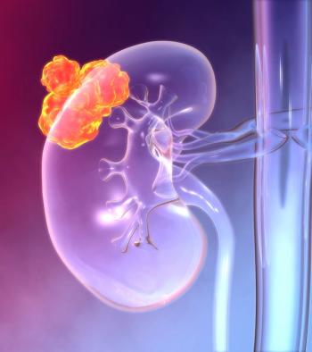
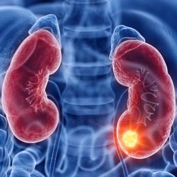
A radiographic measure may help clinicians distinguish between benign renal oncocytoma and chromophobe renal cell carcinoma.
Advertisement
Researchers have identified a radiographic measure to help clinicians distinguish between benign renal oncocytoma and chromophobe renal cell carcinoma in patients with renal tumors.
Tumor-to-cortex peak early-phase enhancement ratio (PEER) using multiphase CT accurately distinguished 100% of the tumors in a
“Current inability to reliably differentiate benign renal oncocytomas from renal cell carcinoma using needle core biopsy remains the primary reason behind routine surgical resection of these benign tumors, which is associated with frequent perioperative morbidity and an estimated annual health care cost approaching 100 million dollars in the United States alone,” wrote researchers led by Jay Amin, of the department of urology at Roswell Park Cancer Institute in Buffalo, New York.
Amin and colleagues conducted this study to identify variables that might aid in preoperative differentiation of renal tumors in order to avoid unnecessary resections. Their analysis included 124 patients undergoing nephrectomy at a single institution, 133 tumors were studied. Patient data from 2003–2012 was used for a retrospective cohort (n = 87). The researchers then conducted a prospective validation study among 22 consecutive tumors resected from 2013–2017.
In the retrospective cohort, patients younger than 50 were significantly associated with chromophobe renal cell carcinoma, with an 80% specificity and a 24% sensitivity.
“Although an age cutoff of [50 years old] had high specificity for [chromophobe renal cell carcinoma], the optimal age cutoff to discern these two subtypes needs to be better defined in larger studies with statistical analyses designed to identify this cutoff,” the researchers wrote.
Additionally, multifocality was 100% specific for renal oncocytoma, but only 24% sensitivity.
“The variable most reliably associated with renal oncocytoma vs chromophobe renal cell carcinoma diagnosis was the CT peak signal intensity within a tumor, particularly when expressed as a tumor-to-cortex ratio,” the researchers wrote.
There were 54 PEER-evaluable tumors in the retrospective cohort. PEER classified each tumor correctly as either renal oncocytoma (PEER > .50) or chromophobe renal cell carcinoma (PEER .50 or greater), except for four misclassified CD117-negative chromophobe renal cell carcinoma variants.
In the prospective cohort, PEER confirmed 100% accuracy for both tumor types among 22 additional tumors.
“Goals of this future research should include verification of the optimal PEER cutoff value for chromophobe renal cell carcinoma vs renal oncocytoma differentiation,” the researchers wrote. “The cutoff PEER value of .50 in our prospective cohort was arbitrarily selected on the basis of our retrospective observations; however, a cutoff PEER value of .55 would have achieved 100% diagnostic accuracy in both the retrospective and prospective cohorts.”
Advertisement
Related Content
Advertisement


Advertisement
Advertisement
Trending on CancerNetwork
1
GLP-1 Receptor Agonists Show Modest Reduction in HR+ Breast Cancer Incidence
2
FDA Grants Priority Review to Giredestrant in ER+/HER2– Early Breast Cancer
3
Radiation Oncology Trials You May Have Missed at ASCO 2026
4
Camizestrant Extends PFS and PFS2 in ESR1-Mutated Advanced Breast Cancer
5