|Articles|January 1, 1999
Oncology NEWS International
- Oncology NEWS International Vol 8 No 1
- Volume 8
- Issue 1
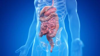
FDG-PET Used to Evaluate Colorectal Cancer Recurrences
TORONTO-Several studies presented at the Society of Nuclear Medicine’s 45th annual conference support the use of positron emission tomography (PET) with fluorine-18-fluorodeoxyglucose (FDG) to evaluate patients with recurrent colorectal cancer.
Advertisement
TORONTOSeveral studies presented at the Society of Nuclear Medicines 45th annual conference support the use of positron emission tomography (PET) with fluorine-18-fluorodeoxyglucose (FDG) to evaluate patients with recurrent colorectal cancer.
FDG-PET is more accurate than the conventional diagnostic modalities for staging patients with recurrent colorectal carcinoma, and has a significant positive impact on patient management in this setting, said Patrick Flamen, MD, of the Department of Nuclear Medicine, KU Leuven-UZ Gasthuisberg, Leuven, Belgium. The advantage of whole-body FDG-PET is that screening is performed in one examination and that unsuspected metastatic sites can be detected, he added.
The Belgium researchers retrospectively reviewed 103 patients with suspected recurrent colorectal cancer who underwent whole body FDG-PET in addition to conventional staging (CEA, endoscopy, CT of the chest and abdomen, MRI, and ultrasound). The PET studies were interpreted with full knowledge of the conventional staging findings.
Sensitivity Results
Of the 37 patients with pelvic recurrence, PET detected 30 cases (81%) whereas CT detected only 22 (59%). Of the 48 patients with liver metastases, PET detected 96%, which was slightly better than CT at 90%.
The sensitivity to the detection of retroperitoneal lymph node involvement was similar for both PET and conventional staging methods (73%). Neither PET nor CT was able to accurately detect peritoneal involvement. PET located all 14 extraab-dominal lesions whereas conventional staging missed 4 such lesions.
As for patient management, conventional staging had categorized 64 patients as operable and 22 as having extended disease. The addition of PET findings correctly downstaged 5 patients and upstaged 11 patients. However, 2 patients were incorrectly overstaged and 5 were under-staged with PET scanning.
In 9 patients with elevated serum CEA who had negative or equivocal conventional staging results, PET correctly detected relapse in 5 patients and excluded disease in 2 patients.
Researchers from the University of Frankfurt, Main, Germany, Medical Center emphasized the role of FDG-PET in detecting extrahepatic metastases before liver surgery in patients with recurrent colorectal cancer.
The goal of the study was to determine a way to select patients for curative hepatic resection and avoid surgery in patients with extrahepatic lesions, said Dr. Andreas Hertel, of the Department of Nuclear Medicine at University Hospital, Frankfurt. We believe that FDG-PET may be a cost-effective way to screen patients with recurrent colorectal cancer, Dr. Hertel added.
The Frankfurt study included 36 colorectal cancer patients with suspected or known liver lesions. Prior to surgery, all underwent CT and FDG-PET imaging. The sensitivity of PET was superior to CT for the identification of metastatic lesions and led to changes in management in 39% of patients, Dr. Hertel said. PET imaging allowed surgeons to perform curative surgery in 4 patients with local recurrence only and avoid surgery in 10 patients with previously undetected multiple extrahepatic metastases.
Lead author Richard P. Baum, MD, chair of the Bad Berka PET Center, said: Our prospective data clearly indicate that whole body FDG-PET is the most accurate noninvasive method for restaging colorec-tal cancer patients before liver surgery. FDG-PET had a decisive influence on the therapeutic strategy in more than one-third of our patients.
Dr. Baum recommends FDG-PET for certain clearly defined situations in the evaluation of colorectal cancer:
Patients to be treated with regional chemotherapy of the liver.
Patients with normal CT, MRI, and/or ultrasound tests and elevated CEA levels.
Patients diagnosed with a primary tumor when thoracic x-ray and abdominal ultrasound are normal but CEA levels are elevated.
Dr. Baum cited an example in which FDG-PET changed one patients planned surgery. The patient was scheduled for surgery to remove a single liver mass located with conventional imaging methods. However, FDG-PET imaging located an unknown lung metastasis. The surgeon then removed both masses, and the patient remains tumor free at 18 months post-surgery.
Dr. Baum also described a patient who had a single lung metastasis. But because the presurgical PET scan also detected mediastinal metastasis (an indication for chemotherapy, not surgery), the operation was canceled.
Articles in this issue
over 27 years ago
Pittsburgh to Build New Cancer Centerover 27 years ago
NCI Initiates Two High-Priority Tobacco Research Programsover 27 years ago
Breast Cancer Stamp Sells Wellover 27 years ago
Hospital Strategies To Prevent Invasive Aspergillosis Spreadover 27 years ago
‘Cancer Patients Should Be Assertive, Know Their Rights’over 27 years ago
EBCTCG Update of Adjuvant Treatment for Early Breast Cancerover 27 years ago
Younger Breast Cancer Patients at Increased Risk of Recurrenceover 27 years ago
Six Named to National Cancer Advisory Boardover 27 years ago
Trial Uses Vitamin A To Prevent Lung Cancer in Former Smokersover 27 years ago
‘Medical School Curriculum Must Include Palliative Care’Advertisement
Related Content
Advertisement


Advertisement
Advertisement
Trending on CancerNetwork
1
Risvutatug Rezetecan Improves Overall Survival in Relapsed SCLC Trial
2
EMA’s Review of Daraxonrasib in PDAC Accelerated Under Phased Review Process
3
FDA OKs Pembrolizumab/EV Combo in Muscle Invasive Bladder Cancer
4
Orca-T Improves Chronic GVHD-Free Survival in Hematologic Cancers
5