
News|Articles|August 22, 2019
Gal9/TIM-3 Expression Levels Affects Chemotherapy in Leukemia Patients
Author(s)John Schieszer
A new report suggests targeting the Gal9/TIM3-axis could help boost chances of complete remission in patients with acute myeloid leukemia.
Advertisement
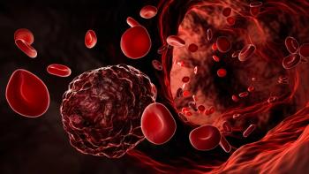
Gal9/TIM -3 expression levels are higher in acute myeloid leukemia (AML) patients who fail chemotherapy according to a new
The authors found the interruption of PD-1/PD-L1 axes by itself does not usually completely restore T-cell function, suggesting other negative regulatory pathways are promoting T-cell exhaustion. The team conducted a study with 26 patients with AML and examined expression of CD47, PD-L1, PD-L2 and Gal9 on CD34+ AML blasts. They also looked at CD34−cell populations, and evaluated expression of inhibitory (PD1, CTLA4, LAG3, TIM-3) and stimulatory (CD28, ICOS, CD137, OX40, CD40L, HLA-DR) co-receptors on CD4+ and CD8+ T-cell subsets.
The researchers found that the frequency of Gal9+ CD34− cells was significantly higher in patients with treatment failure than in those who had CR. Additionally, these findings correlated with increased TIM-3 expression on bone marrow-resident T-cells in patients with treatment failure. The researchers measured the expression level of PD-1 and TIM-3 in bone marrow samples and compared them to peripheral blood samples and found that TIM-3 was significantly higher in the bone marrow specimens.
“Our findings suggest that the Gal9/TIM3 pathway may play a role in patients in remission by subverting ongoing immune surveillance, and suggests that T cells in AML patients, even those who achieve CR to therapy, are likely exhausted or dysfunctional,” wrote study authors led by Paola Dama of the University of Chicago in Chicago.
Naval Daver, MD, an associate professor in the department of leukemia at MD Anderson Cancer Center, Houston, Texas, said this study adds vital information to the growing knowledge regarding the role of T-cell fitness and checkpoint pathway expression in AML at baseline. as well as after different types of AML-directed therapy.
“Our group was one of the first to report that expression of certain checkpoint costimulatory receptors such as PD1, OX40 was increased on CD4 and CD8 bone marrow resident T-cells in patients with AML as compared to healthy donor bone marrows,” Dr. Daver told Cancer Network. “In this manuscript, the authors focus on expression of checkpoint ligands on bone marrow blasts and costimulatory receptors on bone marrow resident T-cells before and after chemotherapy.”
The study found that among non-responders there appeared to be particular upregulation of the GAL9-TIM3 nexus, suggesting this may be associated with impaired response to chemotherapy.
“The weakness of the study is that we do not know whether the increase in GAL9-TIM3 is non-specific phenomenon occurring in non-responders to any therapies or specifically related to non-responders to the mitoxantrone+Arac+selinexor combination (used in this study) and which component of this therapy (mitoxantrone, AraC, selinexor) causes this up-regulation, said Dr. Daver.
Advertisement
Related Content
Advertisement


Advertisement
Advertisement
Trending on CancerNetwork
1
Obesity and Cancer Risk: The Evolving Role of GLP-1s in Oncology
2
FDA Approves Afami-cel in Metastatic Synovial Sarcoma
3
EC Approves Encorafenib/Cetuximab/FOLFOX in 1L BRAF V600E–Mutant mCRC
4
The Moonlight Shift: Dr Joshua Richter on Myeloma, Myths, and the C-Word
5