|Articles|February 11, 2021
- ONCOLOGY Vol 35 Issue 2
Pediatric Locally Advanced Synovial Sarcoma: What Would Be the Best Treatment Option?
KEY POINTS • Synovial sarcomas are often mistreated with unplanned tumor resection. • Attention from specialists early in the course of SS can minimize the risk of recurrence, metastases, and the necessity for resurgery, all of which are increased with unplanned tumor resection. • Chemotherapy alone does not provide sufficient local control of the tumor. • Resurgery, in conjunction with radiotherapy and chemotherapy, is the best choice of management for this patient.
Advertisement
The Case
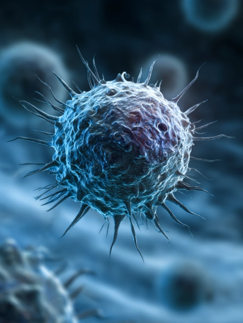
A female patient, age 11 years, presented with a firm, palpable mass in the left lower quadrant of the abdomen. Upon ultrasound imaging, the 3.5 cm × 5 cm lesion was assessed as a benign cyst and a laparoscopic resection of the mass was performed. The initial pathological evaluation reported a diagnosis of fibromyxoid sarcoma. However, after an additional immunohistochemical examination, the diagnosis was changed to grade III synovial sarcoma (CD45–, Bcl+, Desmin–, WT1–, SMA–, Ki65-15%) (Figure 1). Detection of t(X;18) (p11.2; q11.2) translocation was not yet available in Armenia at that time. The pathology report did not include the state of margins. CT and bone scans revealed no metastatic disease at the time.
Oncology (Williston Park). 2021;35(2):57-60.
DOI: 10.46883/ONC.2021.3502.0057
What would be the best choice for further management of this patient?
A. Observation
B. Radiotherapy
C. Resurgery, with further treatment including radiotherapy and chemotherapy
D. Chemotherapy
Discussion
Despite the fact that synovial sarcoma (SS) is a rare disease, it is the second most common type of soft tissue sarcoma (STS) in children and adolescents after rhabdomyosarcoma. This subtype of neoplasms affects both children and adults, with its peak incidence occurring in the third
decade of life.1 Despite its name, SS can develop anywhere in the body, not just the joints. The most common location is the soft tissue of the extremities, especially in the large joints, followed by the upper extremities, trunk, abdomen, head, and neck.
Specific detection of a t(X;18) (p11.2; q11.2) translocation is an important diagnostic tool for SS, but it was not available in Armenia at that time. However, given that, radiologically, the tumor is usually well defined, it is often misdiagnosed as a benign lesion. Tumors of this type tend to be overlooked by general physicians or surgeons because they can be surgically removed as a benign tumor or inflammatory lesion without the need for wide surgical margins.2-4 According to results of a study performed by St Jude Children’s Research Hospital, 81% of patients with a diagnosis of nonrhabdomyosarcoma STSs underwent unplanned resections before being referred to their institution. According to results of another study, residual disease was present in nearly half of the cases (48%) in patients who underwent such unplanned surgery.5,6
In 2017, Gingrich et al performed a prospective study including 76 patients diagnosed with primary STS located in the trunk or extremities who had undergone unplanned excision of the tumor. They concluded that 70% of the patients who underwent repeat excision after the initial unplanned tumor resection indeed had residual tumor.7
Commonly, a patient with STS is referred to an oncologist if a postoperative pathological evaluation reveals a malignancy, or if a local recurrence or distant metastases are detected, to plan reexcision with wider margins, adjuvant chemotherapy, or radiotherapy. Microscopic residual disease is often the result of the surgical removal of soft tissue sarcoma without proper preoperative imaging, biopsy, or correct execution of wide (R0) resection margins; it leads to high risk of local recurrence for patients with STS.5
In 2013, Umer et al performed a retrospective cohort study consisting of 135 patients in2 groups: those who had planned tumor excisions (group 1; n = 84) and those who had unplanned tumor excisions (group 2; n = 51). Mean followup in group 1 (starting from the date of surgery) was 52.6 plus 39.8 months (median, 36 months). Minimum follow-up duration in group 2 (starting from the date of revision surgery) was 36 months. The incidences of local recurrence in group 1 and group 2 were 14.3% (n = 12) and 21.4% (n = 11), respectively. Distant metastasis occurred in 7.3% and 13.7% of the patients, respectively.8
Grimer et al concluded that unplanned surgery leads to higher rates of recurrence, metastasis, and overall fatality; they recommended that soft tissue masses be assessed and managed by specialized oncology teams.9 Similarly, Bhangu et al (2004) concluded that patients who were treated outside a specialized tumor center had a higher rate of local recurrence (39%) compared with those who were treated at specialized centers (19%).10
Therefore, answer A (observation) is not the preferred path for management for this patient, because there is supporting evidence that unplanned surgical removal of STS often leads to probable recurrence (both local and distant) and the inevitable need for more extensive measures.
The most important prognostic factors in STS are the presence of any metastases and the tumor’s resectability, size, and location. The optimal approach to treat a localized synovial sarcoma includes wide excision of the tumor with clear margins, resulting in no loss of function. Wide reexcision, which is ideally done within 3 weeks of unplanned surgery, is the standard treatment in most hospitals.11 In cases when reexcision is not possible, radiotherapy alone is the preferred treatment option, but some research results indicate that it has inferior local control compared with surgical treatment. Even in the case of positive margins, the necessary dosage of radiation is high and increases the risk of complications. Therefore, radiation alone (answer B) would have been a viable option only if reexcision would not have been possible to perform.6,7
The choice of an adjuvant treatment regimen is based on factors including tumor size, grade, and margins. For high-risk tumors, the treatment includes radiotherapy in combination with chemotherapy. SS seems to have relatively more sensitivity to chemotherapy than other STSs. The most commonly used chemotherapeutic agents in such a regimen are ifosfamide and doxorubicin, which showed higher response in SS than in other nonrhabdomyosarcoma soft-tissue sarcomas (NRSTSs).12 Chemotherapy alone, without proper local control, cannot be curative. In the majority of studies, it is reserved as adjuvant treatment or in the palliative setting. Hence, performing chemotherapy without adequate surgical and/or radiotherapy will lead to the failure to control local disease, making answer D a poor choice.13
Pediatric NRSTSs have a poor prognosis without multimodal treatment. Ingley et al have recently outlined that for patients with localized disease who received multimodal therapy, including chemotherapy and radiotherapy, 5-year overall survival is 90.7% and event-free survival is 80.7%, based on risk classification.14 Given this information, the best course of action for this case is answer C, resurgery, along with further treatment including radiotherapy
and chemotherapy.
Outcome
The patient’s parents initially refused further treatment. One year later, the patient noticed a painless mass beneath the postoperative scar on the anterior abdominal wall (Figure 2). The MRI revealed a grape-shaped, well-defined soft tissue mass in the left lower quadrant of the abdominal wall, measuring 15 cm (Figure 3). The CT showed left inguinal lymphadenopathy without signs of distant metastases. A bone scan revealed no abnormalities. A tumor biopsy confirmed disease relapse.
Overall, the child received 7 courses of chemotherapy with ifosfamide and doxorubicin (according to COG-ARST0332; outside of a clinical trial) and underwent 3D radiotherapy
(70 Gy).12
Subsequently, a PET-CT was performed, showing lesions (ranging in size from 50 mm x 62 mm to 16 mm x 24 mm) on the anterior abdominal wall, extending into the left inguinal region with moderate metabolic activity (SUVmax, 2.84-3.03). Because of logistical issues, wide re-resection of the previous surgical scar, tumor bed, and the new palpable mass was performed in Armenia
4 months after the neoadjuvant treatment. Soft tissue reconstruction consisted of a rectus femoris pedicled musculocutaneous flap. Final pathology showed no viable tumor.
As of October 2020, the child was disease free. She had painless lymphedema of the involved extremity but was able to participate in daily life activities.
Financial Disclosure: The authors have no significant financial interest in or other relationship with the manufacturer of any product or provider of any service mentioned
in this article.
Mesrobian is a visiting scientist at the Pediatric Cancer and Blood Disorders Center of Armenia.
Avagyan is a pediatric oncology and hematology fellow at the Yerevan State Medical University and Pediatric Cancer and Blood Disorders Center
of Armenia.
Petrosyan is a pediatric oncology and hematology fellow at the Yerevan State Medical University and Pediatric Cancer and Blood Disorders Center
of Armenia.
Papyan is an assistant professor at the Department of Pediatric Oncology and Hematology, Yerevan State Medical University and chair of the Musculoskeletal Tumors Working Group, Pediatric Cancer and Blood Disorders Center of Armenia.
Hovsepyan is a pediatric oncology and hematology fellow at the Yerevan State Medical University and Pediatric Cancer and Blood Disorders Center
of Armenia.
Koloyan is an orthopedic surgeon and the director of the Wigmore Clinic.
Tamamyan is the chairman of the Department of Pediatric Oncology and Hematology, Yerevan State Medical University and the head of the Pediatric Cancer and Blood Disorders Center of Armenia, Hematology Center after Prof. R.H. Yeolyan. He is also the chairman of the ASCO’s IDEA Working Group.
REFERENCES
1. Wolden SL, Alektiar KM. Sarcomas across the age spectrum. Semin Radiat Oncol. 2010;20(1):45-51. doi:10.1016/j.semradonc.2009.09.003
2. Kerouanton A, Jimenez I, Cellier C, et al. Synovial sarcoma in children and adolescents. J Pediatr Hematol Oncol. 2014;36(4):257-262. doi:10.1097/MPH.0000000000000154
3. Speth BM, Krieg AH, Kaelin A, et al. Synovial sarcoma in patients under 20 years of age: a multicenter study with a minimum follow-up of 10 years. J Child Orthop. 2011;5(5):335-342. doi:10.1007/s11832-011-0360-4
4. Andrassy RJ, Okcu MF, Despa S, Raney RB. Synovial sarcoma in children: surgical lessons from a single institution and review of the literature. J Am Coll Surg. 2001;192(3):305-313. doi:10.1016/s1072-7515(00)00806-1
5. Chui CH, Spunt SL, Liu T, et al. Is reexcision in pediatric nonrhabdomyosarcoma soft tissue sarcoma necessary after an initial unplanned resection? J Pediatr Surg. 2002;37(10):1424-1429. doi:10.1053/jpsu.2002.35405
6. Qureshi SS, Prabhu A, Bhagat M, et al. Re-excision after unplanned resection of nonmetastatic nonrhabdomyosarcoma soft tissue sarcoma in children: comparison with planned excision. J Pediatr Surg. 2017;52(8):1340-1343. doi:10.1016/j.jpedsurg.2017.01.006
7. Gingrich AA, Elias A, Lee C-YM, et al. Predictors of residual disease after unplanned excision of soft tissue sarcomas. J Surg Res. 2017;208:26-32. doi:10.1016/j.jss.2016.08.096
8. Umer HM, Umer M, Qadir I, Abbasi N, Masood N. Impact of unplanned excision on prognosis of patients with extremity soft tissue sarcoma. Sarcoma. 2013;2013:498604. doi:10.1155/2013/498604
9. Grimer R, Parry M, James S. Inadvertent excision of malignant soft tissue tumours. EFORT Open Rev. 2019;4(6):321-329. doi:10.1302/2058-5241.4.180060
10. Bhangu AA, Beard JAS, Grimer RJ. Should soft tissue sarcomas be treated at a specialist centre? Sarcoma. 2004;8(1):1-6. doi:10.1080/13577140410001679185
11. Haas RL, Gronchi A, van de Sande MAJ, et al. Perioperative management of extremity soft tissue sarcomas. J Clin Oncol. 2018;36(2):118-124. doi:10.1200/JCO.2017.74.7527
12. Spunt SL, Million L, Chi YY, et al. Risk-based treatment for nonrhabdomyosarcoma soft tissue sarcomas (NRSTS) in patients under 30 years of age: Children’s Oncology Group study ARST0332. J Clin Oncol. 2014; 32:S15, 10008-10008. doi:10.1200/jco.2014.32.15_suppl.10008
13. Ramu EM, Houdek MT, Isaac CE, Dickie CI, Ferguson PC, Wunder JS. Management of soft-tissue sarcomas; treatment strategies, staging, and outcomes. SICOT J. 2017;3:20. doi:10.1051/sicotj/2017010
14. Ingley KM, Cohen-Gogo S, Gupta AA. Systemic therapy in pediatric-type soft-tissue sarcoma. Curr Oncol. 2020;27(Suppl 1):6-16. doi:10.3747/co.27.5481
Articles in this issue
over 5 years ago
ASCO GI Round upover 5 years ago
Redefining Prostate Cancer Careover 5 years ago
WANTED: An “All-Out” National Vaccination ProgramAdvertisement


Advertisement
Advertisement
Trending on CancerNetwork
1
FDA OKs Regulatory T-Cell Immunotherapy in Hematologic Malignancies
2
What Does The Future Hold for Thoracic Oncology and AI Use in Cancer?
3
Illuminating Optimal CAR T-Cell Therapies Across Large B-Cell Lymphoma Groups
4
Relapsed/Refractory Multiple Myeloma: CARTITUDE-4 and the Evolving Role of BCMA CAR T-Cell Therapy
5