
|Articles|October 15, 2011
- ONCOLOGY Vol 25 No 11
- Volume 25
- Issue 11
Mucocutaneous Paraneoplastic Syndromes Associated With Hematologic Malignancies
A case report is followed by a review of the diagnosis and treatment of other cutaneous paraneoplastic syndromes that are associated with hematologic malignancies.
Advertisement
Cutaneous paraneoplastic syndromes are a group of dermatoses that demonstrate a range of morphological and pathological findings. These syndromes may precede, be concurrent with, or follow the diagnosis of an underlying malignancy. Treatment of the malignancy is often associated with improvement in or resolution of the mucosal and cutaneous disease; however, this is not the case with paraneoplastic pemphigus (PNP). PNP is a rare syndrome that was first described in 1990, and it occurs almost exclusively in patients with lymphocytic neoplasms. Pulmonary manifestations occur in 30% to 40% of cases, and it is the only form of pemphigus that attacks epithelium other than squamous epithelium in an antibody-mediated fashion. The mortality rate for PNP associated with malignancy is greater than 90%. Treatment guidelines are not available, but case series point to the use of rituximab (Rituxan) as well as corticosteroids and various other immunomodulating agents. Here we present a diagnostic and treatment dilemma in a 39-year-old active-duty male who developed PNP in the setting of treatment with rituximab, cyclophosphamide, doxorubicin, vincristine, and prednisone (R-CHOP) for grade 3 follicular lymphoma. This case report is followed by a review of the diagnosis and treatment of other cutaneous paraneoplastic syndromes that are associated with hematologic malignancies.
Introduction
Paraneoplastic pemphigus (PNP) is an autoimmune-mediated, blistering, erosive, and painful mucocutaneous disease that is diagnosed almost exclusively in patients with lymphocytic neoplasms. The most commonly associated neoplasms are, in decreasing order of frequency, non-Hodgkin lymphoma, chronic lymphocytic leukemia, Castleman disease, thymoma, retroperitoneal sarcoma, and Waldenstrm macroglobulinemia.[1] When PNP is associated with an underlying malignancy, it is extremely resistant to treatment and carries a 90% mortality rate within a few months following diagnosis, independent of the course of malignancy.[2] PNP is characterized by the presence of IgG autoantibodies that react to desmosomal and hemidesmosomal plakin proteins as well as to desmosomal transmembrane proteins called desmogleins, and these reactions result in the loss of cell adhesion of epithelial cells.
The original diagnostic criteria for PNP were devised in 1990 based on initial case reports; these criteria included:
TABLE 1
Proposed Minimal Criteria for the Diagnosis of Paraneoplastic Pemphigus
1. Mucosal ulcerations and blisters, and polymorphous skin lesions in the context of an underlying neoplasm.
2. Histological findings of vacuolar interface change, keratinocyte necrosis, and intraepidermal acantholysis.
3. Epidermal intercellular deposition of IgG and C3, with or without linear deposition at the basement membrane zone, as demonstrated by direct immunofluorescence (DIF).
4. Serum autoantibodies that bind to the cell surface of skin mucosa in a pattern typical for pemphigus-but that also bind to simple, columnar, and transitional epithelia, as demonstrated by indirect immunofluorescence (IIF) in murine esophageal and bladder tissue.
5. Serum autoantibodies that immunoprecipitate a complex of high–molecular weight proteins from keratinocytes, with relative molecular weights of 250 kDa (desmoplakin 1), 230 kDa (bullous pemphigoid antigen [BP Ag 1]), 210 kDa (desmoplakin 2 and envoplakin), 190 kDa (periplakin), and 170 kDa (possible novel epidermal adhesion molecule antibody).[3]
FIGURE 1
Largest Mass on Presentation in a 39-Year-Old Patient With Grade 3 Follicular Lymphoma
With additional experience, it became obvious that not all persons with PNP would meet all these criteria.[1] Subsequently, minimal criteria for the diagnosis of PNP were proposed (
PNP is generally resistant to therapy, especially with regard to stomatitis and lung involvement. Although there are no available treatment guidelines, and although PNP response may not follow the course of tumor resolution, initial treatment is aimed at the underlying malignancy in an attempt to slow the autoantibody production.[5] Numerous immunosuppressive agents have been tried, with varying results. Systemic corticosteroids are used the most frequently, often in combination with other immunomodulatory modalities, such as rituximab, azathioprine, cyclosporine, methotrexate, mycophenolate mofetil, cyclophosphamide, and IVIg.
Case Report
FIGURE 2
Apparent Mucositis That Developed in a Patient Receiving R-CHOP Chemotherapy for Grade 3 Follicular Lymphoma
A 39-year-old active-duty male with grade 3 follicular lymphoma was transferred from Germany for therapy. He was treated with and responded to 2 cycles of R-CHOP (rituximab, cyclophosphamide, doxorubicin, vincristine, and prednisone), with a 30% decline in the size of his largest mass (
It was not until progression of his mucosal manifestations that a larger left buccal surgical biopsy was performed and the specimen sent for direct immunofluorescence (DIF), which strongly favored PNP (
FIGURE 3
Purple, Polygonal, Pruritic, Skin Papules
Within three days of initiation of systemic corticosteroids, the patient experienced a notable improvement in his oral and dermal lesions. However, one week later, he was admitted with shortness of breath and a new oxygen requirement. Bronchoscopy demonstrated bronchiolitis obliterans with no underlying infections. Oral cyclosporine was started.
Worsening of the oral lesions was noted seven weeks after prednisone was started, so a difficult multidisciplinary decision was made to start the patient on IVIg in the setting of ongoing CHOP therapy, oral cyclosporine, and a corticosteroid taper. Rituximab was not reinitiated because of the agent’s black-box warning regarding severe mucocutaneous reactions, limited data on its use in the setting of pulmonary involvement, and the fact that the patient began experiencing PNP-related symptoms while undergoing lymphoma treatment with rituximab. The addition of IVIg led to a complete resolution of the patient’s cutaneous lesions and a substantial improvement in his mucosal and ocular lesions, which allowed him to eat solid foods and to visualize objects clearly and without photophobia. One month later, the patient was admitted for fever and worsening shortness of breath; the repeat bronchoscopy demonstrated aspergillosis, and voriconazole (Vfend) was started. During the same hospital stay, a CT scan demonstrated stable lymphomatous masses after cycles 5 and 6 of chemotherapy, with an overall response of 39%. A repeat pulmonary evaluation one month later showed no signs of infection. However, the patient’s respiratory status declined over the next few months, and he experienced multiple admissions, increasing oxygen requirements, and the need for mechanical ventilation. He passed away 11 months after being diagnosed with follicular lymphoma and 9 months after recognition of the associated PNP.
Discussion
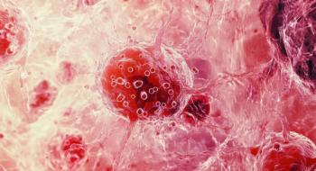
FIGURE 4
Shave Biopsy Revealed Lichenoid Interface Dermatitis, Numerous Eosinophils, and Prominent Pseudoepitheliomatous Hyperplasia
PNP is a rare syndrome that was first described by Anhalt in 1990; approximately 150 cases have been reported to date.[6] The clinical features vary greatly from patient to patient, but in all reported cases the patients developed severe treatment-resistant stomatitis. Unlike other types of pemphigus, PNP may involve not only stratified epithelium but also the columnar epithelium of the lungs. Pulmonary involvement, which is seen in approximately 30% of cases, manifests as obstructive lung disease, progresses to bronchiolitis obliterans, and almost always results in death.[7] Thus, shortness of breath and hypoxia even in the setting of a clear chest X-ray are ominous signs and may be the first indication of pulmonary involvement.
Skin findings are polymorphous in nature and appear to change as the disease progresses.[1] The skin lesions can appear as blisters that rupture and leave erosions, like those seen in bullous pemphigus, or they may appear as lichenoid eruptions. An important difference between PNP and pemphigus vulgaris is that PNP lesions are often seen on the palms and soles, while it would be very unusual to see lesions in these locations in pemphigus vulgaris.
FIGURE 5
Direct Immunofluorescent Staining for IgG Is Appreciated Along the Surface of the Epidermal Keratinocytes
The histopathology of PNP lesions is as varied as the clinical features of the disease. Biopsy specimens from areas of stomatitis are often misleading, since the mucosal disease is typically severe but yields nonspecific inflammatory changes, as seen in our patient. In such cases, it is helpful to biopsy perilesional epithelium, as this is likely to demonstrate suprabasilar acantholysis of the oral mucosa. Suprabasilar acantholysis is also seen in biopsy specimens of intact cutaneous blisters, while keratinocyte necrosis and lymphocytic infiltration into the epidermis can be seen in non-blistering lesions. The diverse pathological findings are likely secondary to the fact that PNP is presumed to be the result of an antitumor immune response involving a combination of both humoral and cell-mediated immunity.[1] It is this spectrum of clinical findings and histopathology that leads to a broad differential diagnosis as well as a delay in diagnosis-and subsequently a delay in treatment, due to the need to wait for the more definitive immunofluorescence results.
Indirect immunofluorescence (IIF) assists in differentiating PNP from pemphigus vulgaris and pemphigus foliaceous. The pemphigus vulgaris antigen and the pemphigus foliaceous antigen are only expressed in stratified squamous epithelia. Patients with PNP have antibodies present in their serum that cross-react with desmoplakins, which are present in all epithelial structures and even in some nonepithelial structures. Therefore, the serum of a patient with PNP will react via IIF with both the stratified squamous epithelium of monkey esophagus and the transitional epithelium of mouse bladder. It is estimated that this serological test is 75% sensitive and 83% specific.[1]
There are no treatment guidelines for PNP, but theoretically three treatment strategies are available.[5] These options include: treatment of the underlying malignancy, treatment of the autoimmune process by reducing serum levels of autoantibodies, and suppression of the immune system. When PNP is associated with benign tumors such as Castleman tumors and thymoma, surgical resection is indicated and is associated with an improvement or complete resolution of symptoms, as well as a decline in the titer of circulating autoantibodies.[4] However, resection is not possible in most cases that are driven by malignancy, and the course of the disease is progressive and independent of the underlying malignancy. In most cases, PNP progresses and leads to death from sepsis, gastrointestinal bleeding, multiorgan failure, or respiratory failure within weeks to two years of diagnosis.
While corticosteroids are the mainstay of therapy, additional immune-modulating agents are added for patients who do not respond to corticosteroids alone. Multiple agents, including gold, azathioprine, cyclosporine, methotrexate, mycophenolate mofetil, cyclophosphamide, intravenous immunoglobulins, and rituximab, have been utilized with varying results. No therapeutic regimen has shown consistent efficacy. One case series demonstrated some response to rituximab in five out of six cases, with the most improvement seen in skin lesions but only minimal response in oral lesions.[8] Additional case reports show a 50% success rate with rituximab.[5] However, it is important to keep in mind that rituximab also carries a black-box warning for severe, sometimes fatal, mucocutaneous reactions, including Stevens-Johnson syndrome, which can appear similar to the lesions seen in PNP. Rituximab has also been reported to cause lung injury. Our case and treatment dilemma are unique in that the patient began to experience oral, ocular, and dermal lesions, along with shortness of breath, while receiving rituximab.
The biggest medical-legal pitfall in the management of PNP is failure to make a prompt diagnosis. Our case highlights the need for continuous scrutiny of diagnoses, especially when treatments expected to be beneficial yield a suboptimal clinical response; the case also showcases the difficult decisions regarding treatment of PNP in a patient receiving rituximab.
Review of Mucocutaneous Paraneoplastic Syndromes
TABLE 2
Mucocutaneous Paraneoplastic Syndromes Associated With Hematologicc Malignancies
PNP is an example of a mucocutaneous paraneoplastic syndrome (MCPS). These syndromes comprise a group of dermatoses that exhibit variable morphology, pathology, and etiology, and that may be associated with solid tumors or hematological malignancies. Here we focus on the MCPS associated with hematological malignancies (
Dermal depositions
Amyloidosis is characterized by purpura, papules, hyperpigmentation, macroglossia, tongue papules, periocular purpura, and alopecia. The papules are formed from dermal depositions of immunocyte amyloid.[10] Of patients with primary amyloidosis, 13% to 26% have or will develop multiple myeloma. A biopsy of clinically involved skin will yield a diagnosis in almost 100% of patients. Paraneoplastic amyloidosis carries a poor prognosis, with a mean survival of less than 1 year.[9]
Neutrophilic dermatoses
Pyoderma gangrenosum and Sweet syndrome are paraneoplastic dermatoses that are associated with hematological malignancy. Infiltration of the dermis by mature neutrophils is the key pathological change in these syndromes. Pyoderma gangrenosum starts as a painful papule or pustule that develops into a nodule; this then ulcerates and forms violaceous borders with purulent exudates and a necrotic base. Bullous or atypical pyoderma gangrenosum is associated with an underlying hematological malignancy (acute myelogenous leukemia, myelodysplasia, myeloproliferative disorders, and multiple myeloma) in 7% of cases.[11] Treatment is aimed at the underlying malignancy; systemic corticosteroids have also been efficacious.[12]
Paraneoplastic Sweet syndrome is most commonly associated with acute myelogenous leukemia, but it has also been reported with solid tumors of the genitourinary organs, the breast, and the gastrointestinal tract.[11] Much as with idiopathic Sweet syndrome, manifestations may include erythematous, painful, raised cutaneous plaques that occur primarily on the upper extremities, face and neck; pyrexia; neutrophilia; and a variety of extracutaneous findings. Cytopenias and immature cells in the peripheral blood may be seen with paraneoplastic Sweet syndrome. It is thought to be secondary to hypersensitivity, and many patients respond promptly to corticosteroid therapy.[13]
Papulosquamous disorders
Papulosquamous disorders are characterized by large (plaques) and small (papules) raised skin lesions.[9] This type of paraneoplastic dermatosis is commonly seen in the setting of solid tumors; however, acquired ichthyosis and pruritis have been reported in patients with hematological malignancies.
Acquired ichthyosis involves the skin of the extensor surfaces of the extremities and the trunk. The keratinized skin lesions are rhomboid scales with free edges. Ichthyosis is most commonly seen in male patients with Hodgkin lymphoma,[10] but it has been described in patients with carcinoma of the breast, lung, or cervix; Kaposi sarcoma; leiomyosarcoma; and multiple myeloma.[9] The diagnosis is made with a skin biopsy, which demonstrates hyperkeratosis with a decreased or absent granular layer, similar to what is seen in inherited forms of ichthyosis vulgaris. The paraneoplastic skin lesions tend to parallel the course of the underlying neoplasm, so the best treatment is that aimed at the underlying malignancy. Topical lubricating agents and keratolytics are generally only helpful with symptom control and not with lesion regression.[14]
Pruritis of unknown origin is a persistent, daily itchiness that can lead to excoriations, prurigo nodularis, and lichen simplex chronicus. An underlying malignancy is the etiology in 11% of pruritis patients. Polycythemia vera is the malignancy most commonly associated with pruritus, followed by Hodgkin lymphoma, cutaneous T-cell lymphoma, Szary syndrome, leukemia, and a variety of solid tumors. In 40% of patients with meningioma, astrocytoma, and cerebral glioma, pruritis localized to the nares is seen.[10]
Reactive erythemas
Reactive erythemas are a group of skin conditions that are characterized by red skin, which may appear as flat macules and patches, expanding annular lesions, or erythema with marked scaling. Erythroderma and exfoliative dermatitis is associated with malignancy in 8% to 23% of cases.[14] It is most often linked to lymphoma (cutaneous T-cell and Hodgkin) or leukemia, but it has also been seen in cases of cancer of the uterus, lung, stomach, prostate, thyroid, liver and larynx.[11] The manifestations can include generalized erythema with or without scaling, alopecia, nail dystrophy, lymphadenopathy, pruritis, fever, eosinophilia, and leukocytosis. Skin changes are typically seen prior to the diagnosis of malignancy. Repeat skin biopsies are likely, since the findings are frequently nonspecific, but biopsy specimens can show a dense perivascular lymphocytic infiltrate.[15] It is thought that the interactions of cytokines with cellular adhesion molecules are related to the formation of erythroderma.[9] Treatment is targeted at the underlying malignancy, since the course of erythroderma parallels the course of the malignancy. Lukewarm soaks, narrow-band ultraviolet B (UVB) phototherapy, topical emollients and corticosteroids, as well as antihistamines, are also indicated.
Erythromelalgia is another paraneoplastic reactive erythema. Adult-onset erythromelalgia is related to a myeloproliferative disorder with thrombocytosis in 20% of cases,[10] and its onset is generally several months prior to the diagnosis of malignancy. Erythromelalgia is defined by severe attacks of burning pain, erythema, and warmth of the distal extremities that can be relieved by cold exposure and/or elevation of the extremities. As a consequence of intravascular platelet activation and aggregation, the distal arterioles become occluded, which is why a daily 500-mg dose of acetylsalicylic acid can alleviate symptoms.[9]
Vasculitis
Inflammation of blood vessels, which may lead to necrosis, defines vasculitis. The most common paraneoplastic vasculitis is leukocytoclastic vasculitis. Associated cutaneous lesions are polymorphous and include nonblanchable purpura, erythematous papules and nodules, ulcers, and pruritic plaques. Patients may also present with abnormal blood counts, lymphadenopathy, splenomegaly, and joint involvement. Approximately 18% of patients have an underlying malignancy. Leukocytoclastic vasculitis involves the small vessels of the upper dermis, and it is associated with hairy cell leukemia, chronic myelogenous leukemia, myeloma, lymphoma, myelodysplastic syndrome, and non–small-cell lung cancer.[9] Skin biopsies demonstrate leukocytoclasia, endothelial swelling, fibrinoid necrosis of the vessels, and extravasation of erythrocytes into the surrounding dermis. Paraneoplastic vasculitis tends to mirror the state of the underlying malignancy, so initial treatment should target the driving process. Improvement has also been seen with the use of methylprednisolone, prednisone, dapsone, colchicine, methotrexate, azathioprine, and IVIg[15]; however, patients who are over 50 years of age and who present suddenly do not tend to respond to therapy.[10]
Vesiculobullous disorders
REFERENCE GUIDE
Therapeutic Agents
Mentioned in This Article
Acetylsalicylic acid
Azathioprine
Colchicine
Cyclophosphamide
Cyclosporine
Dapsone
Prednisone
Gold
Methotrexate
Methylprednisolone
Mycophenolate mofetil
Rituximab (Rituxan)
R-CHOP (rituximab, cyclophosphamide, doxorubicin, vincristine, and prednisone)
Voriconazole (Vfend)
Brand names are listed in parentheses only if a drug is not available generically and is marketed as no more than two trademarked or registered products. More familiar alternative generic designations may also be included parenthetically.
While the primary cutaneous lesions in vesiculobullous disorders are blisters, the appearance of these disorders is varied. PNP typically resembles erythema multiforme, with polymorphous pruritic papules and blisters as well as painful cutaneous and mucosal erosions. The details of PNP are described in the above case report. Pemphigus vulgaris, on the other hand, manifests as large intraepidermal bullae of the skin with oral blisters and erosions, and pemphigus foliaceus is characterized by superficial crusted erosions.
Pemphigus vulgaris has been reported in patients with Hodgkin and non-Hodgkin lymphoma, Kaposi sarcoma, chronic lymphocytic lymphoma, breast cancer, and thymic cancer. The palms and soles are typically not involved in paraneoplastic pemphigus vulgaris, which helps to distinguish this syndrome from PNP.[1] The treatment of pemphigus vulgaris is similar to that of PNP in that a wide variety of immune-modulating agents have been utilized with varying effect.
Summary
Mucocutaneous paraneoplastic syndromes can occur before, after, or at the time of the cancer diagnosis; they can also be idiopathic entities. It is our job as physicians to recognize these conditions and to screen for a new or recurrent malignancy when they are identified. Prompt treatment of any underlying malignancy provides the patient with the best chance for improvement in most MCPS-except for PNP, which does not tend to mirror the neoplastic process. Overall, the presence of a MCPS is associated with a poor prognosis. As the general population lives longer and more cancers are diagnosed, it can be expected that the incidence of paraneoplastic conditions will increase.
Financial Disclosure: The authors have no significant financial interest or other relationship with the manufacturers of any products or providers of any service mentioned in this article.
References:
REFERENCES
1. Anhalt GJ. Paraneoplastic pemphigus. Adv Dermatol. 1997;12:77-96; discussion 7.
2. Barnadas M, Roe E, Brunet S, et al. Therapy of paraneoplastic pemphigus with Rituximab: a case report and review of literature. J Eur Acad Dermatol Venereol. 2006;20:69-74.
3. Anhalt GJ, Kim SC, Stanley JR, et al. Paraneoplastic pemphigus. An autoimmune mucocutaneous disease associated with neoplasia. N Engl J Med. 1990;323:1729-35.
4. Zhu X, Zhang B. Paraneoplastic pemphigus. J Dermatol. 2007;34:503-11.
5. Hoque SR, Black MM, Cliff S. Paraneoplastic pemphigus associated with CD20-positive follicular non-Hodgkin's lymphoma treated with rituximab: a third case resistant to rituximab therapy. Clin Experiment Dermatol. 2007;32:172-5.
6. Van Rossum MM, Verhaegen NT, Jonkman MF, et al. Follicular non-Hodgkin’s lymphoma with refractory paraneoplastic pemphigus: case report with review of novel treatment modalities. Leukemia Lymphoma. 2004; 45:2327-32.
7. Nousari HC, Deterding R, Wojtczack H, et al. The mechanism of respiratory failure in paraneoplastic pemphigus. N Engl J Med.1999;340:1406-10.
8. Allen KJ, Wolverton SE. The efficacy and safety of rituximab in refractory pemphigus: a review of case reports. J Drugs Dermatol. 2007;6:883-9.
9. Cohen PR, Kurzrock R. Mucocutaneous paraneoplastic syndromes. Sem Oncol. 1997;24:334-59.
10. Silvestris N, Chetta G, Sasso N, et al. Mucocutaneous paraneoplastic syndromes (MCPS). A clinical-pathologic review. J Exp Clin Cancer Res. 1998;17:453-64.
11. Cohen PR. Cutaneous paraneoplastic syndromes. Am Fam Physician. 1994;50:1273-82.
12. Lamet S, Bracke A, Geluykens E, et al. Medical and surgical management of paraneoplastic pyoderma gangrenosum--a case report and review of the literature. Acta Clinica Belgica. 2010;65:37-40.
13. Devita VT, Hellman S, Rosenberg SA. Cancer principles & practice of oncology. 7th ed. Philadelphia: Lippincott Williams & Wilkins; 2005.
14. Boyce S, Harper J. Paraneoplastic dermatoses. Dermatol Clin. 2002;20:523-32.
15. Pelosof LC, Gerber DE. Paraneoplastic syndromes: an approach to diagnosis and treatment. Mayo Clin Proc. 2010;85:838-54.
16. Cianchini G, Corona R, Frezzolini A, et al. Treatment of severe pemphigus with rituximab: report of 12 cases and a review of the literature. Arch Dermatol. 2007;143:1033-8.
17. Cummins DL, Mimouni D, Tzu J, et al. Lichenoid paraneoplastic pemphigus in the absence of detectable antibodies. J Am Acad Dermatol. 2007;56:153-9.
18. Heizmann M, Itin P, Wernli M, et al. Successful treatment of paraneoplastic pemphigus in follicular NHL with rituximab: report of a case and review of treatment for paraneoplastic pemphigus in NHL and CLL. Am J Hematol. 2001;66:142-4.
19. Kurzrock R, Cohen PR. Cutaneous paraneoplastic syndromes in solid tumors. Am J Med. 1995;99:662-71.
20. Maverakis E, Goodarzi H, Wehrli LN, et al. The etiology of paraneoplastic autoimmunity. Clin Rev Allergy Immunol. 2011 Jan 19. [Epub ahead of print]
21. McLean DI. Cutaneous paraneoplastic syndromes. Arch Dermatol. 1986;122:765-7.
Articles in this issue
over 14 years ago
Recommendations for Women With Lobular Carcinoma In Situ (LCIS)over 14 years ago
PARP Inhibitors in Breast Cancer: BRCA and Beyondover 14 years ago
Obesity and Breast Cancerover 14 years ago
The Obesity and Breast Cancer Connection: Advancing the Agendaover 14 years ago
PARP Inhibitors: the Story is Still Unfoldingover 14 years ago
The Challenges of Treating Lobular Carcinoma In Situover 14 years ago
Lobular Neoplasia: How to Manage With Partial Understandingover 14 years ago
PARP Inhibitors and Their Evolving Role in Breast CancerAdvertisement
Related Content
Advertisement


Advertisement
Advertisement
Trending on CancerNetwork
1
Sigvotatug Vedotin Does Not Significantly Improve Survival in NSCLC Trial
2
Cellular Therapy and Antibody-Drug Conjugates Shape Day 1 of ASCO Breakthrough 2026
3
Offering Hope and Support Toward the Cancer Survivorship Journey
4
AI and MRD Take Center Stage on Day 2 of ASCO Breakthrough 2026
5