
|Articles|April 1, 1997
- ONCOLOGY Vol 11 No 4
- Volume 11
- Issue 4
Researchers Corral Millions of Microscopic Membranes
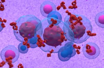
Scientists at Stanford University have done the next best thing to packaging living cells in individual boxes for study.
Advertisement
Scientists at Stanford University have done the next best thing to packagingliving cells in individual boxes for study.
Borrowing microfabrication techniques from electrical engineering, theresearchers have created a specially prepared surface that holds millionsof cell-sized squares, or "corrals," composed of an artificialmembrane that closely mimics the surface of living cells. According tothe researchers, the ability to work with these independent membranes thatare uniform in size and fixed in space makes many new experiments possible.
The work, coauthored by graduate student Jay T. Groves, chemistry ProfessorSteven G. Boxer, and Ginzton Laboratory research associate Nick Ulman,is reported in the January 31st issue of Science.
According to the scientists, not only are the micro-membranes likelyto become an important research tool but the system also could serve asthe basis for improved cell and drug screening methods because it is ideallysuited for automation.
Determining the Structure of Membrane Proteins
One possible application of these micromembranes is as a tool for determiningthe structure of membrane proteins.
One powerful method for determining the structure of proteins is x-raycrystallography. For this method to work, however, researchers must purifyand crystallize the material. This has proven very difficult for proteinsassociated with membranes because they cannot be easily separated fromthe membrane material.
According to Boxer, the new method may help overcome this difficulty.Many associated proteins can move freely on a membrane's surface. In previouswork, the researchers demonstrated that they can use electric fields toconcentrate such proteins against two-dimensional membrane boundaries.It may be possible to both concentrate and crystallize such proteins byapplying an intense electrical field. If so, the method could be used withexisting techniques to determine the two-dimensional structure of groupsof membrane proteins and with x-ray crystallography to identify the three-dimensionalstructures of some of these compounds.
Potential Biomedical Application
A potential biomedical application is cell screening of the type requiredfor leukemia patients. Doctors must closely monitor the different kindsof cells in a leukemia patient's blood. Using a small wafer holding millionsof micropatches that have been seeded with proteins that bind to differentkinds of cells, it should be possible to obtain measurements of the relativenumbers of different cell types by simply covering the wafer with blood,washing it off, and counting the cells that remain stuck to different patches.Not only could this method identify and separate different kinds of cells(like current methods), but also it potentially could measure how wellthe cells are functioning, Groves suggested.
In a similar fashion, the technique might be used to screen for drugs,such as channel blockers, that interact with receptors on a cell's surfaceand interior membranes, the researchers said.
"We have developed something new at the interface between cellscience, chemistry and electrical engineering that has widespread potentialin many areas," Boxer said.
Articles in this issue
about 29 years ago
Book Review: Physicians' Guide to the Internetabout 29 years ago
Study of Protein Blocking Normal Cell Death May Improve Cancer Drugsabout 29 years ago
An Overview of Adenocarcinoma of the Small Intestineabout 29 years ago
Roswell Park to Administer "Old" Drug in a New Wayabout 29 years ago
NY Lt. Governor Calls for a Halt to HMO AbusesAdvertisement
Related Content
Advertisement


Advertisement
Advertisement
Trending on CancerNetwork
1
Analyzing ADC Options Across Metastatic TNBC
2
Arlo-Cel Produces Deep Responses in Pretreated R/R Multiple Myeloma
3
From Conference to Practice: Top 5 Takeaways From EHA 2026
4
Pembrolizumab Combo Shows Efficacy in Pediatric High-Risk Hodgkin Lymphoma
5