|Articles|March 1, 1996
- ONCOLOGY Vol 10 No 3
- Volume 10
- Issue 3
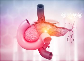
Treatment of Pancreatic Cancer: Current Limitations, Future Possibilities
Author(s)Harold J. Wanebo, MD
Drs. Blackstock, Cox, and Tepper have outlined some salient aspects of the management of pancreatic cancer. I agree with most of their comments, and will address some issues from my own perspective, colored largely by a symposium on cancer of the pancreas held in Newport, Rhode Island, in July 1994. This gathering of a large nucleus of investigators with a major interest in pancreatic cancer provided some additional insights that I will explore in my commentary and that largely complement the points made by Blackstock et al. Among other issues, my remarks will focus on: (1) the use of molecular markers for diagnosis and treatment, (2) preoperative chemoradiation, and (3) some surgical considerations that still generate controversy; ie, the extent of resection.
Advertisement
Drs. Blackstock, Cox, and Tepper have outlined some salient aspectsof the management of pancreatic cancer. I agree with most of theircomments, and will address some issues from my own perspective,colored largely by a symposium on cancer of the pancreas heldin Newport, Rhode Island, in July 1994. This gathering of a largenucleus of investigators with a major interest in pancreatic cancerprovided some additional insights that I will explore in my commentaryand that largely complement the points made by Blackstock et al.Among other issues, my remarks will focus on: (1) the use of molecularmarkers for diagnosis and treatment, (2) preoperative chemoradiation,and (3) some surgical considerations that still generate controversy;ie, the extent of resection.
The survival rate from cancer of the pancreas is 5% overall [1],and even in patients with locally resectable pancreatic cancer,surgical resection has been reported to yield 5-year survivalrates no better than 10% to 25% [2]. In the National Cancer DataBase report involving 17,970 patients with pancreatic cancer,the 5-year survival rate was 12% in patients with resectable pancreaticcancer, opposed to 4% in those with unresectable disease [2].Although several groups have recorded higher survival rates (19%to 24%) [3,18-21], these are still exceptions and not therule. The higher survival rates may reflect more selective stagingand resectability criteria at some centers, among other factors.
In resected patients, factors associated with a significantlyshorter median survival include tumor size more than 2 cm, bloodvessel invasion, and lymph node metastasis [3]. The absence orpresence of nodal metastasis is the single best predictor of survival.Patients without nodal metastases have a median survival of 56months, as compared with 11 months for those with metastatic disease(P less than .05) [3].
Molecular Pathology and Chromosomal Abnormalities
Recent studies of molecular pathology and chromosomal abnormalitiesin pancreatic cancer have provided additional information aboutthe pathogenesis of this cancer beyond that afforded by classicalpathology. Allison and colleagues [4] measured the DNA contentof pancreatic cancers in patients who had undergone pancreaticoduodenectomyand determined that 60% of primary tumors were aneuploid. Ploidywas the single most important predictor of long-term survival(10.5 months for aneuploid tumors vs 25 months for diploid tumors)[4].
Direct chromosomal analysis has yielded additional clues to thespecific genes involved in the pathogenesis of pancreatic cancer.Studies using a panel of molecular probes specific for each chromosomearm have provided techniques for precise allotype mapping, allowingone to survey gene pairs for allelic loss. Studies by Seymouret al [5] and Hahn et al [6] showed high frequencies of loss at1p, 6p, 6q, 8p, 9p, 10p, 10q, 12p, 12q, 17p, 18q, 21q, and 22q. The loss of one allele of the tumor- suppressor gene resultsin the loss of gene function if the second copy is mutated. Thus,the tumor-suppressor gene p53, located on the short arm of chromosome17 (17p), was lost in 95% of the cancers studied by Hahn et al[6]. The "deleted in colon cancer" tumor-suppressorgene (DCC) located on the long arm of chromosome 18 (18q) anda recently characterized multiple tumor suppressor 1 (MTS 1) gene,which resides on chromosome 9p, were lost in 88% and 76% of thecancers, respectively.
In addition to loss of tumor-suppressor genes, K-ras abnormalitiesand related oncogenes are commonly expressed in pancreatic cancer.K-ras, H-ras, and N-ras encode G proteins, which are involvedin signal transduction [7]. Point mutations in codons 12, 13,and 61 of K-ras inactivate the protein product by interferingwith its GTPase activity [7]. Approximately 80% of pancreaticcancers contain activating point mutations in codon 12 [8]. Themutated K-ras expressed in pancreatic cancer was more prevalentin tumors obtained from smokers than from nonsmokers (88% vs 68%),suggesting that carcinogens in cigarette smoke may be relatedto the K-ras mutations in these patients. The finding of K-rasmutations in hyperplastic "pancreatic duct lesions"appears to be analogous to the increased prevalence of K-ras mutationsamong intermediate adenomas in the model of colorectal tumor progression[9]. This finding suggests that these duct lesions may be precursorsto pancreatic cancer.
Screening for K-ras Mutations Has Potential
It may be possible to take advantage of these abnormalities inscreening for early disease because K-ras mutations are virtuallyrestricted to a single codon (codon 12). An assay based on identificationof K-ras mutations, which appears to occur prior to the developmentof invasive cancer, should be able to detect pancreatic cancersand offer sensitive methods for screening.
Tada et al collected pancreatic juice endoscopically from patientswith pancreatic cancer and from control patients with chronicpancreatitis or choledocholithiasis [10]. K-ras mutations weredetected in the pancreatic juice from all six cancer patientsstudied but from none of the controls. Of interest, K-ras mutationscould also be detected in circulating cells in the peripheralblood of some patients with pancreatic cancer [10].
Caldas et al have extended these studies and screened for K-rasmutations in the stools of patients with clinical pancreatitis,cholangiocarcinoma, and pancreatic cancer [11]. They found K-rasmutations in stool specimens from nine patients, six of whom hadpancreatic cancer. In five cases, mutations found in the pancreatictumor itself were the same as those found in the stool. In theremaining four patients, the mutations identified were identicalto those isolated from intraductal lesions present in the resectedpancreatic specimen. These data suggest that mutations found inpancreatic cancers and intraductal pancreatic lesions may be detectedin the stool [11]. These researchers are currently testing forK-ras in stool specimens from a large number of patients; thisapproach may have potential as a screening test for early pancreaticcancer.
As Blackstock et al discuss, K-ras may represent a target fortherapeutic intervention using farnesyl transferase inhibitors(FTIs). These compounds inhibit attachment of the 15-carbon isoprenylgroup, which is necessary for attachment of ras with the cellmembrane and for ras-mediated transformation. By preventingthe joining of the mutant K-ras with the cell membrane, FTIs mayinterrupt signals leading to cell proliferation in establishedpancreatic cancer.
Relationship of p53 Mutations to Pathogenesis
Tumor-suppressor gene abnormalities also play an important rolein the pathogenesis of pancreatic cancer. Mutations of p53 arefound in over half of primary pancreatic cancers [12,13] and correlatewell with allelic loss of 17p. Thus, p53 mutations may precedethe development of invasive cancer. Several groups have foundoverexpression of p53 product, a marker for p53 mutations, inintraductal lesions in the pancreas [14,15]. The expression ofmutated p53 and K-ras in the same tumors suggests some associationbetween these two gene abnormalities. There also appears to bea correlation of mutated p53 with tumor grade and decreased survivalin patients with pancreatic cancer [15].
Potential Impact of New Diagnostic Technologies
It is obvious that the greatest impact on survival in pancreaticcancer will probably come from earlier diagnosis. New technology,such as fast spiral CT using dynamic intravenous contrast, hasresulted in high-resolution images of small masses, which hasimproved the accuracy of vascular staging [16]. Endoscopic retrogradecholangiopancreatography (ERCP) with improved cytology, brushes,and biopsy forceps should enhance preoperative diagnosis of thismalignancy. Endoscopic ultrasound also may help detect small lesionsand determine the depth of invasion and vascular involvement.The potential for vastly improved MRI images with three-dimensionalreconstruction (MRI with cholangiopancreatography) may permitearlier diagnosis and management of these patients.
Unfortunately, screening for pancreatic cancer is not yet feasible.A Japanese screening study of 10,162 persons over 40 years ofage with Ca 19-9 and elastase-1 or ultrasonography revealed onlyfour cases (P = 0.042) of pancreatic cancer, includingonly one patient who was operated on for cure [17].
When pancreatic cancer is suspected, imaging with CT or the moresensitive spiral CT, plus CT-directed FNA cytology and ERCP withbrushings should result in a definitive diagnosis. Although CT-guidedFNA is probably now the most common method of performing the biopsy,it is controversial because of the potential risk of peritonealcontamination [30].
Evans et al have examined the effect of preoperative CT-guidedFNA on the cytologic washings obtained a median of 3 weeks later[30]. Among 60 consecutive patients who had peritoneal lavagefor cytology, 49 (82%) had CT-guided FNA, and 11 did not. Peritonealwashings yielded four patients (6.6%) with positive cytology;3 of 49 patients (6.1%) had undergone preoperative CT-FNA and1 of 11 (9.1%) did not. All four patients developed metastaticdisease about 5 months later, suggesting that this was simplydue to advanced disease rather than being secondary to the FNA.
The value of laparoscopy as a staging technique has long beenchampioned by Warshaw et al [32], as well by Evans and the M.D.Anderson group [30]. Laparoscopy permits direct visualizationof the liver, the serosal surface, and provides opportunity forperitoneal cytology. With care, one can examine the porta hepatis,the peripancreatic region, and in some cases examine and biopsyaccessible peripancreatic nodes. Laparoscopy also permits placementof a tube jejunostomy and cholecystojejunostomy as needed, ifpreoperative therapy is planned.
Extent of Resection Still Debated
Currently, surgical resection is the only potentially curativetreatment for pancreatic cancer. The actuarial 5-year survivalrate at Johns Hopkins was 26% in 195 patients who underwent pancreaticoduodectomyfor carcinoma of the head of the pancreas [3,18]. This is similarto survival rates reported in other series, including those ofGeer and Brennan [19] at Memorial Sloan-Kettering and Trede etal [20] in Germany.
Some of the factors that have been shown to influence survivalinclude:
- Tumor size (median survival, 30 months for tumors less than2 cm vs 11 months for those more than 2 cm) [18]
- Vessel invasion (median survival, 39 months in patients withvessel invasion vs 11 months in those without invasion)
- Lymph node involvement (median survival, 56 months if negativevs 12 months if positive).
- Blood loss also appears to have an impact on survival. Inone study, median survival was 25 months if fewer than 3 unitsof blood were given, as compared with 10 months if 3 or more unitswere transfused [18].
Mortality from the Whipple procedure has improved markedly. Inthe Hopkins experience, mortality has changed from 20% over 2decades ago to 2.5% in the last 326 patients. In the more recentseries, no deaths occurred in 145 consecutive operative procedures[3,18,21].
The question of the extent of resection--ie, whether to performa total or subtotal Whipple procedure or a more aggressive regionalresection--is still a matter of debate. The Mayo Clinic foundno survival difference between patients treated with partial vstotal pancreatectomy [22]. In contrast, a review by Farley etal found that the long-term survival rate following total pancreatectomyin 11 series involving 643 patients was 8.5%, as compared witha rate of 11.5% in 1,491 patients treated with partial pancreatectomy[22].
Studies of more extended resections (ie, the regional pancreatectomyof Fortner [23] and a similar procedure by Sindelar [24]) havefailed to confirm a true benefit from these more aggressiveprocedures [22]. However, Japanese authors, such as Ishikawa etal [25], Nakagawa [26], and Manabe et al [27], have claimed thatregional pancreatectomy prolongs survival, and Kairaluoma et al[28] of Finland have asserted that extended pancreatic resectionaffords some benefit. On the other hand, other researchers,such as Satake and colleagues [29], have found that extended lymphadenectomyconfers no survival advantage in patients with T1 cancers.
One further point about the extent of surgery (radical surgeryvs pylorus-preserving pancreaticoduodenectomy) deserves comment.The Lahey group have demonstrated no difference in the tumor involvementof resection margins in patients undergoing pylorus-preservingsurgery vs those undergoing proximal or total pancreatectomy [35].Moreover, the incidence of marginal ulceration and bleeding gastritisafter pylorus-preserving procedures was lower than with standardpancreaticoduodenectomy with hemigastrectomy or antrectomy andvagotomy. The 5-year survival rate was 6.1% in patients who underwentpylorus-preserving resection for pancreatic duct cancer and mediansurvival was 18 months [35]. This is in contrast to the 19% to26% recorded survival rate for the Whipple procedure or more extendedresections [3,18,21,36]. Again, such differences may reflect patientstage and selection rather than something unique to the surgicalprocedure per se.
Preoperative Chemoradiation and Intraoperative External-BeamRadiation
Because of the high rate of potential nodal involvement (up to50% risk of finding involved nodes within the resected specimen),the addition of therapy to augment surgical resection would beappear to be of value. Several authors have addressed the potentialbenefits of these approaches [30,31].
Evans et al at M.D. Anderson have explored the value of preoperativechemoradiation prior to pancreatic resection and intraoperativeexternal-beam radiotherapy [30,33]. An updated study encompassed51 patients who completed chemoradiation [30]. At the restagingof 13 patients, 3 (25%) were found to have metastatic diseaseand did not undergo surgery. A total of 38 patients underwentlaparotomy, and resection was not performed in 8 patients becauseof metastases (3 patients) or locally unresectable disease (5patients). Of 30 patients undergoing potentially curative pancreaticoduodenectomy,26 received external-beam intraoperative radiotherapy (IORT) followingtumor removal; there were no operative deaths from myocardialinfarction.
Median survival for the entire group of 51 patients was 11.7 months(18.6 months for the 30 resected patients and 6.7 months for theunresected patients). At a median follow-up of 15 months, 22 patientshad died (18 from their cancer), 1 was alive with disease, and7 (23%) showed no evidence of disease. Of the patients who diedof disease, 83% had metastases to the liver or lung as the firstsign of failure [33]. The median tumor sizes prior to treatmentwere 3 cm and 3 cm (anteroposterior [AP] and lateral diameters,respectively) and the median tumor shrinkage following chemoradiationwas .5 cm and .4 cm, respectively.
There were no measurable differences in tumor size between thepatients who were able to undergo resection and those who werenot (AP tumor diameter was 3 cm and lateral, 3 cm). In none ofthe patients was preoperative radiation associated with a completeresponse; however, a grade 2B (destruction of 51% to 90% of tumorcells) or grade 3 pathologic response (more than 90% tumor celldestruction) was seen in 40% of the resected patients. There wereno positive margins at the resection line, although there wasmicroscopic evidence of cancer in the retroperitoneal margin inapproximately 15% to 20% of patients [30,33].
A similar approach has been used by Hoffmann and colleagues atthe Fox Chase Cancer Center [31]. They treated 63 patients withpreoperative radiation therapy and two 96-hour infusions of fluorouracil(1,000 mg/m²/d on days 2 to 5 and days 29 to 32) and a singlebolus of mitomycin (Mutamycin; 10 mg on day 2). Patients who hadno evidence of distant disease and were considered resectableunderwent pancreatic resection (16 Whipple procedures, 7 totalpancreatectomies, and 2 distal pancreatectomies).
Thirty-eight percent of the patients had grade 3 or 4 toxicityfrom chemoradiation, and one patient died during chemoradiationof biliary sepsis. Operative mortality in 25 patients with potentiallycurative resection was 4%; one of four patients who underwentpalliative resection died. Median survival for those undergoingpancreatic resection was 22 months and the 5-year survival ratewas 20%. Recurrence was noted in 20 patients and was primarilyin the liver in 50% of the patients.
In this protocol, 37 patients made up the Fox Chase pilot studyand 26 patients were part of a larger phase III trial run underthe auspices of the Eastern Cooperative Oncology Group (ECOG).A recent report from the senior author updated the actual ECOGphase II study [34]. This included 53 eligible patients who wereaccrued to the ECOG trial examining the efficacy of preoperativetherapy with radiation (180 cGy in 28 fractions) and two 96-hourinfusions of fluorouracil (1,000 mg/m²/d on days 2 to 5 anddays 29 to 32) and bolus mitomycin (10 mg on day 2).
There were two treatment-related deaths, and 18 patients (34%)experienced grade 4 toxicity. The incidence of toxicity was higherin those who did not undergo previous surgical staging than inthose who did. Forty-one patients underwent surgery and 23 (56%)had resection. Four patients (17%) had positive surgical margins.Median survival was 16.6 months from the time of registrationfor the patients who underwent resection. The 2-year survivalrate was 30% in the group as a whole and 33% in the 19 patientswith N0 disease. Nine of the 41 resectable patients are aliveat 30 to 33 months' follow-up.
These survival results were thought to be less favorable thanthose reported from a single institution (Fox Chase Cancer Center)using a similar preoperative regimen (P = 0.085). However,preoperative chemoradiation was considered to be a safe intereventionin the cooperative study setting.
Conclusion
This article by Blackstock, Cox, and Tepper is an informativechapter in the continuing saga of research, diagnostic, and interventionaldevelopments being made to address a formidable disease process,which is only recently beginning to yield ground. I would anticipate(and hope) that a similar chapter 3 to 5 years hence would bemore optimistic regarding outcome.
References:
1. Boring CC, Squires TS, Tong T, et al: Cancer Statistics 1994.Atlanta, American Cancer Society, 1994.
2. Niederhuber JE, Brennan MF, Menck HR: The National Cancer DataBase report on pancreatic cancer. Cancer 76:1671-1677, 1995.
3. Cameron JL, Crist DW, Sitzmann JV, et al: Factors influencingsurvival after pancreaticoduodenectomy for pancreatic cancer.Am J Surg 161:120-125, 1991.
4. Allison DC, Kallol KB, Hruban RH, et al: Pancreatic cancercell DNA content correlates with long-term survival after pancreatoduodenectomy.Ann Surg 214:648-656, 1991.
5. Seymour AB, Hruban RH, Redston M, et al: Allelotype of pancreaticadenocarcinoma. Cancer Res 54:2761-2764, 1994.
6. Hahn SA, Seymour AB, Hruban RH, et al: Multiple sites of allelicloss in pancreatic carcinoma. Gastroenterology 106:A390, 1994.
7. Barbacid M: Ras genes. Ann Rev Biochem 56:779-827, 1987.
8. DiGiuseppe JA, Hruban RH, Offerhaus GJA, et al: Detection ofK-ras mutations in mucinous pancreatic duct hyperplasia from apatient with a family history of pancreatic carcinoma. Am J Pathol144:889-895, 1994.
9. Fearon ER, Vogelstein B: A genetic model for colorectal tumorigenesis.Cell 61:759-767, 1990.
10. Tada M, Omata M, Kawai S, et al: Detection of ras gene mutationsin pancreatic juice and peripheral blood of patients with pancreaticadenocarcinoma. Cancer Res 53:2472-2474, 1993.
11. Caldas C, Hahn SA, Hruban RH, et al: Detection of K-ras mutationsin the stool of patients with pancreatic adenocarcinoma and pancreaticductal hyperplasia. Cancer Res 54:3568-3573, 1994.
12. Scarpa A, Capelli P, Mukai K, et al: Pancreatic adenocarcinomasfrequently show p53 tumor-suppressor gene. Am J Pathol 142:1534-1543,1993.
13. Redston M, Caldas C, Seymour AB, et al: p53 mutations in pancreaticcarcinoma and evidence of common involvement of homocopolymertracts in DNA microdeletions. Cancer Res 54:3025-3033, 1994.
14. Barton CM, Staddon SL, Hughes CM, et al: Abnormalities ofp53 tumor suppressor gene in human pancreatic cancer. Br J Cancer64:1076-1082, 1991.
15. DiGiuseppe JA, Hruban RH, Goodman SN, et al: Overexpressionof p53 protein in adenocarcinoma of the pancreas. Am J Clin Pathol101:684-688, 1994.
16. Malik A: Early diagnosis of pancreatic carcinoma: Realityor oxymoron? Semin Surg Oncol 11:97-102, 1995.
17. Homma T, Tsuchiya R: The study of the mass screening of personswithout symptoms and of the screening of outpatients with gastrointestinalcomplaints or isterus for pancreatic cancer in Japan using CA19-9 and Elastase-1 or ultrasonography. Int J Pancreatol 9:119-124,1991.
18. Cameron JL: Pancreaticoduodenectomy: The Johns Hopkins experience.Semin Surg Oncol 11:114-116, 1995.
19. Geer RJ, Brennan MF: Prognostic indicators for survival afterresection of pancreatic adenocarcinoma. Am J Surg 165:68-72, 1993.
20. Trede M, Schwall G, Saeger HD: Survival after pancreatoduodenectomy.Ann Surg 211:447-458, 1990.
21. Cameron JL, Pitt HA, Yeo CJ, et al: One hundred and forty-fiveconsecutive pancreaticoduodenectomies without mortality. Ann Surg217:430, 1993.
22. Farley DR, Sarr MC, vanHeerden JA: Pancreatic resection forductal adenocarcinoma: Total pancreatectomy versus partial pancreatectomy.Semin Surg Oncol 11:124-131, 1995.
23. Fortner JG: Regional resection of the pancreas: A new surgicalapproach. Surgery 73:307-320, 1973.
24. Sindelar WF: Clinical experience with regional pancreatectomyfor adenocarcinoma of the pancreas. Arch Surg 124:127-132, 1989.
25. Ishikawa O, Ohhigashi H, Sasaki Y, et al: Practical usefulnessof lymphatic and connective tissue clearance for the carcinomaof the pancreas head. Ann Surg 208:215-220, 1988.
26. Nagakawa T: Extended radical pancreatectomy-translateral retroperitonealapproach. Presented at Cancer of the Pancreas: A Challenge ofthe Nineties, Newport, Rhode Island, July 1994.
27. Manabe T, Ohshio G, Baba N, et al: Radical pancreatectomyfor ductal cell carcinoma of the head of the pancreas. Cancer64:1132-1137, 1989.
28. Kairaluoma MI, Stahlberg M, Kiviniemi H: Pancreatic resectionfor carcinoma of the pancreas and the periampullary region. Atwenty-year experience. HPB Surg 2:57-67, 1990.
29. Satake K, Nishiwaki H, Yokomastu H, et al: Surgical curabilityand prognosis for standard versus extended resection for T1 carcinomaof the pancreas. Surg Gynecol Obstet 175:259-265, 1992.
30. Evans DB, Abbruzzese JL, Lee JE, et al: Preoperative chemoradiationfor adenocarcinoma of the pancreas: M.D. Anderson experience.Semin Surg Oncol 11:132-140, 1995.
31. Hoffman JP, Weese JL, Ahmad N, et al: Preoperative chemotherapyand radiation therapy for patients with pancreatic carcinoma withoutdemonstrable metastatic disease: The Fox Chase experience. SeminSurg Oncol 11:141-148, 1995.
32. Warshaw AL: Implications of peritoneal cytology for stagingof early pancreatic cancer. Am J Surg 161:26-30, 1991.
33. Evans D, Abbruzzese J, Lee J, et al: Preoperative chemoradiationand pancreaticoduodenectomy for the pancreas (abstract). Am SocClin Oncol 13:226, 1994.
34. Hoffman JP, Weese JL, Lipsitz S, et al: Preoperative chemoradiationfor patients with resectable pancreatic adenocarcinoma: An EasternCooperative Oncology Group (ECOG) Phase II study. Am Soc ClinOncol 14:482, 1995.
35. Leslie KA, Rossi RL, Hepp J: Pylorus-preserving pancreatoduodenectomyfor carcinoma of the pancreas. Semin Surg Oncol 11:117-123, 1995.
36. Wanebo HJ, Vezeridis M: Pancreatic cancer: An overview. SeminSurg Oncol 11:168-180, 1995.
Articles in this issue
over 30 years ago
Trends in Cancer Screening-United States, 1987 and 1992over 30 years ago
Book Review: Dr. Susan Love's Breast Book, 2nd Editionover 30 years ago
DaunoXome Shows Promise as Breast Cancer Treatmentover 30 years ago
New Agent Blocks HIV Integrase, Another Target for Anti-AIDS Therapyover 30 years ago
Xerostomia as a Complication of Cancer Treatmentover 30 years ago
Irinotecan Shown to Be Effective Against Colorectal CancerAdvertisement
Related Content
Advertisement


Advertisement
Advertisement
Trending on CancerNetwork
1
FDA Approves Afami-cel in Metastatic Synovial Sarcoma
2
The Moonlight Shift: Dr Joshua Richter on Myeloma, Myths, and the C-Word
3
Binimetinib Tablets Receive Tentative FDA Approval in Melanoma/NSCLC
4
How Are Key Lymphoma Data From ASCO and EHA 2026 Shaping Treatment?
5