

|Articles|June 2, 2008
Oncology NEWS International
- Oncology NEWS International Vol 17 No 6
- Volume 17
- Issue 6
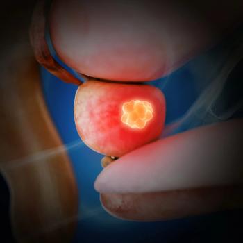
MRI can monitor the prostate for local progression after ultrasonic ablation
Author(s)Shalmali Pal, Shalmali Pal
High-intensity focused ultrasonic (HIFU) ablation is used to manage localized prostate cancer after external beam radiation therapy. But post-treatment alterations to prostate zonal anatomy hamper the assessment of local tumor progression in order to make decisions about second-line treatment. An interdisciplinary group from South Korea tested two MRI techniques for predicting local tumor progression.
Advertisement
ABSTRACT: Better sensitivity with dynamic contrast-enhanced MRI, better specificity with T2-weighted MRI with diffusion weighted imaging.
High-intensity focused ultrasonic (HIFU) ablation is used to manage localized prostate cancer after external beam radiation therapy. But post-treatment alterations to prostate zonal anatomy hamper the assessment of local tumor progression in order to make decisions about second-line treatment. An interdisciplinary group from South Korea tested two MRI techniques for predicting local tumor progression.
“Prostate cancer has a lower apparent diffusion coefficient (ADC) than noncancerous prostate tissue,” explained Chan Kyo Kim MD, and colleagues from Samsung Medical Center, Seoul. “The purpose of this study was to compare the diagnostic performance of dynamic contrast-enhanced MRI (DCE-MRI) with that of T2-weighted MRI with diffusion weighted imaging (DWI)” (Am J Roentgenol 190:1180-1186, 2008).
Between 2004 and 2006, 27 patients underwent transurethral prostate resection and endorectal HIFU. These patients had clinical stage T1-2 disease, no evidence of metastases, and a pre-ablation mean PSA of 7.25 ng/mL. PSA levels continued to rise after treatment.
Based on biopsy results, local tumor progression was found in 33% of the 162 prostate sectors. The diagnostic performance of DCE-MRI and DWI varied, with DCE-MRI delivering better
sensitivity and DWI better specificity. Accuracy rates for both readers and techniques were similar. False-positive results were higher for DCE-MRI vs DWI.
In an interview with ONI, Dr. Kim said that “DCE-MRI and DWI are part of the routine protocol for evaluating post-HIFU local tumor progression.” He added that the urologists who performed the ablation procedures did rely on imaging information as well as clinical and histopathological results.
Commenting on the study, Robert Bard, MD, associate professor of radiology, of New York Medical College, Valhalla, praised the researchers for highlighting the differences between the two MRI techniques. But despite the results in favor of MRI, the modality is not adept at distinguishing between treated and untreated tissue as well as inflammation and cancer, he said.
3D Doppler ultrasound an option
Dr. Bard said he preferred 3D Doppler ultrasound imaging in the post-ablation prostate. Sonographic signs to look for are capsule penetration and lack of abnormal vascularity for separating treated from untreated tissue, he said.
Articles in this issue
over 17 years ago
Who’s responsible for safety of outsourced drugs?over 17 years ago
New GnRH blocker degarelix quickly suppresses levels of testosteroneover 17 years ago
Evaluating lung cancer response to therapy: Thinking beyond RECISTover 17 years ago
NELSON trial sails on toward final results in 2015over 17 years ago
Novel breast probe reduces repeat surgeriesover 17 years ago
Radioactive microspheres benefit liver met ptsover 17 years ago
INTORACT trial of Torisel/Avastin in RCC is initiatedover 17 years ago
New codes needed for chronically ill ptsNewsletter
Stay up to date on recent advances in the multidisciplinary approach to cancer.
Advertisement
Related Content
Advertisement

Latest CME


Advertisement
Advertisement
Trending on CancerNetwork
1
Modifiable Risk Factors Suggest Potential for Improving Cancer Prevention
2
2026 Tandem Meetings: What’s the Latest Research in Multiple Myeloma?
3
Dato-DXd Receives Priority Review in Unresectable/Metastatic TNBC
4
Barriers to CAR T-Cell Referral and Center Access in Multiple Myeloma
5