
|Articles|April 24, 2009
- ONCOLOGY Vol 18 No 4
- Volume 18
- Issue 4
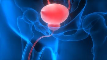
Evidence grows for value of high-field MRI in prostate cancer Rx strategy
Author(s)Karen Sandrick
VIENNA-Image-guided intensity-modulated radiotherapy, high-intensity focused ultrasound, and cryotherapy are increasing the curative treatment options for men with prostate cancer. The problem is how to determine which patients are most suitable for these therapies.
Advertisement
ABSTRACT: New treatments for prostate cancer require precise localization of disease, but how to determine which patients are most suitable for these therapies? High-field MRI could be the answe
VIENNA-Image-guided intensity-modulated radiotherapy, high-intensity focused ultrasound, and cryotherapy are increasing the curative treatment options for men with prostate cancer. The problem is how to determine which patients are most suitable for these therapies.
The new treatments for prostate cancer require precise localization of disease, but routinely used imaging techniques are not sensitive or specific enough for accurate staging. Gray-scale transrectal ultrasound has a sensitivity of 30% to 50% and specificity of 77% to 90% for local staging and tumor localization, and 1.5T MRI has a joint sensitivity and specificity of 71% to 74% for conventional anatomical prostate imaging, according to recent research.
High-field MRI could be the answer, according to a special focus session at the European Congress of Radiology 2009 (ECR SF1B).
“MR is a powerful method for imaging the prostate gland because its high spatial resolution provides excellent soft-tissue contrast, which may aid the detection and localization of malignant lesions, the evaluation of disease stage based on the extraprostatic extension of cancer, and the detection of local or distant disease recurrence after treatment,” said Jurgen J. Ftterer, MD, PhD, of the department of radiology at Radboud University Nijmegen Medical Centre in the Netherlands.
What 3T MRI adds
The signal-to-noise ratio increases almost linearly with magnetic field strength, whereas the noise remains nearly unchanged with 3T MRI. Thus, the SNR is approximately two times higher at 3T compared with 1.5T. The most significant benefit of this difference is the increase of spatial resolution, which improves the depiction of anatomical details and/or shortens acquisition time, thereby increasing patient throughput, Dr. Ftterer explained.
The chemical shift effect also increases linearly with magnetic field strength. Spectral resolution at 3T is therefore enhanced compared with 1.5T. Signal intensity and frequency dispersion are higher, which may lead to better tumor characterization. In dynamic contrast-enhanced MR imaging, the increase in SNR can be used to improve the temporal resolution of dynamic measurements. Increased temporal resolution may improve the accuracy of measurement of pharmacokinetic parameters.
Endorectal 3T T2-weighted and high spatial resolution dynamic contrast-enhanced MRI are able to identify morphological and vascular details, which may guide not only the detection and staging of disease but the direction of biopsies and treatment.
Signal intensity changes obtained during dynamic image acquisition provide an estimate of the amount of contrast material that has accumulated in lesions.
The passage of contrast into and out of lesions reflects the kinetic properties of tissue physiology, such as Ktrans and Ve values, and provides an indication of microvascular permeability and angiogenetic potential, according to B. Nicolas Bloch, MD, who discussed MR perfusion and high-resolution imaging. Dr. Bloch is an instructor in radiology at Beth Israel Deaconess Medical Center in Boston.
The high field strength of 3T MRI is particularly useful in dynamic contrast-enhanced studies of the prostate gland. “Dynamic contrast-enhanced MRI makes it possible to combine fast imaging-less than three seconds per image set-with good spatial resolution. This technique directly benefits from the increased SNR at 3T,” Dr. Ftterer said.
Drawbacks
High-field MRI has its drawbacks and challenges, however, including a quadrupling of radiofrequency power deposition compared with 1.5T, shorter T2- and longer T1-relaxation times, increased susceptibility differences, dielectric effect, and signal heterogeneity from larger B1 field variations. Using thinner slices and increasing the spatial resolution or bandwidth at a higher field may partially circumvent some of these issues, but these changes will offset some of the SNR increase gained with 3T imaging.
In addition, the loss in SNR is less than linear with voxel size due to the decreasing line width (e.g., when resolution is increased from 0.075 to 0.094 cm3, the SNR decreases between 44% and 60%). Due to radiofrequency penetration effects, it can be difficult to achieve good RF field homogeneity. Parallel imaging techniques, increased TR, reduced flip angle, and increased RF pulse duration can be used to overcome these problems, but at the expense of SNR.
Despite these difficulties, the increase in SNR provided by 3T MRI is likely to expand the potential clinical applications for evaluating the prostate, Dr. Ftterer said.
“Endorectal 1.5T MRI is a common method of evaluating men with suspected prostate cancer. But in the next few years, 3T MRI will become the standard. An SNR at 3T that is equal to the SNR at 1.5T with an endorectal coil is a big advantage,” he said.
Articles in this issue
about 22 years ago
Myelomaabout 22 years ago
Proteomics to Diagnose Human Tumors and Provide Prognostic Informationabout 22 years ago
Proteomics to Diagnose Human Tumors and Provide Prognostic Informationabout 22 years ago
Proteomics to Diagnose Human Tumors and Provide Prognostic Informationabout 22 years ago
MyelomaAdvertisement
Related Content
Advertisement


Advertisement
Advertisement
Trending on CancerNetwork
1
FDA Approves Afami-cel in Metastatic Synovial Sarcoma
2
The Moonlight Shift: Dr Joshua Richter on Myeloma, Myths, and the C-Word
3
Binimetinib Tablets Receive Tentative FDA Approval in Melanoma/NSCLC
4
How Are Key Lymphoma Data From ASCO and EHA 2026 Shaping Treatment?
5