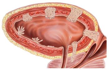
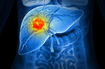
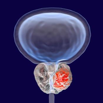
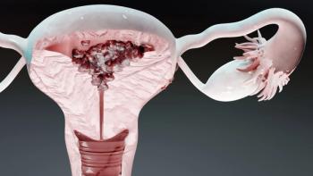
Extra-CNS imaging is most often performed using PET or PET/CT, which have been shown to be more sensitive than nonmetabolic imaging studies.[17] Similar to the CNS, the testicles are another relatively immune-privileged location. Thus, in male patients, testicular ultrasound can be considered-although it is uncertain what additional sensitivity or specificity this study adds.[15] In our practice, we limit its use to clinical scenarios where there is suspicion because of findings on radiographic imaging or clinical examination.
Bone marrow biopsy is also often performed to search for occult lymphoma. Many clinical trials require its inclusion. The literature is not clear regarding its benefit for detecting lymphoma not identified via other staging evaluations. Although a bone marrow aspiration and biopsy are commonly performed in our own practice and in other academic settings, this practice can arguably be challenged. However, the risks of inadvertently moving forward with an inappropriate therapeutic regimen may be viewed as greater than the risks of the bone marrow biopsy.
To summarize, the essential components of staging include HIV status evaluation, ophthalmologic examination, and whole-body PET/CT. Staging in immunocompetent and immunocompromised patients is essentially the same; however, in the immunocompromised population, there will be greater concern about the possibility of there being more than one underlying cause for clinical/radiographic findings, particularly if such findings are multiple. Additional studies in both immunocompetent and immunocompromised patients may be considered for either staging or prognostication; these include CSF analysis, serum LDH level, bone marrow biopsy, and testicular ultrasound. Which of these studies are utilized varies among practitioners, institutions, and countries. While not a component of diagnostic or staging studies, the creatinine clearance is an essential parameter that helps establish the feasibility of utilizing certain therapeutic regimens, including high-dose methotrexate, the cornerstone of primary CNS lymphoma treatment (since methotrexate is renally cleared).
Prognosis
Contemporary studies of large patient cohorts and epidemiologic studies have shed additional light on outcomes in patients with primary CNS lymphoma. Neither the epidemiologic reports from the Central Brain Tumor Registry of the United States (CBTRUS) nor the two most frequently utilized prognostic systems address potential differences in outcomes between immunocompetent and immunocompromised patients. The CBTRUS reveals that young age is associated with superior outcomes. In the pediatric population (≤ 19 years old), which has a very low incidence of primary CNS lymphoma, survival rates at 1, 5, and 10 years range from 83% to 88%, 74% to 78%, and 66% to 73%, respectively.[1] Among patients aged 20 to 64 years, survival rates range from 40% to 60%, 29% to 37%, and 23% to 27%.[1] In elderly patients (65 years of age or older), outcomes are even worse, with fewer than half of patients alive at 1 year. In population-based studies, estimated rates for survival at 1, 5, and 10 years in this age cohort are 33% to 48%, 13% to 24%, and 10% to 13%, respectively.[1] Retrospective evaluation of a large multi-institutional cohort of patients treated for primary CNS lymphoma demonstrated that response rate and overall survival were only affected by the treatments used and not by factors intrinsic to the patients.[18]
Two widely accepted prognostic classification systems are used in primary CNS lymphoma. These employ various criteria (Table 3); however, age and performance status are included in both scoring systems. The International Extranodal Lymphoma Study Group (IELSG) developed a prognostication system in which a number of factors significantly associated with survival on multivariate analyses are awarded points (1 point for each unfavorable factor). The aggregate score can then be used to estimate prognosis. Unfavorable factors are: age > 60 years; impaired Eastern Cooperative Oncology Group performance status of 2–4; elevated serum LDH level; elevated CSF protein level; and involvement of deep brain structures, specifically the periventricular regions, basal ganglia, corpus callosum, brainstem, and/or cerebellum. Two-year survival estimates for all patients are divided into three groups and range from 15% to 80%. In a subgroup analysis of patients who received high-dose methotrexate, the outcomes improved-ranging between 24% and 85%, depending on prognostic factors-reflecting in part the selection bias inherent in an analysis of the influence of treatment choices in a retrospective cohort of patients.[19] A second easy-to-use system, developed at Memorial Sloan Kettering Cancer Center, incorporates only age (< 50 years vs ≥ 50 years) and Karnofsky Performance Scale score (< 70 vs ≥ 70). As with the IELSG system, patients are classified into three categories, with median overall survival ranging from 8.5 years in the best category to 1.1 years in the worst. Median failure-free survival ranges from 2.0 years to 0.6 years.[20]
KEY POINTS
- Primary central nervous system (CNS) lymphoma is a malignancy most frequently encountered in elderly, HIV-positive, and posttransplant populations.
- Prebiopsy administration of corticosteroids may limit the diagnostic yield of the biopsy and thus should be avoided.
- Staging is performed to evaluate for the presence of extra-CNS and ocular involvement of lymphoma.
- Age and performance status are the most reliable prognostic markers across prognostic systems.
Although in extra-CNS lymphoma, protein expression as detected via immunohistochemistry can be predictive of outcome, no clear histologic biomarker has yet been established for primary CNS lymphoma. BCL-2 is present in most primary CNS lymphomas.[21] BCL-6 is also found in many primary CNS lymphomas, although reports on its prognostic value are conflicting.[22,23] The serum LDH level is a validated prognostic marker and serves as a component of the IELSG prognostication classification. Other serum biomarkers, such as absolute lymphocyte count, are under investigation.[24] The presence of programmed death ligand 1 (PD-L1) expression on tumor-infiltrating lymphocytes and tumor-associated macrophages has been reported in primary CNS lymphoma; however, the clinical significance of this finding has yet to be elucidated.[25] In EBV-positive primary CNS lymphoma, increased expression of PD-L1 and PD-L2 has also been noted within tumor cells. Novel PD1 and PD2 gene rearrangements have been described in primary CNS lymphoma.[26]
In addition to tumor-specific factors, markers of the overall systemic immune profile can also serve prognostic purposes. In multivariate analyses, low absolute lymphocyte count at diagnosis is associated with shorter overall survival.[24] A high neutrophil:lymphocyte ratio is associated with similar findings.[27] Although these markers are not included in formal prognostication systems, they can influence clinicians’ perceptions of prognosis. The prognosis of immunocompromised patients with primary CNS lymphoma has long been thought to be worse than that of immunocompetent patients. Most clinical trials have excluded immunocompromised patients, which has limited our understanding of this population. Retrospective studies, however, have recently provided support for similar outcomes in the HIV-negative and HIV-positive primary CNS lymphoma populations in the modern era.[18]
Conclusions
Primary CNS lymphoma is a rare primary CNS tumor associated with substantial morbidity and mortality. With an increasing incidence in the growing elderly population, its prevalence may grow. Familiarity with the clinical and radiographic presentation will help facilitate the initial diagnostic workup, decreasing the potential for nondiagnostic biopsies. It will also allow for rapid evaluation for evidence of extra-CNS disease-which, if present, would classify the lymphoma’s CNS involvement as secondary, altering therapeutic management. While the risk of relapse is thought to diminish with longer disease-free intervals, there are reports of recurrence many years out.[28,29] A firm understanding of prognosis will allow for optimal counseling of patients and will influence clinical decision making.
Financial Disclosure:The authors have no significant financial interest in or other relationship with the manufacturer of any product or provider of any service mentioned in this article.
References:
1. Ostrum QT, Gittleman HR, Xu J, et al. CBTRUS statistical report: primary brain and central nervous system tumors diagnosed in the United States in 2009-2013. Neuro Oncol. 2016;18(suppl 5):v1-v75.
2. Wu X, Andrews P, Chen VW, et al. Incidence of extra-nodal non-Hodgkin lymphomas among whites, blacks, and Asians/Pacific Islanders in the United States: anatomic site and histology differences. Cancer Epidemiol. 2009;33:337-46.
3. Shiels MS, Pfeiffer RM, Besson C, et al. Trends in primary central nervous system lymphoma incidence and survival in the U.S. Br J Haematol. 2016;174:417-24.
4. Gopal S, Patel MR, Yanik EL, et al. Temporal trends in presentation and survival for HIV-associated lymphoma in the antiretroviral therapy era. J Nat Cancer Inst. 2013;105:1221-9.
5. O’Neill BP, Decker PA, Tieu C, et al. The changing incidence of primary central nervous system lymphoma is driven primarily by the changing incidence in young and middle-aged men and differs from the time trends in systemic diffuse large B-cell non-Hodgkin’s lymphoma. Am J Hematol. 2013;88:997-1000.
6. Martinella A, Lanzafame M, Bonometti MA, et al. Neurological complications of HIV infection in pre-HAART and HAART era: a retrospective study. J Neurol. 2015;262:1317-27.
7. Penn I, Porat G. Central nervous system lymphomas in organ allograft recipients. Transplantation. 1995;59:240-4.
8. Dharnidharka VR, Webster AC, Martinez OM, et al. Post-transplant lymphoproliferative disorders. Nat Rev Dis Primers. 2016;2:15088.
9. Lukas RV, Riedell P, Horowitz PM, et al. Diagnostic evaluation in primary CNS lymphoma. Neurologist. In press.
10. Bathla G, Hegde A. Lymphomatous involvement of the central nervous system. Clin Radiol. 2016;71:602-9.
11. Partovi S, Karimi S, Lyo JK, et al. Multimodality imaging of primary CNS lymphoma in immunocompetent patients. Br J Radiol. 2014;87:20130684.
12. Nicholas MK, Collins J, Lukas RV. AIDS. In: Winn HR, editor. Youmans and Winn neurological surgery. 7th ed. Amsterdam: Elsevier; 2016.
13. Koubska E, Weichet J, Malikova H. Central nervous system lymphoma: a morphological study. Neuro Endocrinol Lett. 2016;37:318-24.
14. Malikova H, Koubska E, Weichet J, et al. Can morphological MRI differentiate between primary central nervous system lymphoma and glioblastoma? Cancer Imaging. 2016;16:40.
15. O’Neill BP, Dinapoli RP, Kurtin PJ, et al. Occult systemic non-Hodgkin’s lymphoma (NHL) in patients initially diagnosed as primary central nervous system lymphoma (PCNSL): how much staging is enough? J Neurooncol. 1995;25:67-71.
16. Grimm SA, McCannell CA, Omoru AM, et al. Primary CNS lymphoma with intraocular involvement: International PCNSL Collaborative Group Report. Neurology. 2008;71:1355-60.
17. Mohile NA, DeAngelis LM, Abrey LE. The utility of body FDG PET in staging primary central nervous system lymphoma. Neuro Oncol. 2008;10:687-93.
18. Karmali R, Nabhan C, Petrich A, et al. Impact of treatment variability on survival in immune-competent and immune-compromised patients with primary central nervous system lymphoma. Br J Haematol. 2017;177:72-9.
19. Ferreri AJ, Blay JY, Reni M, et al. Prognostic scoring system for primary CNS lymphomas: the International Extranodal Lymphoma Study Group experience. J Clin Oncol. 2003;21:266-72.
20. Abrey LE, Ben-Porat L, Panageas KS, et al. Primary central nervous system lymphoma: the Memorial Sloan-Kettering Cancer Center prognostic model. J Clin Oncol. 2006;24:
5711-5.
21. Kreher S, Johrens K, Strehlow F, et al. Prognostic impact of B-cell lymphoma 6 in primary CNS lymphoma. Neuro Oncol. 2015;17:1016-21.
22. Braaten KM, Betensky RA, de Leval L, et al. BCL-6 expression predicts improved survival in patients with primary central nervous system lymphoma. Clin Cancer Res. 2003;9:1063-9.
23. Rubinstein JL, Hsi ED, Johnson JL, et al. Intensive chemotherapy and immunotherapy in patients with newly diagnosed primary CNS lymphoma: CALGB 50202 (Alliance 50202). J Clin Oncol. 2013;31:3061-8.
24. Jang JE, Kim YR, Kim SJ, et al. A new prognostic model using absolute lymphocyte count in patients with primary central nervous system lymphoma. Eur J Cancer. 2016;57:127-35.
25. Berghoff AS, Ricken G, Widhalm G, et al. PD1 (CD279) and PDL1 (CD274, B7H1) expression in primary central nervous system lymphomas. Clin Neuropathol. 2014;33:42-9.
26. Chapuy B, Roemer MG, Stewart C, et al. Targetable genetic features of primary testicular and primary central nervous system lymphomas. Blood. 2016;127:869-81.
27. Jung J, Lee H, Yun T, et al. Prognostic role of the neutrophil-to-lymphocyte ratio in patients with primary central nervous system lymphoma. Oncotarget. 2017;8:74975-86.
28. Nayak L, Hedvat C, Rosenblum MK, et al. Late relapse in primary central nervous system lymphoma: clonal persistence. Neuro Oncol. 2011;13:525-9.
29. Steinbeck JA, Stuplich M, Blasius E, et al. Relapse of primary central nervous system lymphoma 13 years after high-dose methotrexate-based polychemotherapy. J Clin Neurosci. 2011;18:1554-5.