
|Articles|March 1, 2000
Oncology NEWS International
- Oncology NEWS International Vol 9 No 3
- Volume 9
- Issue 3
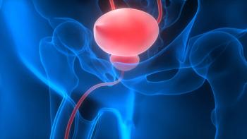
3D CT-Guided Seminal Vesicle Biopsy for Staging
CHICAGO-Three-dimensional, CT-guided transischiorectal biopsy of the seminal vesicles in patients with biopsy-proven prostate cancer resulted in upstaging of 10% of patients, according to a report at the Radiological Society of North America (RSNA) annual meeting.
Advertisement
CHICAGOThree-dimensional, CT-guided transischiorectal biopsy of the seminal vesicles in patients with biopsy-proven prostate cancer resulted in upstaging of 10% of patients, according to a report at the Radiological Society of North America (RSNA) annual meeting.
Standard ultrasound-guided biopsy of the seminal vesicles is not considered to be practical by some clinicians because it requires six needles to be placed through the bowel. However, the 3D CT-guided transischiorectal biopsy procedure can be performed in 3 to 5 minutes with a minimum of discomfort to patients and little danger to the rectum, coccyx, and bowel, said Panos G. Koutrouvelis, MD, of the Uro-Radiology Prostate Institute (URPI), Vienna, Virginia.
The study included 303 men with localized prostate cancer who underwent 3D CT-guided biopsy of the seminal vesicles (before or during brachytherapy) between June 1995 and January 1999. The biopsy disclosed that prostate cancer had invaded one or both seminal vesicles in 30 men, which changed pathologic staging of their disease from T1 or T2 to T3c.
The biopsy was performed at the beginning of brachytherapy so that anesthesia was administered only once. The stereotactic template for the 3D system was placed on the ischiorectal area with the patient in the prone position. To ensure that the biopsy needle would not penetrate the rectum, the stereotactic system was adjusted to correspond with the angle of the gantry, which usually is kept at negative 26 degrees.
The biopsy needle was passed through the pararectal fat to avoid the rectum and coccyx, and three biopsies were taken of the seminal vesicles. One was obtained near the base of the prostate, one in the midportion of the gland, and one in the distal region.
Twenty-four of the 30 men with a positive seminal vesicle biopsy were not considered to be at high risk for seminal vesicle involvement because they had a Gleason score of 7 or less. Thirteen had PSA levels of 10 ng/mL or below, which is not consistent with spread of prostate cancer to the seminal vesicles.
Based on these findings, Dr. Koutrouvelis concluded that after a positive diagnosis of prostate cancer and prior to implementation of any treatment optionradical prostatectomy, brachytherapy, or external beam radiationbiopsy of the seminal vesicles, whether it is guided by CT or ultrasound, is recommended in all patients to rule out seminal vesicle invasion.
Articles in this issue
over 26 years ago
IL-13 Used to Deliver Bacterial Toxin to Brain Tumors in Miceover 26 years ago
Rituximab/CHOP Combo Effective in Low-Grade NHLover 26 years ago
Elderly Patients Tolerate Breast Cancer Therapyover 26 years ago
Coaxial Breast Biopsy Device Provides Diagnostic Specimensover 26 years ago
3D Digital Camera Accurately Calculates Breast Shape, Volumeover 26 years ago
Thermoacoustic CT Under Development for Breast Imagingover 26 years ago
Early Local Recurrence After Lumpectomy Predicts Metastasisover 26 years ago
Panel Recommends Listing 9 Substances in Carcinogen Reportover 26 years ago
State of the Union on Youth Smoking ‘Clearly Not Good’Advertisement
Related Content
Advertisement


Advertisement
Advertisement
Trending on CancerNetwork
1
FDA Approves Afami-cel in Metastatic Synovial Sarcoma
2
The Moonlight Shift: Dr Joshua Richter on Myeloma, Myths, and the C-Word
3
Current Treatment Landscape of HER2-Positive Metastatic Breast Cancer and Phase 3 Trial Overview
4
Zanzalintinib/Atezolizumab Misses Survival End Point in Non-MSI-H mCRC Subgroup
5