
|Articles|December 15, 2014
- Oncology Vol 28 No 12
- Volume 28
- Issue 12
Base of the Skull Metastases in Metastatic Castration-Resistant Prostate Cancer
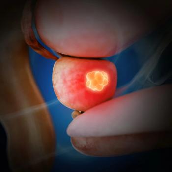
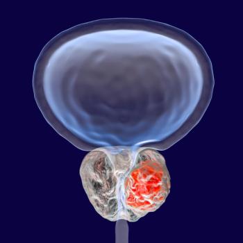
A 63-year-old man with no family history of prostate cancer has prostate biopsy that revealed 9 out of 12 cores involved with prostatic adenocarcinoma, mostly Gleason score 5+4=9.
Advertisement
The Case: A 63-year-old man with no family history of prostate cancer underwent annual screening with serum prostate-specific antigen (PSA) testing. His PSA level was under 3 ng/mL 2 years earlier. A year and a half ago he developed lower urinary tract symptoms (urgency and frequency) and had a PSA level of 18 ng/mL. A prostate biopsy revealed 9 out of 12 cores involved with prostatic adenocarcinoma, mostly Gleason score 5+4=9. On rectal examination he had a fixed prostate; he was not a candidate for surgery. A bone scan revealed no evidence of metastatic disease. Magnetic resonance imaging (MRI) of the pelvis showed involvement of the prostate and three lymph nodes.
He was initially treated with image-guided radiation therapy to the prostate and lymph nodes, and was started on androgen deprivation therapy (ADT). After 4 months of treatment, his PSA level was < 0.3 ng/mL. Six months ago he presented with numbness on the chin and in the perioral area. His PSA level was at that point 29.5 ng/mL. A brain CT scan and MRI found no evidence of disease, and he received no further treatment. Four months later he had worsening of neurologic symptoms, with increased chin and lower lip numbness, as well as a right eye droop, consistent with facial nerve involvement. He was started on sipuleucel-T and referred to our institution for further management. His PSA level was rising despite ADT and reached 45 ng/mL. A new brain MRI showed base of the skull involvement, with a mass in the right petrous apex and a component extending to the prepontine cistern, but no evidence of parenchymal brain metastases (Figure). He received radiation to the base of the skull, with little improvement in speech or eye droop. A post-therapy brain MRI showed a decrease in the size of the right petrous apex mass and the prepontine cistern lesion. He subsequently had a positive bone scan, showing rib and vertebral involvement. Despite treatment, his PSA level continued to rise to 135 ng/mL and he had unintended weight loss (30 pounds in 30 days). He was referred for treatment evaluation of aggressive metastatic castration-resistant prostate cancer (mCRPC), with a PSA doubling time of less than a month and a PSA level of 145 ng/mL. After evaluation of the available treatment options, it was recommended that he start treatment with docetaxel chemotherapy, abiraterone, and denosumab.
Discussion
As with other osseous sites, the skull is a common site of metastases from systemic cancers.[1] Skull base metastases from distant tumors occur in 4% of cancer patients.[1] These metastases are most commonly secondary to breast, lung, and prostate cancers. Prostate cancer is the most common cause of skull base metastases in men.[2] This type of metastasis has been reported as the first sign of neoplastic disease, but it generally occurs as a late event in the course of disease.[3,4]
Cranial nerve palsies due to prostate cancer metastases are an uncommon discovery in advanced disease, but constitute a significant clinical finding.[5] Metastatic disease to the skull can lead to debilitating neurologic symptoms-such as cranial nerve palsies-that result from tumor growth or new bone formation compromising the neural foramina. Several clinical syndromes associated with skull base metastases have been described according to the metastatic site, including orbital, parasellar, middle fossa, jugular foramen, and occipital condyle syndromes.[1] Subtle complaints such as facial paresthesia or numb chin should alert physicians to look for a skull base metastasis.[6]
Currently, T2- and T1-weighted MRI before and after intravenous gadolinium administration is the best method for detecting skull base metastases.[7] Lesions to the calvaria are easy to detect with current radiologic images.[8] However, skull base metastases that involve the ethmoid and sphenoid bones or part of the frontal, temporal, or occipital osseous architecture remain hard to identify with current techniques and are difficult to explore even at autopsy.[8] Perineural invasion occurs in advanced prostate cancer and is an under-recognized route of metastatic spread, a fact that may explain the lack of correlation between imaging and clinical findings in some patients, and that makes clinical diagnosis imperative.[9] While radiologic confirmation has been reported in around 70% of patients,[1] normal images do not exclude the diagnosis, and clinical acumen leading to early radiation therapy may prevent further neurologic dysfunction.[10]
Treatment is mainly palliative, usually using external beam radiation therapy in conjunction with corticosteroids. Radiation is the standard treatment, although some patients with hormone-sensitive or chemosensitive disease states may benefit from hormone therapy or chemotherapy. Surgical intervention is reserved for select patients, given that it does not produce better control than radiation and creates unwanted risks, such as cerebrospinal fluid leakage and meningitis.[11] Radiotherapy can provide pain relief and regression of cranial nerve dysfunction in up to 90% of cases.[12]
The prognosis for skull base metastases is poor, with a median survival of about 1 year. In some series, the presence of cranial nerve palsies was associated with an average survival of 5 months.[13] However, more recent series have suggested a median survival of approximately 30 months, most likely related to the availability of better treatments for metastatic disease.[12]
In this day and age, an array of drugs with different mechanisms of action is available for patients with mCRPC. These treatments have changed the natural history of mCRPC, as well as patient survival.[14] The current drug arsenal includes immunotherapy (sipuleucel-T), radiopharmaceuticals (radium-223), highly effective antiandrogen therapy (HEAT-ie, abiraterone, enzalutamide), and chemotherapy (docetaxel, cabazitaxel).[15] However, there is a lack of useful correlative biomarkers to help physicians choose among these options. Therefore, therapeutic decisions are made on clinical grounds.
Sipuleucel-T is an approved autologous cellular immunotherapy for minimally symptomatic patients with mCRPC, designed to stimulate an immune response.[16] Given the advanced, symptomatic, and rapidly progressive status of our patient, we discontinued the treatment.[17] Radium-223 (an alpha-emitting radionuclide) provides another mechanism of action for the treatment of mCRPC that has spread to the bones. Besides conferring a 3-month survival benefit, it offers palliation for many of the bone-related symptoms (time to first skeletal-related event is 13.6 vs 8.4 months with placebo).[18] However, this therapy might not target nonosseous sites of metastatic disease.
Ruling out parenchymal brain metastases was also important for treatment purposes in this case. Docetaxel is a substrate of P-glycoprotein, a blood–brain barrier (BBB) efflux transporter; it cannot cross the BBB or control metastatic foci.[19] Thus, in the setting of parenchymal brain metastases, use of an agent capable of crossing the BBB, such as cabazitaxel [20] or abiraterone, might be warranted. However, cabazitaxel is only approved in the second-line setting, and no data that focus on the ability of abiraterone to control brain metastases are available, since trials in the advanced setting excluded patients with brain metastases.[21,22]
The possibility of using combination therapy to achieve the greatest clinical benefit was appealing in this case. Thus, combining docetaxel with one of the HEAT agents such as abiraterone or enzalutamide was a priority. The evidence surrounding this practice is scarce but emerging.[23] Recently, a phase Ib safety and pharmacokinetic study (N = 22) assessed the safety of docetaxel plus abiraterone. The presentation at the 2014 American Society of Clinical Oncology Annual Meeting revealed that the combination was well tolerated and that 90% of the treated population achieved a ≥ 50% decrease in PSA level at a median follow-up of 11.8 months (70% had a ≥ 90% response).[24] While further study of this approach is under way, we felt it was appropriate in our patient.
Outcome of This Case
After 3 cycles of docetaxel, our patient felt less fatigued and stopped losing weight, and his PSA level achieved stabilization at around 140 ng/mL. As for his symptoms of skull base involvement, he has experienced decreased numbness in the chin and perioral area, improved swallowing, and decreased right eye droop. He has, however, developed facial asymmetry due to loss of right masseter muscle bulk from denervation. He has experienced dryness and irritation of the right cornea, which is being treated by an ophthalmologist. Maintaining good nutrition has been challenging, given his cranial nerve involvement, and dietician support has been implemented. Since his response to docetaxel alone was insufficient, abiraterone was added. We have not yet noted any relevant side effects, and we are awaiting the clinical response to this combination. No skeletal-related events have been documented.
Conclusion
Maintaining a high index of suspicion for skull base metastases in patients with cancer is important for early diagnosis and treatment, which will prevent further neurologic sequelae. An increasing incidence of skull base metastases is likely, given that patients with advanced prostate cancer are living longer due to the development of more active drugs. Skull base metastases are an entity for which aggressive treatment is indicated upon careful clinical assessment alone. Treatment selection in the setting of mCRPC and skull base metastases should be individualized.
Acknowledgment: The authors would like to acknowledge the “Canales de Ayuda A.C.” Foundation for their support in Dr. Bourlon’s visiting fellowship in urologic oncology at the University of Colorado Cancer Center.
Financial Disclosure:Dr. Glodé has served as an investigator and/or on an advisory board for Janssen and Exelisis. Dr. Bourlon has no significant financial interest in or other relationship with the manufacturer of any product or provider of any service mentioned in this article.
References:
1. Greenberg HS, Deck MD, Vikram B, et al. Metastasis to the base of the skull: clinical findings in 43 patients. Neurology. 1981;31:530-7.
2. Gupta SR, Zdonczyk DE, Rubino FA. Cranial neuropathy in systemic malignancy in a VA population. Neurology. 1990;40:997-9.
3. Long MA, Husband JE. Features of unusual metastases from prostate cancer. Br J Radiol. 1999;72:933-41.
4. Svare A, Fossa SD, Heier MS. Cranial nerve dysfunction in metastatic cancer of the prostate. Br J Urol. 1988;61:441-4.
5. O’Sullivan JM, Norman AR, McNair H, Dearnaley DP. Cranial nerve palsies in metastatic prostate cancer-results of base of skull radiotherapy. Radiother Oncol. 2004;70:87-90.
6. Posner JB. Cancer involving cranial and peripheral nerves. In: DeAngelis LM, Posner JB, editors. Neurologic complications of cancer. Philadelphia: FA Davis; 1995.
7. Johnston JL. Parasellar syndromes. Curr Neurol Neurosci Rep. 2002;2:423-31.
8. Stark AM, Eichmann T, Mehdorn HM. Skull metastases: clinical features, differential diagnosis, and review of the literature. Surg Neurol. 2003;60:219-25; discussion 225-6.
9. Liebig C, Ayala G, Wilks JA, et al. Perineural invasion in cancer: a review of the literature. Cancer. 2009;115:3379-91.
10. Tejani N, Cooper A, Rezo A, et al. Numb chin syndrome: a case series of a clinical syndrome associated with malignancy. J Med Imaging Radiat Oncol. 2014 Jun 26. [Epub ahead of print]
11. Yi HJ, Kim CH, Bak KH, et al. Metastatic tumors in the sellar and parasellar regions: clinical review of four cases. J Korean Med Sci. 2000;15:363-7.
12. Laigle-Donadey F, Taillibert S, Martin-Duverneuil N, et al. Skull-base metastases. J Neurooncol. 2005;75:63-9.
13. Ransom DT, Dinapoli RP, Richardson RL. Cranial nerve lesions due to base of the skull metastases in prostate carcinoma. Cancer. 1990;65:586-9.
14. Kessler ER, Flaig TW. Sequencing medical therapy in prostate cancer: not as easy as 1-2-3. Oncology (Williston Park). 2013;27:1158-60.
15. Hurwitz M, Petrylak DP. Sequencing of agents for castration-resistant prostate cancer. Oncology (Williston Park). 2013;27:1144-9, 1154-8.
16. Kantoff PW, Higano CS, Shore ND, et al. Sipuleucel-T immunotherapy for castration-resistant prostate cancer. N Engl J Med. 2010;363:411-22.
17. Kessler ER, Flaig TW. Geriatric considerations in the treatment of advanced prostate cancer. F1000Prime Rep. 2014;6:33.
18. Parker C, Nilsson S, Heinrich D, et al. Alpha emitter radium-223 and survival in metastatic prostate cancer. N Engl J Med. 2013;369:213-23.
19. Caffo O, Veccia A, Russo L, Galligioni E. Brain metastases from prostate cancer: an emerging clinical problem with implications for the future therapeutic scenario. Future Oncol. 2012;8:1585-95.
20. Dykes DJ, Sarsat JP, Bissery M-C: Efficacy evaluation of TXD258, a taxoid compound, against orthotopic and subcutaneous glioblastomas. Presented at the 91st Annual Meeting of the American Association for Cancer Research; 2000; San Francisco, CA.
21. Oudard S. TROPIC: phase III trial of cabazitaxel for the treatment of metastatic castration-resistant prostate cancer. Future Oncol. 2011;7:497-506.
22. de Bono JS, Logothetis CJ, Molina A, et al. Abiraterone and increased survival in metastatic prostate cancer. N Engl J Med. 2011;364:1995-2005.
23. From ASCO-prostate cancer: docetaxel and abiraterone combination therapy. Nat Rev Urol. 2014;11:364.
24. Tagawa ST, Posadas EM, Bruce JY, et al. Phase 1b study of abiraterone acetate (AA) and docetaxel (D) in patients (pts) with metastatic castration-resistant prostate cancer (mCRPC). J Clin Oncol. 2014;32(suppl 5s):abstr 5025.
Articles in this issue
over 11 years ago
Chemotherapy for Low-Grade Gliomas: Lessons and Questionsover 11 years ago
Chemotherapy for Treatment of Grade II Gliomasover 11 years ago
Tailoring Chemotherapy for Low-Grade Gliomasover 11 years ago
What We Can Learn From Our Patientsover 11 years ago
Who Should-or Should Not-Receive RT for DLBCL?over 11 years ago
Grade II Gliomas-Not So Low Gradeover 11 years ago
Cancer Stem Cells: Implications for Cancer Therapyover 11 years ago
Cancer Stem CellsAdvertisement


Advertisement
Advertisement
Trending on CancerNetwork
1
FDA Accepts NDA for Daraxonrasib in Pretreated Metastatic Pancreatic Cancer
2
CAR T-Cell Expansion Predicts Toxicity, But Not Always Efficacy
3
Mapping the Full CAR T-Cell Journey in DLBCL From Referral to Survivorship
4
FDA Approves Zidesamtinib in Pretreated ROS1+ NSCLC
5