|Poll|September 14, 2017
Chest X-Ray Reveals Large Pleural Mass
Author(s)Mehmet Sitki Copur, MD, Kris Mleczko, MD
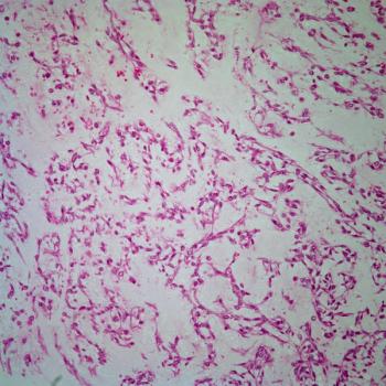
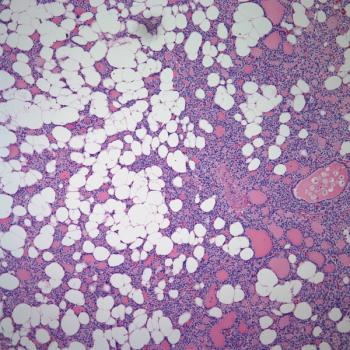
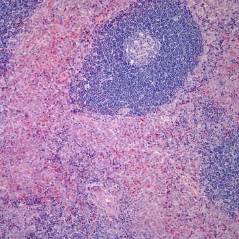
A 70-year-old woman with no history of smoking or asbestos exposure presented with dyspnea on exertion, nonproductive cough, left-sided pleuritic chest pain, and fatigue. Chest radiography revealed a large left pleural effusion and a mass in the left lower lobe. What is the diagnosis?
Advertisement
A 70-year-old woman with no history of smoking or asbestos exposure presented with dyspnea on exertion, nonproductive cough, left-sided pleuritic chest pain, and fatigue. Chest radiography revealed a large left pleural effusion and a mass in the left lower lobe. Chest CT confirmed a pleural-based mass of 7.8 × 5.5 cm invading the anterior chest wall. A left-sided thoracentesis revealed a bloody, lymphocyte-predominant exudative pleural effusion, with a white blood cell count of 8,144/μL (26% lymphocytes, 2% neutrophils, 3% monocytes, and 69% other cell-line types). A cytologic examination of the pleural fluid and a biopsy of the pleural mass were performed, followed by aspiration and biopsy of the bone marrow.
What is the diagnosis?
Advertisement
Related Content
Advertisement


Advertisement
Advertisement
Trending on CancerNetwork
1
Where Does LAG-3/PD-1 Inhibition Fit In PD-1–Refractory Hodgkin Lymphoma?
2
VS-7375 Exhibits Activity in KRAS G12D–Mutated Solid Tumors
3
Offering Hope and Support Toward the Cancer Survivorship Journey
4
Sigvotatug Vedotin Does Not Significantly Improve Survival in NSCLC Trial
5