|Articles|August 15, 2011
- ONCOLOGY Vol 25 No 9
- Volume 25
- Issue 9
Surgical Management of Neuroendocrine Tumors of the Gastrointestinal Tract
This article reviews the surgical management of gastrointestinal neuroendocrine tumors, including the preoperative control of hormonal symptoms, extent of resection required, postoperative outcomes, and differing management strategies as determined by whether the tumor has arisen sporadically or as part of a familial disorder, such as multiple endocrine neoplasia type 1.
Advertisement
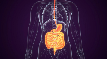
Neuroendocrine tumors of the pancreas (islet cell tumors) and of the luminal gastrointestinal tract (carcinoids) are a heterogeneous group of epithelial neoplasms that share certain common characteristics. First, they are similar histologically and are difficult to distinguish under light microscopy. Second, they can be associated with hypersecretory syndromes. Third, they are generally slow-growing and have a better prognosis than adenocarcinomas at the same site; however, they do become incurable when they progress to unresectable metastatic disease. Surgery is the only curative treatment and is recommended for most patients for whom cross-sectional imaging suggests that complete resection is possible. This article reviews the surgical management of gastrointestinal neuroendocrine tumors, including the preoperative control of hormonal symptoms, extent of resection required, postoperative outcomes, and differing management strategies as determined by whether the tumor has arisen sporadically or as part of a familial disorder, such as multiple endocrine neoplasia type 1 (MEN1).
Neuroendocrine tumors (NETs) are a diverse group of epithelial tumors that can originate from almost any organ derived from the primitive endoderm, including pancreatic islet cells (pancreatic NETs), diffuse neuroendocrine cells distributed throughout the gut (gastrointestinal carcinoids), the respiratory epithelium (bronchial carcinoids), the thymus (thymic carcinoids), and parafollicular cells distributed within the thyroid gland (medullary thyroid carcinoma). Despite their different sites of origin, however, these tumors can be considered as a common entity since they are very difficult to distinguish under the microscope and can share certain common biological features, such as an association with hormonal hypersecretory syndromes. The purpose of this article is to review the important aspects of the diagnosis, preoperative treatment, and operative treatment of neuroendocrine tumors of the pancreas and the luminal gastrointestinal tract. Specific management strategies, which are dependent on whether the tumor has arisen sporadically or as part of a familial disorder, such as multiple endocrine neoplasia type 1 (MEN1), are also discussed.
Pancreatic Neuroendocrine Tumors
Pancreatic NETs (PNETs) account for 2% of all pancreatic neoplasms.[1] Their annual incidence in the United States is estimated to be approximately three cases per million persons but appears to be increasing.[2] The majority of these tumors can be associated with the hypersecretion of detectable hormones, such as insulin, gastrin, glucagon, vasoactive intestinal polypeptide (VIP), or somatostatin, leading to specific clinical syndromes. PNETs generally arise sporadically. However, 80% to 100% of patients with MEN1 will develop a PNET, most commonly a gastrinoma, with insulinoma the next most common type.[3]
Insulinoma
Insulinoma is the most common functional sporadic PNET. Clinical symptoms result from episodes of hypoglycemia; patients can present with neuroglycopenia (headaches, blurred vision, confusion) or sympathoadrenal stimulation (tachycardia, diaphoresis).[4] Symptoms can be insidious and are often prevented or ameliorated by increased caloric intake, sometimes leading to weight gain. Although Dr. Alan Whipple's diagnostic triad of symptomatic hypoglycemia, low blood glucose level, and relief of symptoms with glucose administration is still valid,[5] most patients today are diagnosed using a monitored fast and serial serum measurements of glucose and insulin every 4 to 6 hours. The fast is terminated after 72 hours-or sooner if the patient develops neuroglycopenic symptoms.[4] Measurement of serum proinsulin and C-peptide levels is useful in ruling out surreptitious insulin administration (the condition is ruled out if these values are elevated).
The first step in the management of insulinomas consists of dietary changes and medical control of the symptoms. Adding cornstarch to the diet can slow the absorption of food, while diazoxide (Proglycem), an antihypertensive, can suppress insulin secretion in approximately 60% of patients.[6] For patients unable to tolerate the nausea, hirsutism, and sodium retention associated with diazoxide, other options include calcium channel blockers, beta-adrenergic antagonists, and octreotide (Sandostatin).
FIGURE 1
Pancreatic Insulinoma
The only potentially curative therapy for insulinomas is surgical removal. The cure rate and disease-specific survival can be as high as 100% after resection of sporadic insulinomas, as these tumors are generally (> 90%) benign.[7,8] Insulinomas are typically small (< 2 cm) and can be difficult to localize preoperatively. In addition, they lack a high density of type 2 somatostatin receptors, making somatostatin receptor scintigraphy (SRS, or octreotide scan) rarely helpful (in only 10% of cases). For most patients, pancreatic protocol computed tomography (CT) (
The management of insulinomas in patients with MEN1 is complicated by the presence of multiple tumors in 89% of cases.[11] A single large (2- to 3-cm) dominant tumor can usually be identified in this setting, and removal will lead to control of the hypoglycemic episodes and long-term cure in 86% of patients.[11] However, some surgeons have advocated subtotal distal pancreatectomy with enucleation of head tumors as an alternative approach; this approach has a lower risk of recurrence but a higher rate of iatrogenic diabetes mellitus.[12,13]
Gastrinoma
FIGURE 2
Distribution of Gastrinomas
Patients with Zollinger-Ellison syndrome (ZES) present with severe peptic ulcer disease and secretory diarrhea caused by gastric acid hypersecretion secondary to gastrin-secreting tumors. Gastrinomas arise sporadically in 70% to 80% of patients and are the second most common functional PNET. However, 20% to 30% of affected patients have MEN1, and gastrinomas are the most common PNET in these patients.
ZES is a rare cause of peptic ulcer disease (0.1% to 1% of cases) but should be suspected in patients with recurrent or multiple peptic ulcers or ulcers located in the distal duodenum or jejunum. The diagnosis is made based on elevated fasting serum gastrin levels (> 100 pg/mL) and basal acid output > 15 mEq/hr when patients abstain from antisecretory medications, such as histamine (H2) antagonists and proton pump inhibitors. An additional useful test is the secretin stimulation test; approximately 90% of patients with ZES will have a paradoxical increase (> 200 pg/mL over baseline) in serum gastrin in response to intravenously administered secretin.
The first step in the management of ZES consists of controlling acid hypersecretion. Modern proton-pump inhibitor drugs can control acid secretion in almost all patients. Dose titration should be driven by measurement of basal acid output since the relief of symptoms is an unreliable indicator of effective acid control. The typical dose of omeprazole to control acid hypersecretion in ZES is 40 mg twice daily.
FIGURE 3
Localization of Duodenal Gastrinomas
The best modality for visualizing gastrinomas is SRS in conjunction with CT and magnetic resonance imaging (MRI). Using the radiolabeled somatostatin analog 111In-pentetreotide, SRS has a sensitivity of 80% and a specificity of 100% in this setting. Approximately 80% of gastrinomas are found within the gastrinoma triangle, an anatomic area that includes the first, second, and third portions of the duodenum and the head of the pancreas (
The most common site for a gastrinoma is not the pancreas but the duodenum (
FIGURE 4
Impact of Surgery on Disease-Specific (A) and Liver Metastasis–Free Survival (B) in Patients With Gastrinoma
For patients with sporadic gastrinomas, surgery can offer biochemical and oncologic cure. The majority of gastrinomas are malignant, and while most tumors are slow-growing, there appears to be a more aggressive type that is found in approximately a quarter of patients.[21] These more aggressive gastrinomas are commonly associated with pancreatic tail primary tumors outside the gastrinoma triangle. Patients who undergo surgical resection have improved 20-year survival compared with those not undergoing surgery (98% vs 74%,
The role of surgery in patients with gastrinoma and MEN1 is more controversial. Because primary hyperparathyroidism resulting from multiple gland disease is usually present, subtotal (3 1/2 gland) parathyroidectomy is advocated first as it lessens the end-organ effects of hypergastrinemia and markedly improves the management of ZES (hypercalcemia acts as a secretagogue for gastrin).[23] However, unlike with sporadic gastrinomas, the long-term (biochemical and radiographic) cure rate in patients with MEN1 who undergo surgery is low, mainly because of gastrinoma multifocality (
FIGURE 5
Duodenectomy Specimens of a Sporadic, Solitary Gastrinoma (A) and Multiple Familial Gastrinomas in the Setting of MEN1(B)
The arguments against routine pancreaticoduodenectomy, however, are (1) that patients with MEN1 already have excellent overall survival outcomes (86% at 15 years) with less aggressive surgery (enucleation),[23] (2) that these patients may develop and die from other neuroendocrine neoplasias, and (3) that the Whipple operation may further complicate future liver-directed therapy (resection or embolization) for metachronous hepatic metastases because of the increased risk of liver abscess and ascending infection.[16] Because patients who have MEN1 have a high incidence of disease recurrence following surgery, these are important considerations that must be taken into account when planning surgical intervention.
Glucagonoma
Glucagonomas are classically associated with necrolytic migratory erythema and the “4Ds” (diabetes mellitus, dermatitis, deep vein thrombosis, and depression).[28] The characteristic rash (
VIPoma
FIGURE 6
Typical Skin Rash (Necrolytic Migratory Erythema) in a Patient With a Pancreatic Glucagonoma
Vasoactive intestinal peptide–secreting tumors (VIPomas) are associated with “WDHA” syndrome: watery diarrhea, hypokalemia, and achlorhydria. The tumors are typically solitary pancreatic lesions > 3 cm in size, with 70% to 80% metastatic at presentation.[30] Although typically intrapancreatic, about 10% can be found in the colon, bronchus, adrenals, liver, or sympathetic ganglia. Initial management consists of correcting the dehydration and metabolic abnormalities with IV fluid and electrolyte supplementation, but this can be challenging. Octreotide is often useful in this setting and for long-term control in patients unable to undergo resection.
Somatostatinoma
Somatostatinomas are the rarest of the PNETs and are often discovered only incidentally. No specific syndrome exists, but suggestive symptoms include diabetes mellitus, cholelithiasis, weight loss, steatorrhea, and anemia.[30] Patients may also present with duodenal obstruction from duodenal tumors. Approximately 50% of tumors arise in the pancreas (with two-thirds located in the head of the pancreas); the remainder are found in the small intestine.[30] Most are malignant and large (> 5 cm) at presentation. Diagnosis is made with elevated somatostatin levels. Surgical resection, if feasible, is the first-line treatment.
Nonfunctional PNETs
Recent series report that 35% to 50% of PNETs are nonfunctional, that is, they do not secrete detectable levels of functional hormones. Patients with nonfunctional tumors often present at a later stage, with large (> 5 cm) tumors and liver metastases, although as a result of the widespread use of cross-sectional imaging, a growing number of these tumors are being identified incidentally at an earlier stage. These tumors can be differentiated from pancreatic adenocarcinoma on CT, since they are more likely to be enhancing or associated with cystic degeneration or calcifications; however, they are less likely to encase vascular structures or obstruct the pancreatic duct. Nonfunctional tumors secrete a variety of peptides, such as pancreatic polypeptide (PP) and chromogranin A, which can serve as tumor markers. A positive SRS scan can confirm the diagnosis and reveal occult metastatic disease. PNETs are classified by the World Health Organization as low-grade if the mitotic count is < 2/10 high-power fields (HPF) and the Ki-67 proliferation index is < 3%, as intermediate-grade if the mitotic count is 2/10 to 20/10 HPF or Ki-67 is 3% to 20%, and as high-grade if the mitotic count is > 20/10 HPF or the Ki-67 is > 20%.[1] For low- and intermediate-grade tumors, upfront surgical resection is undertaken, with 10-year disease-specific survival rates of 80% and 30%, respectively.[31] Small superficial tumors can be enucleated, but larger tumors may require pancreatic resection with pancreaticoduodenectomy, or central or distal pancreatectomy.[30] High-grade PNETs (poorly differentiated neuroendocrine carcinomas, large-cell or small-cell) are very aggressive malignancies that should be treated upfront with platinum-etoposide combination chemotherapy, potentially followed by a form of local therapy (surgery or radiotherapy) if localized tumor persists.[32,33]
Neuroendocrine Tumors of the Luminal Gastrointestinal Tract
NETs of the luminal gastrointestinal tract (carcinoid tumors) are derived from chromaffin or Kulchitsky cells, which can be found in the gastrointestinal, urogenital, and bronchial epithelia. Approximately 70% arise in the gastrointestinal tract, with an annual incidence of about two to three cases per 100,000 persons.[2,34] They most commonly arise in the small bowel (29%), appendix (19%), and rectum (12.6%).[34] There are a number of similarities between carcinoid tumors and other neuroendocrine tumors; carcinoids thus require specific histologic and cytochemical staining in order to be identified under light microscopy.[35] Some experts have advocated moving away from use of the term “carcinoid”; however, it remains in widespread use.[1,36]
The signs and symptoms of carcinoid tumors can be highly variable. The classic carcinoid syndrome consists of facial flushing, diarrhea, diaphoresis, weight loss, endocardial thickening of the right-sided heart valves, and bronchoconstriction.[37] The occurrence and severity of symptoms is directly related to whether the bulk of tumor drains into the systemic circulation (liver or retroperitoneal nodal metastasis) and bypasses the liver. Most patients with the carcinoid syndrome will have an ileal primary tumor with nodal and liver metastases. The most common diagnostic test for symptomatic carcinoid tumors is measurement of the serotonin metabolite 5-hydroxyindoleacetic acid (5-HIAA) in a 24-hour urine sample.[38] The best agent for medical management of symptomatic carcinoid disease is the somatostatin analogue octreotide. Most patients respond to octreotide treatment and have significant improvement in their symptoms.[37] Octreotide should also be used preoperatively and intraoperatively to minimize the risk of a carcinoid crisis (unresponsive hypotensive episodes), which can be precipitated in patients with carcinoid syndrome by stressful situations, such as induction of anesthesia. Long-acting octreotide (Sandostatin LAR) may also be useful to inhibit tumor growth and decrease tumor progression postoperatively.[39]
FIGURE 7
Coronal Computed Tomography Image of a Patient With a Neuroendocrine Tumor of the Ileum
Imaging of carcinoids is typically done using CT or MRI. Carcinoids generally express somatostatin receptors, making SRS a fairly sensitive adjunct for evaluating the primary tumor and metastatic disease. Gastric, duodenal, and rectal carcinoids are usually evaluated using endoscopy. However, localization of small-bowel carcinoids can be difficult. Abdominal CT imaging does not always locate the primary tumor but may detect liver metastases, regional lymph nodes, or bowel wall thickening and cicatrization consistent with mesenteric nodal metastases from carcinoid tumors (
Small bowel
Approximately one-third of all small- bowel tumors are carcinoids, and they most commonly arise in the distal ileum.[37,13] Patients may present with carcinoid syndrome, have nonspecific symptoms such as abdominal pain or bowel obstruction, or be asymptomatic. Encasement of the mesenteric vasculature resulting from fibrotic changes at the root of the mesentery (see
Appendix
Appendiceal carcinoids are usually found incidentally once every 200 to 300 appendectomies. Patients are typically young adults, and the tumors are usually < 1 cm in size. Appendectomy is sufficient treatment for all patients with tumors < 2 cm in size that are not present at the base of the appendix (resection margin). In one series with a median follow-up of 26 years, no patients with tumors < 2 cm developed nodal or distant metastatic disease.[43] In contrast, metastases were found in 21% of patients with tumors 2 to 3 cm in size and in 44% of patients with tumors > 3 cm. Thus, a right hemicolectomy is appropriate for patients with tumors > 2 cm, positive resection margins, or mesoappendiceal invasion. In addition, patients whose tumors show both neuroendocrine and glandular differentiation (often classified as goblet cell carcinoids, adenocarcinoids, or mixed adenocarcinoma carcinoids) should also undergo a right hemicolectomy because the tumors behave similarly to adenocarcinomas.[44] Appendiceal carcinoids do not cause the carcinoid syndrome.
Rectum
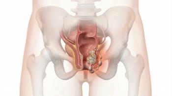
Carcinoid tumors of the rectum are found once every 2,500 sigmoidoscopies.[37] Virtually all are found in the mid-rectum between 4 and 13 cm from the dentate line. Unlike ileal carcinoids, they do not show histological evidence of serotonin production and thus do not cause carcinoid syndrome, even in the presence of metastatic disease. Patients with tumors < 1 cm in size can safely undergo wide local transanal or endoscopic excision. No additional preoperative staging or disease-specific follow-up appears to be necessary.[37,45] Patients with tumors between 1 and 2 cm can generally undergo wide local excision as well, provided there is no invasion into the muscularis propria, no lymphovascular invasion, nor a high mitotic rate (> 2/50 HPF).[45] If any of these factors is present or if the tumor is > 2 cm, a more radical procedure (abdominoperineal or low anterior resection) is strongly, but not routinely, recommended. It is important to recognize that for rectal carcinoids that are > 2 cm in size, the risk of distant metastasis is so high that the marginal benefit of local control obtained from radical surgery may become less important than sphincter preservation.
Duodenum
Special mention should be made of duodenal carcinoid tumors. Patients with small nonampullary duodenal carcinoids < 1 cm in size have been successfully treated using endoscopic excision alone, while tumors between 1 and 2 cm can be treated with transduodenal excision.[46] Patients with tumors > 2 cm will generally require a more extensive procedure. However, the appropriate surgical approach is controversial. A recent surgical series of 24 patients with duodenal carcinoid tumors (gastrinomas and ampullary carcinoids were excluded) reported that, although the majority (89%) of tumors measured less than 2 cm, and most (85%) were limited to the mucosa or submucosa, lymph node metastases were identified in the surgical specimen in 7 of 13 patients (54%) in whom lymph nodes were examined, including 2 patients with tumors smaller than 1 cm and limited to the submucosa. Therefore, the authors concluded that the presence of regional lymph node metastasis cannot be predicted reliably on the basis of tumor size or depth of invasion.[47] In addition, it is now established that carcinoid tumors arising from the ampulla of Vater exhibit considerably more aggressive behavior, with a strong tendency to develop nodal metastasis even at a small size (< 2 cm); pancreaticoduodenectomy is thus recommended irrespective of tumor size.[48-51]
Colon
Carcinoid tumors of the colon are typically diagnosed late in the seventh decade of life and are usually accompanied by abdominal pain, anorexia, and weight loss.[52] The tumors are most often found in the cecum.[34] Unfortunately, most carcinoids are large (> 2 cm) at the time of presentation and metastatic in approximately 60% of cases. Overall 5-year survival- 42%-is the lowest of all the carcinoids arising in the gastrointestinal tract.[34] Surgical resection, usually by hemicolectomy, is the treatment of choice.
Stomach
TABLE 1
Comparison of the Three Types of Gastric Carcinoid Tumors
Gastric carcinoids arise from enterochromaffin-like (ECL) cells that are located in the gastric mucosa. These tumors can be classified into three types (Table). Type I and II carcinoids are believed to develop in the setting of chronic hypergastrinemia (from pernicious anemia and ZES, respectively), which stimulates ECL-cell hyperplasia and leads to gastric carcinoid development. Most of these carcinoids are small (< 1 cm) and 57% to 78% are multicentric, but only about 5% to 10% are metastatic.[53-55] Therefore, the prognosis for type I carcinoids is generally excellent, and these tumors are usually treated with endoscopic resection and surveillance, unless the tumor is too large to be removed endoscopically or is associated with concomitant adenocarcinoma.[53,55] Antrectomy has been proposed as a treatment option for type I gastric carcinoids, since removal of the gastrin-producing G-cells (which stimulate the proliferation of ECL cells) is expected to interrupt the pathogenetic cycle of the disease.[55,56] However, normalization of gastrin levels after antrectomy does not prevent tumor progression in 14% of patients, possibly because of autonomous gastrin-independent ECL hyperplasia or other stimulating factors.[55-57] The treatment of type II carcinoids is the same as that for type I, except that patients with MEN1 should also undergo resection of their gastrinoma(s). However, approximately 5% of these patients with MEN1 develop significant clinical symptoms, such as bleeding, carcinoid syndrome, weight loss, and obstruction caused by aggressive tumor growth; symptoms are likely related to the duration and magnitude of hypergastrinemia.[58] These carcinoids are often diffusely spread throughout the stomach and exhibit a metastatic phenotype, requiring radical surgery. In comparison, type III carcinoids arise sporadically and are diagnosed in 15% to 20% of patients with gastric carcinoids. Patients with type III carcinoids usually have normal levels of gastrin, but their tumors are larger, solitary, invasive beyond the mucosa, and carry a > 50% metastatic potential, requiring formal resection.[54,55] Types II and III gastric carcinoids may cause the carcinoid syndrome without liver metastases, since the serotonin enters the systemic circulation through the coronary vein. In these patients perioperative octreotide is mandatory.
Neuroendocrine Liver Metastases
REFERENCE GUIDE
Therapeutic Agents
Mentioned in This Article
Diazoxide (Proglycem)
Insulin
Long-acting octreotide
(Sandostatin LAR)
Octreotide (Sandostatin)
Omeprazole
Platinum-etoposide chemotherapy
Brand names are listed in parentheses only if a drug is not available generically and is marketed as no more than two trademarked or registered products. More familiar alternative generic designations may also be included parenthetically.
There is no level 1 evidence to guide the optimal management of patients with NET liver metastases. For patients who have potentially resectable disease, surgical resection is associated with 5-year survival rates of 60% to 70% in retrospective series, and can occasionally lead to cure.[59,60] Cytoreductive surgery may help reduce hormonal secretion and prolong survival. Our current approach is to attempt surgical resection in patients in whom it has been determined, on the basis of preoperative imaging studies, that at least 90% of the liver tumors can be safely removed. Residual disease may be effectively controlled postoperatively with long-acting somatostatin analogues.[39] For patients with clearly unresectable disease, transarterial chemo-, radio-, or bland embolization appears to be effective in controlling hormonal symptoms and delaying the development of liver failure.
Summary and Future Directions
Neuroendocrine tumors of the gastrointestinal tract are a heterogeneous group of neoplasms that nonetheless share common biologic characteristics. Their management continues to evolve, but surgery remains the only curative treatment and is recommended for most patients in whom cross-sectional imaging suggests that complete resection is possible. Preoperative control of hormonal symptoms is mandatory. The extent of resection is specific to tumor type. In general, insulinomas can be enucleated, while gastrinomas should be treated with enucleation of pancreatic head tumors, duodenotomy and local resection of duodenal tumors, and removal of the peripancreatic/periduodenal lymph nodes. Patients with MEN1 often have multifocal disease, and while biochemical and radiographic cure rates after surgery are much lower, overall survival is good. The goal in treating patients with MEN1 should be removal of dominant tumors. Most other functional and nonfunctional tumors are usually large at presentation and require formal surgical resection. For carcinoid tumors, the somatostatin analogue octreotide is an important adjunct in managing the carcinoid syndrome and preventing perioperative carcinoid crisis. The traditional "2-cm rule" that guides a more extensive use of resection for appendiceal carcinoids is not applicable to duodenal, small bowel, and rectal tumors, which can be metastatic at smaller sizes. Endoscopic resection is playing an increasing role in the management of type I and type II gastric carcinoids, as well as in small and superficial duodenal and rectal carcinoids. Although NETs generally have a better prognosis than adenocarcinomas at the same site, they are incurable once they advance to unresectable distant metastatic disease. Long-acting octreotide may be useful for inhibiting tumor growth and decreasing tumor progression. New therapeutic approaches for NETs, such as peptide receptor radiotherapy and systemic agents targeting vascular endothelial growth factor (VEGF) and mammalian target of rapamycin (mTOR) are under development.[61]
Financial Disclosure:The authors have no significant financial interest or other relationship with the manufacturers of any products or providers of any service mentioned in this article.
References:
REFERENCES
1. Klimstra DS, Modlin IR, Coppola D, et al. The pathologic classification of neuroendocrine tumors: a review of nomenclature, grading, and staging systems. Pancreas. 2010;39:707-12.
2. Yao JC, Hassan M, Phan A, et al. One hundred years after “carcinoid”: epidemiology of and prognostic factors for neuroendocrine tumors in 35,825 cases in the United States. J Clin Oncol. 2008;26:3063-72.
3. Jensen RT, Berna MJ, Bingham DB, Norton JA. Inherited pancreatic endocrine tumor syndromes: advances in molecular pathogenesis, diagnosis, management, and controversies. Cancer. 2008;113:1807-43.
4. Weber KJ, Lin J, Prinz RA. Endocrine tumors of the pancreas: clinical picture, diagnosis and therapy. In: Blumgart LH, Belghiti J, editors. Surgery of the liver, biliary tract, and pancreas. 4th ed. Philadelphia, PA: Saunders Elsevier; 2007. p. 904-14.
5. Whipple AO. The surgical therapy of hyperinsulinism. J Int Chir. 1938;3:237.
6. Peterson DA, Dolan JP, Norton JA. Neuroendocrine tumors of the pancreas and gastrointestinal tract and carcinoid disease. In: Norton JA, Barie PS, Bollinger RR, et al., editors. Surgery: basic science and clinical practice. 2nd ed. New York, NY: Springer; 2008. p. 1249-84.
7. Hiramoto JS, Feldstein VA, LaBerge JM, Norton JA. Intraoperative ultrasound and preoperative localization detects all occult insulinomas. Arch Surg. 2001;136:1020-5; discussion 1025-6.
8. Nikfarjam M, Warshaw AL, Axelrod L, et al. Improved contemporary surgical management of insulinomas: a 25-year experience at the Massachusetts General Hospital. Ann Surg. 2008;247:165-72.
9. Anderson MA, Carpenter S, Thompson NW, et al. Endoscopic ultrasound is highly accurate and directs management in patients with neuroendocrine tumors of the pancreas. Am J Gastroenterol. 2000;95:2271-7.
10. Doppman JL, Chang R, Fraker DL, et al. Localization of insulinomas to regions of the pancreas by intra-arterial stimulation with calcium. Ann Intern Med. 1995;123:269-73.
11. Norton JA, Fang TD, Jensen RT. Surgery for gastrinoma and insulinoma in multiple endocrine neoplasia type 1. J Natl Compr Canc Netw. 2006;4:148-53.
12. O'Riordain DS, O'Brien T, van Heerden JA, et al. Surgical management of insulinoma associated with multiple endocrine neoplasia type I. World J Surg. 1994;18:488-93; discussion 93-4.
13. Demeure MJ, Klonoff DC, Karam JH, et al. Insulinomas associated with multiple endocrine neoplasia type I: the need for a different surgical approach. Surgery. 1991;110:998-1004.
14. Stabile BE, Morrow DJ, Passaro E, Jr. The gastrinoma triangle: operative implications. Am J Surg. 1984;147:25-31.
15. Zogakis TG, Gibril F, Libutti SK, et al. Management and outcome of patients with sporadic gastrinoma arising in the duodenum. Ann Surg. 2003;238:42-8.
16. Norton JA, Jensen RT. Resolved and unresolved controversies in the surgical management of patients with Zollinger-Ellison syndrome. Ann Surg. 2004;240:757-73.
17. Thom AK, Norton JA, Doppman JL, et al. Prospective study of the use of intraarterial secretin injection and portal venous sampling to localize duodenal gastrinomas. Surgery. 1992;112:1002-8; discussion 8-9.
18. Norton JA, Alexander HR, Fraker DL, et al. Does the use of routine duodenotomy (DUODX) affect rate of cure, development of liver metastases, or survival in patients with Zollinger-Ellison syndrome? Ann Surg. 2004;239:617-26.
19. Norton JA, Alexander HR, Fraker DL, et al. Possible primary lymph node gastrinoma: occurrence, natural history, and predictive factors: a prospective study. Ann Surg. 2003;237:650-7; discussion 7-9.
20. Herrmann ME, Ciesla MC, Chejfec G, et al. Primary nodal gastrinomas. Arch Pathol Lab Med. 2000;124:832-5.
21. Weber HC, Venzon DJ, Lin JT, et al. Determinants of metastatic rate and survival in patients with Zollinger-Ellison syndrome: a prospective long-term study. Gastroenterology. 1995;108:1637-49.
22. Norton JA, Fraker DL, Alexander HR, et al. Surgery increases survival in patients with gastrinoma. Ann Surg. 2006;244:410-9.
23. Norton JA, Fraker DL, Alexander HR, et al. Surgery to cure the Zollinger-Ellison syndrome. N Engl J Med. 1999;341:635-44.
24. Fraker DL, Norton JA, Alexander HR, et al. Surgery in Zollinger-Ellison syndrome alters the natural history of gastrinoma. Ann Surg. 1994;220:320-8; discussion 8-30.
25. Yu F, Venzon DJ, Serrano J, et al. Prospective study of the clinical course, prognostic factors, causes of death, and survival in patients with long-standing Zollinger-Ellison syndrome. J Clin Oncol. 1999;17:615-30.
26. Norton JA, Alexander HR, Fraker DL, et al. Comparison of surgical results in patients with advanced and limited disease with multiple endocrine neoplasia type 1 and Zollinger-Ellison syndrome. Ann Surg. 2001;234:495-505; discussion -6.
27. Stadil F, Bardram L, Gustafsen J, Efsen F. Surgical treatment of the Zollinger-Ellison syndrome. World J Surg. 1993;17:463-7.
28. Chen H. Management of pancreatic islet cell tumors excluding gastrinoma. In: Cameron JL, editor. Current surgical therapy. 9th ed. St. Louis, MO: Mosby; 2008. p. 535-9.
29. Norton JA, Kahn CR, Schiebinger R, et al. Amino acid deficiency and the skin rash associated with glucagonoma. Ann Intern Med. 1979;91:213-5.
30. Kulke MH, Anthony LB, Bushnell DL, et al. NANETS treatment guidelines: well-differentiated neuroendocrine tumors of the stomach and pancreas. Pancreas. 2010;39:735-52.
31. Ferrone CR, Tang LH, Tomlinson J, et al. Determining prognosis in patients with pancreatic endocrine neoplasms: can the WHO classification system be simplified? J Clin Oncol. 2007;25:5609-15.
32. Brenner B, Tang LH, Klimstra DS, Kelsen DP. Small-cell carcinomas of the gastrointestinal tract: a review. J Clin Oncol. 2004;22:2730-9.
33. Strosberg JR, Coppola D, Klimstra DS, et al. The NANETS consensus guidelines for the diagnosis and management of poorly differentiated (high-grade) extrapulmonary neuroendocrine carcinomas. Pancreas. 2010;39:799-800.
34. Modlin IM, Lye KD, Kidd M. A 5-decade analysis of 13,715 carcinoid tumors. Cancer. 2003;97:934-59.
35. Masson P. Carcinoids (argentaffin-cell tumors) and nerve hyperplasia of the appendicular mucosa. Am J Pathol. 1928;4:181-212.
36. Capella C, Heitz PU, Hofler H, et al. Revised classification of neuroendocrine tumours of the lung, pancreas and gut. Virchows Arch. 1995;425:547-60.
37. Moertel CG. Karnofsky memorial lecture. An odyssey in the land of small tumors. J Clin Oncol. 1987;
5:1502-22.
38. Tormey WP, FitzGerald RJ. The clinical and laboratory correlates of an increased urinary 5-hydroxyindoleacetic acid. Postgrad Med J. 1995;71:542-5.
39. Rinke A, Muller HH, Schade-Brittinger C, et al. Placebo-controlled, double-blind, prospective, randomized study on the effect of octreotide LAR in the control of tumor growth in patients with metastatic neuroendocrine midgut tumors: a report from the PROMID Study Group. J Clin Oncol. 2009;27:4656-63.
40. Sugimoto E, Lörelius LE, Eriksson B, Oberg K. Midgut carcinoid tumours. CT appearance. Acta Radiol. 1995;36:367-71.
41. Moertel CG, Sauer WG, Dockerty MB, Baggenstoss AH. Life history of the carcinoid tumor of the small intestine. Cancer. 1961;14:901-12.
42. Bilimoria KY, Bentrem DJ, Wayne JD, et al. Small bowel cancer in the United States: changes in epidemiology, treatment, and survival over the last 20 years. Ann Surg. 2009;249:63-71.
43. Moertel CG, Weiland LH, Nagorney DM, Dockerty MB. Carcinoid tumor of the appendix: treatment and prognosis. N Engl J Med. 1987;317:1699-1701.
44. Tang LH, Shia J, Soslow RA, et al. Pathologic classification and clinical behavior of the spectrum of goblet cell carcinoid tumors of the appendix. Am J Surg Pathol. 2008;32:1429-43.
45. Fahy BN, Tang LH, Klimstra D, et al. Carcinoid of the rectum risk stratification (CaRRs): a strategy for preoperative outcome assessment. Ann Surg Oncol. 2007;14:1735-43.
46. Zyromski NJ, Kendrick ML, Nagorney DM, et al. Duodenal carcinoid tumors: how aggressive should we be? J Gastrointest Surg. 2001;5:588-93.
47. Mullen JT, Wang H, Yao JC, et al. Carcinoid tumors of the duodenum. Surgery. 2005;138:971-7; discussion 7-8.
48. Poultsides GA, Frederick WA. Carcinoid of the ampulla of Vater: morphologic features and clinical implications. World J Gastroenterol. 2006;12:7058-60.
49. Carter JT, Grenert JP, Rubenstein L, et al. Neuroendocrine tumors of the ampulla of Vater: biological behavior and surgical management. Arch Surg. 2009;144:527-31.
50. Clements W. Ampullary carcinoid tumors: rationale for an aggressive surgical approach. J Gastrointest Surg. 2003;7:773-6.
51. Makhlouf HR, Burke AP, Sobin LH. Carcinoid tumors of the ampulla of Vater: a comparison with duodenal carcinoid tumors. Cancer. 1999;85:1241-9.
52. Rosenberg JM, Welch JP. Carcinoid tumors of the colon. A study of 72 patients. Am J Surg. 1985;149:775-9.
53. Gladdy RA, Strong VE, Coit D, et al. Defining surgical indications for type I gastric carcinoid tumor. Ann Surg Oncol. 2009;16:3154-60.
54. Rindi G, Bordi C, Rappel S, et al. Gastric carcinoids and neuroendocrine carcinomas: pathogenesis, pathology, and behavior. World J Surg. 1996;20:168-72.
55. Borch K, Ahrén B, Ahlman H, et al. Gastric carcinoids: biologic behavior and prognosis after differentiated treatment in relation to type. Ann Surg. 2005;242:64-73.
56. Jordan PH, Jr., Barroso A, Sweeney J. Gastric carcinoids in patients with hypergastrinemia. J Am Coll Surg. 2004;199:552-5.
57. Wangberg B, Grimelius L, Granerus G, et al. The role of gastric resection in the management of multicentric argyrophil gastric carcinoids. Surgery. 1990;108:851-7.
58. Norton JA, Melcher ML, Gibril F, Jensen RT. Gastric carcinoid tumors in multiple endocrine neoplasia-1 patients with Zollinger-Ellison syndrome can be symptomatic, demonstrate aggressive growth, and require surgical treatment. Surgery. 2004;136:1267-74.
59. Chen H, Hardacre JM, Uzar A, et al. Isolted liver metastases from neuroendocrine tumors: does resection prolong survival? J Am Coll Surg. 1998;187:88-92; discussion -3.
60. Norton JA, Sugarbaker PH, Doppman JL, et al. Aggressive resection of metastatic disease in selected patients with malignant gastrinoma. Ann Surg. 1986;203:352-9.
61. Yao JC. Neuroendocrine tumors. Molecular targeted therapy for carcinoid and islet-cell carcinoma. Best Pract Res. 2007;21:163-72.
Articles in this issue
almost 15 years ago
Management of DCIS-A Work in Progressalmost 15 years ago
Tailored Strategies for DCIS Managementalmost 15 years ago
Neuroendocrine Tumors: a Heterogeneous Set of Neoplasmsalmost 15 years ago
Can We Know What to Do When DCIS Is Diagnosed?almost 15 years ago
A Rare Case of Metastatic Renal Epithelioid Angiomyolipomaalmost 15 years ago
The Changing Field of Locoregional Treatment for Breast Canceralmost 15 years ago
Cancer and Healthcare Reform: Making the Pieces Fitalmost 15 years ago
An Approach to the Management of Rare TumorsAdvertisement


Advertisement
Advertisement
Trending on CancerNetwork
1
FDA Approves Blood-Based Test for Colorectal Cancer Screening
2
Belzutifan Combo Exhibits Comparable HRQoL Vs Cabozantinib in RCC
3
Steel Particulate Matter Prompts Cyclophosphamide for Injection Recall
4
Optimizing Frontline Therapeutic Strategies in EGFR-Mutated Advanced NSCLC
5