|Articles|February 1, 2005
Oncology NEWS International
- Oncology NEWS International Vol 14 No 2
- Volume 14
- Issue 2
Endorectal Coils, Contrast Media Boost Accuracy of Magnetic Resonance Techniques for Prostate Cancer Diagnosis
This special supplement to Oncology News International comprises expertcommentary and selected reports from the 2004 meetings of RSNA andASTRO about new imaging techniques, with a focus on state-of-the-art magneticresonance imaging, positron emission tomography, computed tomography,and complementary modalities for improving the diagnosis, staging, andtreatment of a variety of cancers. Evident in these reports is the increasingcollaboration between the specialties of radiation oncology and diagnosticradiology as imaging technology continues to evolve.
Advertisement
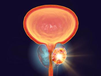
CHICAGO-Endorectal coilstudies presented at the 90th ScientificAssembly and Annual Meeting of theRadiological Society of North Americashow unequivocally that the devicesboost the diagnostic confidence ofimaging to determine management ofprostate disease. Also, new studies ofmagnetic resonance spectroscopicimaging (MRSI), dynamic contrastenhancedMR imaging, and ultrasmallparamagnetic contrast media revealthat these approaches improve diagnosisand provide useful informationfor patient management.Under the guidance of HedvigHricak, MD, PhD, chairman of thedepartment of radiology at MemorialSloan-Kettering Cancer Center(MSKCC), radiologist Liang Wang,MD, confirmed that an endorectal coilboosts the resolution and diagnosticconfidence of MRI and multivoxelMRSI for determining whether prostatecancer is confined within the bordersof the organ (abstract SSA07-01).
Based on experience with 411 patients,including 268 cases in whichboth MRI and MRSI were used, Dr.Wang found that examinations employinga coil enabled the clinician tostage cancer with significantly increasedaccuracy. The average Partinnomogram score for imaging resultsobtained using of a coil was 0.8327,compared with 0.7769 without a coil.Exams performed with MRSI wereeven more accurate. Figure 1 showsuse of an endorectal coil with MRI forassessing organ confinement in a patientwith prostate cancer.Radiologist Joan Vilanova, MD,from Clinica Girona, in Girona, Spain,showed the value of endorectal coilassistedMRI and 3D MRSI for reevaluatingpatients with elevated prostate-specific antigen (PSA) persistingafter a negative biopsy (abstract SSA07-03). Prostate cancer was positivelyidentified in 10 of 27 cases.Cancer was found in the transitionalzone of four patients and theperipheral gland of six. In the positivecancer cases, the choline + creatine/citrate (CC/Ci) ratio and choline/creatine(Ch/Cr) ratios for voxels in thetransitional zone were significantlydifferent (P > .01) from ratios in thevoxels with benign prostatic hyperplasia.For the peripheral gland, theCC/Ci and Ch/Cr ratios for cancervoxels were significantly different fromthe ratios in voxels sampled in normaltissue, Dr. Vilanova said.The protocol's overall accuracy,sensitivity, and specificity were 85%,75%, and 87%, respectively. Dr. Vilanovarecommended that 3D MRSIshould be routinely prescribed forpatients whose PSA levels continue tobe elevated after negative biopsy. Hecautioned radiologists, however, tocarefully survey the entire gland aswell as the transitional and peripheralzones when performing the procedure.Contrast Tools DetectRecurrence, NodalInvolvementDynamic contrast-enhanced (DCE)MRI can assist follow-up therapeuticplanning for recurrent prostate cancerafter radiation therapy, accordingto Masoom A. Haider, MD, an assistantprofessor of radiology at PrincessMargaret Hospital in Toronto, Ontario,Canada (abstract SSA07-04). Astudy of 37 patients demonstrated thatearly intense enhancement on DCEMRIuncovers locally recurrent prostatecancer. The DCE-MRI was performedusing a 1.5T MRI and afour-element torso phased-array coil.Sensitivity and positive predictive valuerates of 72% and 88%, respectively,confirmed that the dynamic contrasttechnique is far superior to conventionalT2-weighted MRI for this role.The enhancement pattern also aids inplanning for follow-up of minimallyinvasive therapy.Ultrasmall iron-oxide contrast continuesto show promise for detectingnodal involvement in prostate cancer.Using ferumoxtran-10, M.G. Harisinghani,MD, a researcher at the Centerfor Molecular Imaging Research atMassachusetts General Hospital, Boston,discovered that metastatic prostatecancer usually first involves lymphnodes located within 2.5 cm of theanterior surface of lumbar vertebralbodies (abstract SSG19-03). Dr. Harisinghani'sstudy of 21 prostate cancerpatients also found that all positivenodes were localized within 1.2 cm ofthe aorta. These results may aid biopsyand therapeutic planning, he said.
Articles in this issue
over 21 years ago
FDA Approves Abraxane for Metastatic Breast Cancerover 21 years ago
FEC Followed by Docetaxel Improves Breast Ca Outcomesover 21 years ago
Follow-up Data Support Use of Stanford V Regimen for HDover 21 years ago
Randomized Trial Validates Advantages of SNBover 21 years ago
70-Gene Signature Predicts Time to Breast Ca Metastasesover 21 years ago
Imaging Guides Efforts to Improve the Therapeutic Ratioover 21 years ago
New 5D Model May Predict Motion of Lung Tumors During RespirationAdvertisement
Related Content
Advertisement


Advertisement
Advertisement
Trending on CancerNetwork
1
FDA Accepts NDA for Mezigdomide Combo in R/R Multiple Myeloma
2
Dostarlimab Yields Sustained Complete Responses in dMMR/MSI-H Rectal Cancer
3
ADCs in the Frontline: Evolving Approaches in Metastatic TNBC
4
Risvutatug Rezetecan Improves Overall Survival in Relapsed SCLC Trial
5