
|Articles|February 1, 2005
Oncology NEWS International
- Oncology NEWS International Vol 14 No 2
- Volume 14
- Issue 2
Imaging Guides Efforts to Improve the Therapeutic Ratio
Author(s)Nora Janjan, MD, MPSA, MBA
This special supplement to Oncology News International comprises expertcommentary and selected reports from the 2004 meetings of RSNA andASTRO about new imaging techniques, with a focus on state-of-the-art magneticresonance imaging, positron emission tomography, computed tomography,and complementary modalities for improving the diagnosis, staging, andtreatment of a variety of cancers. Evident in these reports is the increasingcollaboration between the specialties of radiation oncology and diagnosticradiology as imaging technology continues to evolve.
Advertisement
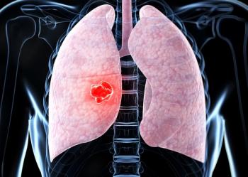
Acontinuing challengefor every specialty inoncology is to improvethe therapeutic ratio, which is thebalance of biological effectivenessof treatment and severity oftreatment-related side effects.Targeted tumor imaging and therapy,based on tumor biology, havethe goal of better identifying areasof tumor to limit surgical andradiation fields, and enablesystemic therapies to limit theireffects to tumor cells. This moretargeted approach has the goal of improving tolerance to cancertreatment.Treatment-Related ToxicitiesTreatment-related toxicities significantly compromise responseto cancer therapy by interrupting and extending thetreatment schedule, reducing the doses administered, or bycausing patients to fail to complete the planned course of therapy.For example, among 273 patients who were randomizedand received postoperative chemoradiotherapy for gastric cancer,41% experienced grade 3 toxicity, 32% had a grade 4toxicity, and three patients (1%) died from toxic effects ofchemoradiotherapy.[1] Only 64% (181 patients) completed theplanned treatment; 17% stopped treatment because of a medicaldetermination about treatment-related toxicity, 8% declinedtreatment after further consideration, and an additional 2%declined to continue treatment. Despite the toxicity and failureto complete treatment in more than one-third of chemoradiotherapypatients, the median overall survival time in the chemoradiotherapyarm of this inter-institutional trial was significantlyhigher than that of the surgery-only group.Unfortunately, inability to control treatment-related symptomsin more than one-third of chemoradiotherapy patientsprobably also significantly impacted survival rates among patients treated with chemoradiotherapy. If symptoms had beenbetter controlled, allowing an additional 36% of patients tocomplete the planned postoperative chemoradiotherapy, thesurvival difference probably would have been even moresignificant. Ironically, the percentage of patients who benefitedfrom the radiotherapy portal quality-assurance program isabout the same as the percentage of patients who did notcomplete treatment because of toxicity from chemoradiotherapy;patients who do not complete therapy because of symptomscannot be cured even if their radiation portals are correct.Horiot emphasized the negative impact of toxicity onsurvival.[2] A radiation treatment interruption of just 1 daymay reduce disease-control rates by 1.4%; a treatment break of1 week will reduce control rates by more than 10%.[2-4] Theimportance and ethics of quality assurance to reduce treatment-related toxicities through aggressive symptom management,using already available therapies, cannot be underscoredenough in the development of clinical trials andstandards of practice.Targeted Tumor Imaging and TherapyRadiation therapy fields historically were large to treattumor, areas of potential microscopic involvement beyondvisible tumor, and regional nodal drainage. Radiation treatmentfields have decreased significantly with the concurrentadministration of chemotherapy during radiation therapy, asclinicians are now depending more on systemic therapy totreat microscopic extensions of disease. Radiation portalsgenerally now encompass the visible tumor and a limitedmargin around it. This evolution in radiotherapy planningalso requires greater dependence on imaging information.Research in the past year has focused on improved imagingtechniques that allow more precise radiation treatment volumes.As reported at the 2004 Radiological Society of NorthAmerica (RSNA) meeting, magnetic resonance spectroscopy,dynamic contrast-enhanced MR imaging, and ultrasmall paramagneticcontrast medium improved the accuracy of diagnosisand staging of prostate cancer, making a significant impacton therapeutic decisions. With endorectal coil-assisted MRIand 3D MR spectroscopy, Vilanova was able to confirm thediagnosis of prostate cancer in 10 of 27 patients with persistentelevations of prostate-specific antigen levels after a negativebiopsy (see report on page 14). Likewise, dynamic contrastenhancedMRI found recurrent prostate cancer earlier afterradiation therapy. The sensitivity rates in all of these studiesare equal to or greater than 75%.Positron emission tomography is like MR spectroscopy inthat it involves imaging based on biological principles. Themost exciting development is the anatomic and functionalfusion of CT, MR, and PET images; fusion of these imagingtechniques overcomes the limitations of each. Specifically, thefusion of CT/MR with PET overcomes the lack of anatomicdetail in PET scans. PET has been shown to have a highersensitivity and at times a higher specificity than CT and/or MRIin staging and post-treatment evaluation for recurrence in awide range of tumors. Multiple studies have consistently shownthat addition of PET to routine CT scanning impacted therapeuticdecisions in almost one out of six cancer patients. Changes intherapeutic decisions were manifested in a variety of ways, froma change in cancer staging to modification of radiation portals.Studies like these are instrumental to match technology withreimbursement. Although PET has a 2% to 3% detection rate forunsuspected malignancies, the cost of PET scanning and theprocedures performed subsequent to false-positive cases make iteconomically unfeasible for cancer screening. Screening issuesaside, Blodgett and colleagues report that Medicare and mostthird-party payers do not reimburse for PET imaging withcarcinoma arising from an unknown primary site, accountingfor 5% of cancer diagnoses (see report on page 3). In addition,Medicare and most third-party payers do not reimburse the useof PET imaging to differentiate whether a nodule in the lung orother visceral structure is benign or malignant.An important distinction needs to be made, however, in theeconomics of procedures done for cancer staging and cancerscreening. Economic models need to incorporate the savings ofaverting the need for biopsy or surgery to determine malignancy,and the cost of less-effective therapeutic approaches taken becausethe primary site is neither detected nor treated. Other costsof cancer and its care include the costs of disability to the patientand his or her family, whether they be temporary expensesrelated to a procedure or cumulative costs incurred over the longterm. These costs are profound in financial and human terms.A Broadened ChallengeMore targeted approaches, which improve the therapeuticratio by improved therapeutic tolerance, have evolved in everyspecialty of oncology. These approaches include receptor-specificsystemic therapies, sentinel lymph node-based dissections,and intensity-modulated radiation therapy (IMRT). IMRT hasallowed more specific radiation dose deposition to spare normalstructures and increase the radiation dose within the tumor. Atthe 2004 RSNA meeting, fused ProstaScint and CT images wereused to deliver 75.6 Gy over 42 fractions to the entire prostate,and the tumor was boosted to 82 Gy using IMRT techniques (seereport on page 15). Doses of this magnitude were not possiblewithout the use of IMRT, but the addition of ProstaScint/CT image fusion reduced side effects even further. At 3 months, only 1 of 38 patients experienced a grade 3 genitourinarytoxicity, which resolved after 1 month of follow-up.Development of respiratory gating during PET/CT canreduce uncertainties about tumor location for radiation treatmentplanning. In the past, large radiation fields were constructedfor lung cancer therapy because of concerns that thetumor would move outside the radiation field during therespiratory cycle. New technology, by precisely accounting formovement of the tumor during the respiratory cycle, hassignificantly reduced the size of the radiation portal (seereports on pages 10 and 11). Respiratory gating during radiationcan reduce the volume of lung included in radiationportals even further, which is especially important for patientswith limited pulmonary reserve.These exciting technological advancements take the nextstep toward tumor-specific therapies that were popularized bythe term,"magic bullet." The goal has always been to eradicatecancer without harm to the patient. Much work still must bedone to achieve that goal. Improved control of disease andtreatment-related symptoms is possible with available meansand must be standardized in protocols and practice. Avoidanceof treatment-related symptoms by more limited surgicalapproaches, and through targeted systemic agents and radiationtherapy, will continue to reduce the burden of cancertreatment and its long-term sequelae, including subsequenttreatment-induced cancer. The study of a new therapeuticapproach should also include a comprehensive cost-benefitanalysis to address the realities of health care demographicsand economics.The challenge of oncology has broadened beyond curingcancer. For almost 2 decades, we have recognized the profoundimpact of the consequences of cancer treatment andcure. The specialty must not only relieve suffering by curingcancer, but it also must relieve suffering from pain, fatigue,and other symptoms while curing cancer. The challenge nowfacing us is to cure cancer without incurring side effects, andto improve the cost-effectiveness of cure. In short, these technologicaladvancements in cancer therapy must provide ameaningful difference in response and minimize treatmentrelatedtoxicity, while reducing the burden of care and cost ofcancer to society as a whole.Nora A. Janjan, MD
Professor of Radiation Oncology
The University of Texas
M. D. Anderson Cancer Center
References:
1.
Macdonald JS, Smalley SR, Benedetti J, et al: Chemoradiotherapyafter surgery compared with surgery alone for adenocarcinoma of thestomach or gastroesophageal junction.
N Engl J Med
345:725-730, 2001.
2.
Horiot JC: Prophylaxis versus treatment: Is there a better way tomanage radiotherapy-induced nausea and vomiting?
Int J RadiationOncol Biol Phys
60:1018-1025, 2004.
3.
Fowler J, Lindstrom M: Loss of local control with prolongation inradiotherapy.
Int J Radiation Oncol Biol Phys
23:457-467, 1992.
4.
Robertson C, Robertson AG, Hendry JH, et al: Similar decreases inlocal tumour control are calculated for treatment protraction and forinterruptions in the radiotherapy of carcinoma of the larynx in fourcentres.
Int J Radiation Oncol Biol Phys
40:319-329, 1998.
Articles in this issue
over 21 years ago
FDA Approves Abraxane for Metastatic Breast Cancerover 21 years ago
FEC Followed by Docetaxel Improves Breast Ca Outcomesover 21 years ago
Follow-up Data Support Use of Stanford V Regimen for HDover 21 years ago
Randomized Trial Validates Advantages of SNBover 21 years ago
70-Gene Signature Predicts Time to Breast Ca Metastasesover 21 years ago
New 5D Model May Predict Motion of Lung Tumors During RespirationAdvertisement
Related Content
Advertisement


Advertisement
Advertisement
Trending on CancerNetwork
1
FDA Accepts NDA for Mezigdomide Combo in R/R Multiple Myeloma
2
Dostarlimab Yields Sustained Complete Responses in dMMR/MSI-H Rectal Cancer
3
ADCs in the Frontline: Evolving Approaches in Metastatic TNBC
4
Risvutatug Rezetecan Improves Overall Survival in Relapsed SCLC Trial
5