Mucinous Adenocarcinoma of the Appendix With Histologic Response to Neoadjuvant Chemotherapy: Review of Histologic and Clinical Spectrum of Epithelial Neoplastic Mucinous Lesions of the Appendix

Mehmet Sitki Copur, MD, and colleagues examine the case of a 65-year-old patient with appendiceal mucinous neoplasms of the appendix who was treated with cytoreductive surgery and hyperthermic intraperitoneal chemotherapy.


Copur is a medical oncologist/hematologist and medical director of oncology at Morrison Cancer Center, Mary Lanning Healthcare in Hastings, Nebraska, and is a professor at the University of Nebraska Medical Center adjunct faculty. He is also an Editorial Advisory Board member at ONCOLOGY®.


Cushman-Vokoun is the medical director of the Molecular Diagnostics Laboratory at University of Nebraska Medical Center, and associate professor at the University of Nebraska Medical Center Department of Pathology and and Microbiology.


Padussis is assistant professor, Department of Surgical Oncology, University of Nebraska Medical Center, Omaha, Nebraska.


Wedel is a pathologist at Mary Lanning Healthcare.


Schroeder is a board certified general surgeon at Morrison Cancer Center, Mary Lanning Healthcare.


Herold is a radiologist at Morrison Cancer Center, Mary Lanning Healthcare.


Lintel is a pathologist at Mary Lanning Healthcare.


Horn is a pathologist at Mary Lanning Healthcare.

Abstract
Appendiceal mucinous neoplasms are a rare and heterogeneous group of diseases with challenging clinical management decisions. They account for less than 1% of all cancers but their incidence is on the rise. Treatment is based on their stage and histology. Appendiceal neoplasms frequently metastasize inside the abdomen; this leads to tumor cell growth in the abdominal cavity, known as peritoneal carcinomatosis, and buildup of mucinous material, known as pseudomyxoma peritonei. While low‐grade, early-stage tumors can be effectively treated with limited surgical resection, patients with low-grade, advanced-stage disease require peritoneal debulking and hyperthermic intraperitoneal chemotherapy. Therapeutic options for high-grade, advanced-stage mucinous tumors of the appendix have not been well established. Debulking surgery with hyperthermic intraperitoneal chemotherapy preceded and/or followed by systemic chemotherapy has been utilized based on some prospective but not randomized data. We present a case of mucinous adenocarcinoma of the appendix treated with neoadjuvant chemotherapy followed by cytoreductive surgery/hyperthermic intraperitoneal chemotherapy and adjuvant chemotherapy. Preoperative chemotherapy provided a favorable histologic response by converting initial mucinous appendiceal adenocarcinoma histology to a high-grade mucinous appendiceal neoplasm.
Keywords: Appendiceal epithelial neoplasms, peritoneal carcinomatosis, hyperthermic intraperitoneal chemotherapy.
Oncology (Williston Park). 2021;35(6):335-340.
DOI: 10.46883/ONC.2021.3506.0335
Case
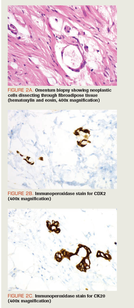
A White woman, aged 65 years, presented with pain and fullness in her lower abdomen. Initial evaluation, including gynecologic exam and a CT scan of the abdomen and pelvis, showed a heterogeneous centrally located uterus with a possible semisolid hematoma, submucosal fibroid, or a polyp. Further evaluation with a transvaginal ultrasound reported a 3.5 × 3.8 × 2.7-cm uterine mass. Endometrial biopsy was negative for hyperplasia or malignancy. She was scheduled for a laparoscopic hysterectomy, which was delayed due to COVID-19 infection and patient nonadherence. Six months from the original presentation, she was taken to the operating room for laparoscopic hysterectomy. During the procedure, she was noted to have carcinomatosis throughout the abdomen with findings of omental caking. The procedure was changed to a diagnostic laparoscopy. The appendix appeared to be obliterated on the tip with inflammatory tissue and inflamed fat around the tip. The base of the appendix and cecum was visualized and looked normal. Peritoneal and omental biopsies were taken. Postprocedure restaging CT scan of the abdomen and pelvis showed induration within the anterior peritoneal fat, representing peritoneal carcinomatosis and prominence of the appendix (Figure 1A, 1B). Upper and lower endoscopies were negative. Pathology of the left peritoneal wall and omentum biopsy was consistent with moderately differentiated adenocarcinoma with mucin present intracellularly. Immunohistochemical stains for pankeratin, cytokeratin 20, and CDX2 were positive(Figure 2A, 2B, 2C), whereas stains for synaptophysin, cytokeratin 7, calretinin, and PAX8 were negative. MLH1, PMS2, MSH2, and MSH6 protein expression was retained. A next-generation sequencing companion diagnostic assay showed no FDA-approved targetable alteration but 1 genomic finding of a pathogenic BRAF p.D594G mutation. PD-L1 immunohistochemistry analysis revealed tumor proportion score of 0% (FoundationOne CDx).


FIGURE 1A. Induration within the anterior peritoneal fat representing peritoneal carcinomatosis
FIGURE 1B. Prominence of appendix possible underlying lesion
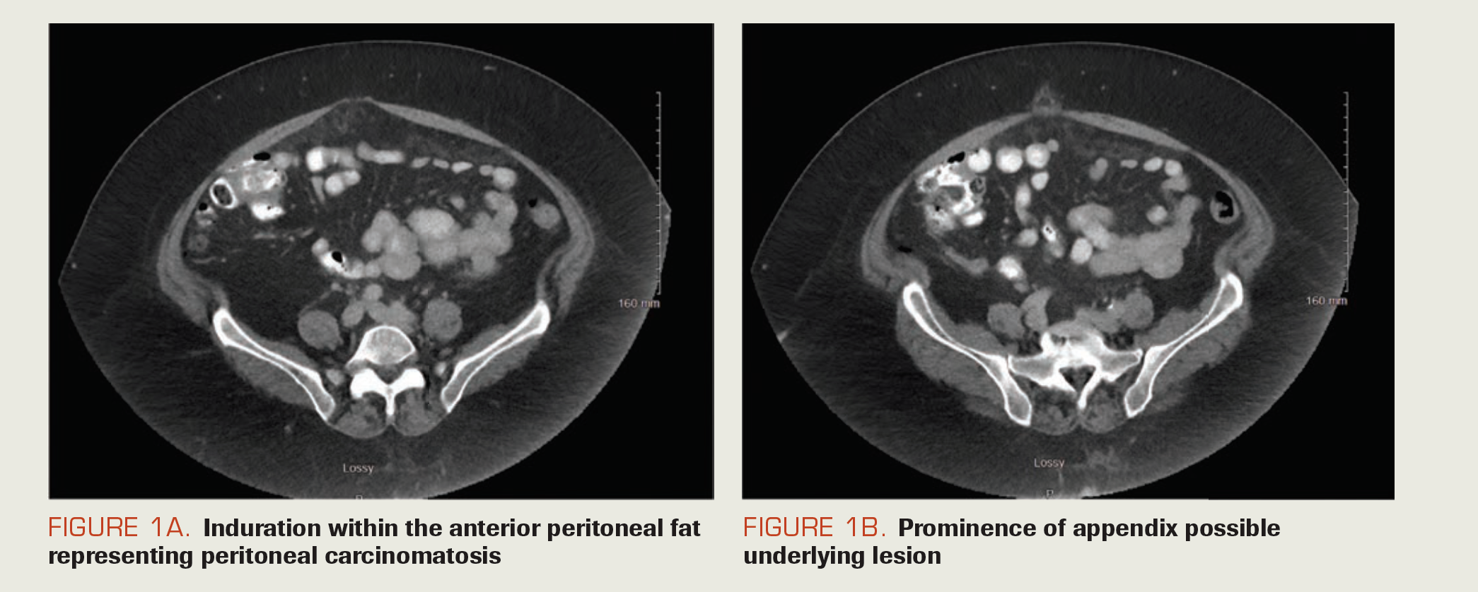


FIGURE 2B. Immunoperoxidase stain for CDX2 (400x magnification)
FIGURE 2C. Immunoperoxidase stain for CK20 (400x magnification)")
FIGURE 2A. Omentum biopsy showing neoplastic cells dissecting through fibroadipose tissue (hematoxylin and eosin, 400x magnification)
FIGURE 2B. Immunoperoxidase stain for CDX2 (400x magnification)
FIGURE 2C. Immunoperoxidase stain for CK20 (400x magnification)
Based on the multidisciplinary tumor board consensus, she was treated with neoadjuvant chemotherapy of 5-fluorouracil (5-FU), folinic acid, and oxaliplatin (FOLFOX) and bevacizumab (Avastin) for 6 cycles, to be followed by cytoreductive surgery and hyperthermic intraperitoneal chemotherapy (CRS/HIPEC). After neoadjuvant therapy, restaging CT scans revealed decreased omental fat stranding and a mildly dilated appendix. She underwent CRS with right hemicolectomy, small bowel resection, cholecystectomy, peritonectomy, total abdominal hysterectomy, bilateral salpingo-oophorectomy, and omentectomy followed by HIPEC. Pathology revealed no evidence of tumor in the ileum or colon but high-grade appendiceal mucinous neoplasm (HAMN) involving the distal half of the appendix with invasion through the muscle wall into the adjoining periappendiceal fibroadipose tissue with extensive acellular mucin areas and focal signet-ring cell differentiation. Twenty-eight regional lymph nodes were negative. There was no lymphovascular or perineural invasion. Uterus revealed focal adenomyosis and atypical hyperplasia of the endometrium but no evidence of mucinous neoplasm. Ovaries showed focal acellular mucin and focal low-grade mucinous neoplasm on the surface without penetration into the ovarian stroma. Pathologic stage was pT4a (tumor invades through the visceral peritoneum), pN0 (no regional lymph node metastasis),pM1b (intraperitoneal metastasis only, including peritoneal mucinous deposits containing tumor cells).
Introduction
Cancers of the appendix are extremely rare, with an estimated incidence of 0.15 to 0.9 per 100,000 people. Appendiceal mucinous neoplasms comprise 0.2% to 0.3% of appendectomy specimens. Affecting men and women equally, the average age of onset is between 50 and 55 years.1,2 Clinical presentation can be an appendicitis, a hernia filled with mucin, an abdominal/pelvic mass, or an incidental finding on some form of imaging study or during a surgical procedure for an unrelated indication. In women, these neoplasms often spread to the ovaries and can easily be confused with ovarian cancer. A majority (65%) of appendiceal tumors are of neuroendocrine origin. Adenocarcinomas (mucinous, nonmucinous, or signet-ring) constitute approximately 20% of these tumors.3 Treatment of appendiceal tumors depends on the extent and the histology of the disease. Systemic chemotherapy and cytoreductive surgery have long been used to treat macroscopic disease. Addition of HIPEC to CRS has become a novel approach to eradicate both macroscopic and microscopic residual tumor tissue in the abdomen. The treatment of appendiceal mucinous neoplasms has not been well defined, with controversies regarding pre- and postoperative chemotherapy, extent of surgery, role of early postoperative intraperitoneal chemotherapy, and HIPEC. Here, we present a review of the histologic and clinical spectrum of epithelial neoplastic mucinous lesions of the appendix.
Histopathology
The appendix can be home to several distinctive benign and malignant pathologies with variable histologic classifications. Accurate histologic diagnosis of appendiceal neoplasms can be quite challenging. A complete examination of the entire specimen and rigorous evaluation of the appendiceal wall for any evidence of epithelial invasion are crucial to distinguish between adenocarcinomas and all other types of mucinous lesions. In 2016, an international panel of pathologists and clinicians—the Peritoneal Surface Oncology Group International (PSOGI)—came together to categorize noncarcinoid epithelial tumors of the appendix.4 The current pathological classification schemes for neoplastic lesions of the appendix take into account both the variations of the histopathological appearance and the pattern of mural involvement in the epithelium. Neoplastic appendiceal mucinouslesions consist of serrated polyps (hyperplastic lesions with or without dysplasia), hyperplastic polyps, low-grade appendiceal mucinous neoplasms, high-grade appendiceal mucinous neoplasms, and mucinous adenocarcinomas with or without signet-ring cell carcinomas (Table).4-6


TABLE. Epithelial Neoplastic Appendiceal Mucinous Lesions
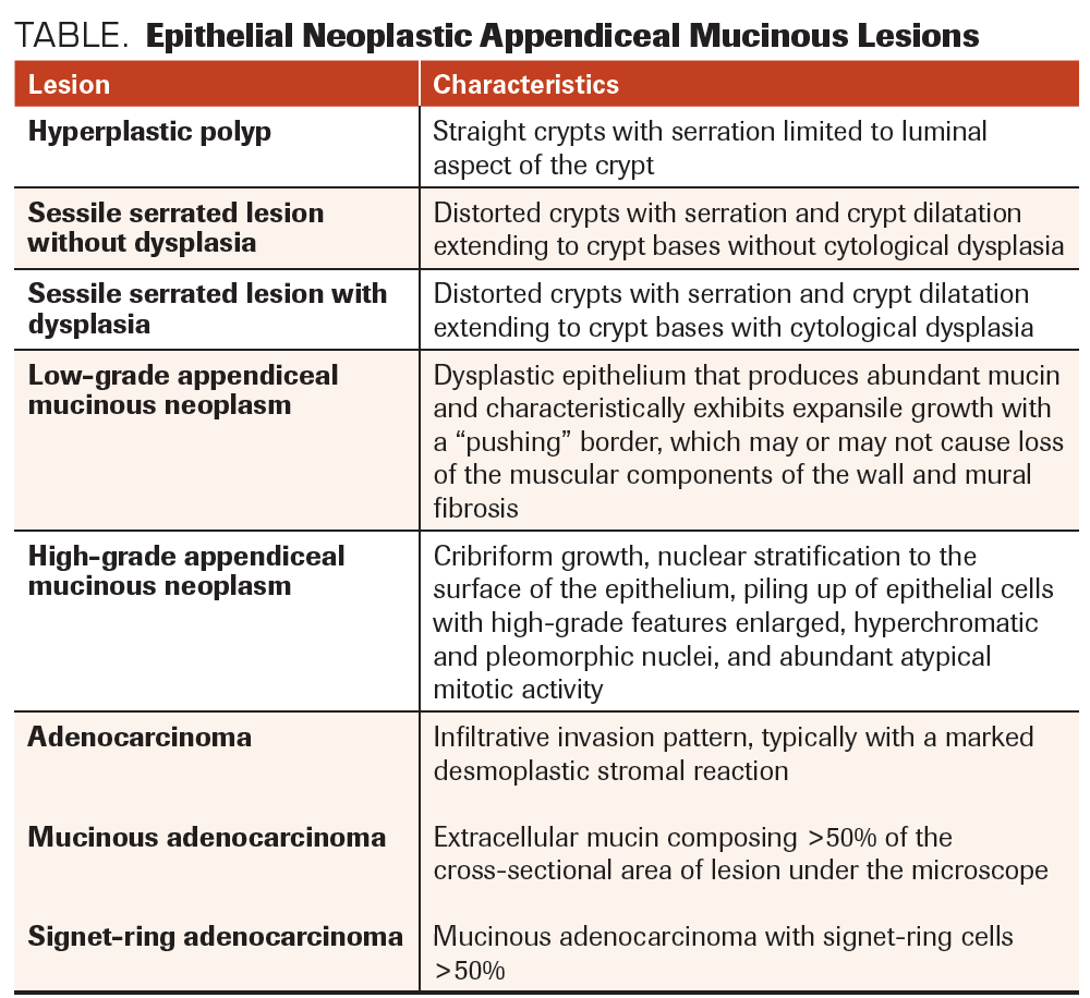
Historically, hyperplastic lesions have been considered to be reactive in nature. Most of them lack cytological atypia/dysplasia and typically involve the mucosa circumferentially. Serrated polyps of the appendix, with or without dysplasia, remain incompletely studied and are not always classified as neoplasms. Due to the lack of certainty about their biological nature, it has been suggested that these lesions should be classified as serrated polyps, either with or without epithelial dysplasia, rather than using the term adenoma, a term that would more specifically classify them as benign neoplasms.4 Low-grade appendiceal mucinous neoplasms (LAMNs) are true neoplasms, with dysplastic epithelium and expansile growth, but they lack overt infiltrative epithelial invasion of the appendiceal wall.5 They remain mostly confined to the muscularis propria. Due to their expansile growth pattern, however, they may push and cause the thinning and rupture of the appendiceal wall; this can result in spillage of cellular or acellular mucin, leading to pseudomyxoma peritonei (PMP). HAMNs are defined by the degree of epithelial dysplasia, including cribriform growth, nuclear stratification to the surface of the epithelium, and hyperchromatic and pleomorphic nuclei with ample atypical mitotic activity.5 Mucinous adenocarcinomas of the appendix are described as infiltrative lesions with invasive pattern, and they typically have a marked desmoplastic stromal reaction. They can be mucinous or nonmucinous.6 Similar to other adenocarcinomas, they are categorized into well-, moderately, or poorly differentiated histologies. The presence of signet-ring cells denotes the poorly differentiated category. While well-differentiated mucinous adenocarcinomas are more likely to produce peritoneal spread with a clinical picture of PMP, the higher-grade tumors present with peritoneal carcinomatosis and distant metastasis.7
Immunohistochemistry and Molecular Diagnostics
Published in 2019, the 5th edition of current World Health Organization classification of appendiceal tumors is largely based on the tumors’ histologic appearance and does not include their immunohistochemistry features or molecular pathology.8 Mucinous neoplasms of various sites, including gastrointestinal, breast, lung, pancreas, and gynecologic, all can present with very similar histomorphologic appearances and frequently metastasize to the peritoneal surface, ovary, liver, or lung, which makes the identification of the primary site rather difficult.9,10 All mucinous neoplasms express low-molecular-weight cytokeratins (CKs), such as CK7, CK8, CK18, and CK20.10-12,13 While most mucinous neoplasms of the lower gastrointestinal tract express CK20, and most nongastrointestinal mucinous neoplasms express CK7, the reverse can be true: CK20 can be expressed in 47% to 83% of ovarian mucinous neoplasms, and CK7 can be expressed in 37% of appendiceal mucinous neoplasms.13-15 CDX2, a transcription factor regulating intestinal epithelial cell differentiation, is strongly expressed in gastrointestinal epithelial tumors, including appendiceal mucinous neoplasms.16,17 PAX8, a very specific ovarian mucinous neoplasm marker, is not commonly expressed in appendiceal neoplasms.18 SATB2, a 733–amino acid human DNA-binding protein involved in transcriptional regulation and chromatin remodeling, has been shown to have highly restricted expression to glandular cells lining the lower gastrointestinal tract, including appendiceal mucinous neoplasms.19,20
Hyperplastic polyps and serrated lesions with or without dysplasia often have KRAS mutations but rarely BRAF mutations. LAMNs and HAMNs share high rates of KRAS and GNAS comutations but are usually wild-type for BRAF, APC, and TP53. Acquisition of TP53 or ATM mutations may drive progression to a more advanced phenotype.21,22 Appendiceal adenocarcinomas frequently harbor mutations in KRAS, GNAS, TP53, PIK3CA, and APC. They can also have significant nuclear expression of β-catenin, loss of nuclear or nuclear and cytoplasmic expression of SMAD4, and loss of cytoplasmic membranous expression of E-cadherin. Recent identification of heterozygous CTNNβ1, NOTCH1, and NOTCH4 germline mutations in LAMNs and HAMNs suggest a hereditary predisposition; however, microsatellite instability does not seem to have a role in the pathogenesis of epithelial appendiceal neoplasms and is rarely detected in these tumors.23-25 This patient’s tumor contained a BRAF p.D594G pathogenic mutation. This BRAF mutation occurs within the highly conserved DFG motif of the kinase domain of the BRAF protein and is a class 3 inactivating mutation (kinase impaired). It is likely insensitive to vemurafenib (Zelboraf).26,27 However, it leads to increased activation of ERK signaling through CRAF-RAS pathways and has been postulated to be potentially responsive to receptor tyrosine kinase or MEK inhibition, although more studies are required.26,27 This mutation has been shown to occur somatically in a diverse set of tumors, including lung, melanoma, and colorectal cancers.28
Clinical Presentation
Clinical presentation is usually asymptomatic or with rather nonspecific symptoms. Acute or chronic right lower quadrant abdominal pain, occasionally a palpable abdominal mass, or, less commonly, intermittent colicky abdominal pain and/or gastrointestinal bleeding associated with intussusception of a mucocele, can be initial presenting symptoms. Intestinal obstruction from mass effect, genitourinary symptoms, or acute abdomen from mucocele rupture or sepsis are other possible presentations.29-31 At the time of diagnosis, appendiceal cancer can be either localized to the appendix or may have already spread to the abdominal cavity and peritoneum. Because of the appendix’s thin-wall anatomy, these neoplasms are more likely to have spread at the time of initial diagnosis. Mucinous lesions due to hyperplasia (serrated polyps) or retention cysts (simple mucoceles) are not associated with recurrence once they rupture. However, when mucinous lesions due to LAMNs, HAMNs, or mucinous adenocarcinomas rupture, they usually present with progressive intraperitoneal spread, resulting in accumulation of both mucinous ascites and neoplastic cells—a clinical syndrome referred to as PMP.2
PMP was originally described as intraperitoneal mucinous spread from a ruptured cystadenoma (now called LAMN), seeding the peritoneum with mucus-producing cells and leading to progressive accumulation of copious amounts of mucinous fluid.32 The recent PSOGI pathologic classification redefined PMP as a complex syndrome with unique biological behavior and described 3 categories: low grade, high grade, and high grade with signet-ring cells. However, acellular mucin was classified separately.4,33 Appendiceal cancers rarely spread outside the abdominal cavity, and when they do, they are most commonly seen in poorly differentiated or signet-ring cell cancers.
Management
Surgical resection is the mainstay of treatment for all localized appendiceal mucinous neoplasms. Depending on the surgeon’s experience, an open or laparoscopic standard appendectomy procedure with the inclusion of the cuff of the cecum, without intruding on the ileocecal valve, is preferable. If the base of the appendix is involved, a partial resection of cecum with preservation of the ileocecal valve, resection of the ileocecal valve, or resection of the entire right colon may be necessary to enable clear surgical margins. While by some groups right hemicolectomy has been proposed for tumors that involve the periappendiceal area, measure 2 cm or larger, have high-grade histology, or present with perforation, we prefer to reserve right colectomy for tumors with high-grade histology or documented lymph node involvement.34 In patients presenting with clinically evident peritoneal disease, a diagnosis can be established by percutaneous biopsy of a peritoneal lesion, and appendectomy can be deferred to the time of definitive cytoreductive surgery.
The initial surgery for ruptured but localized appendiceal mucinous lesions can be limited to an appendectomy/right hemicolectomy with careful inspection of the abdominal cavity and biopsy of any suspicious peritoneal lesions. During the surgery, the goal should be to minimize tumor cell implantation of the abdomen and surgical wounds by thorough washing, cleaning, and irrigation.35 Advanced-stage, ruptured, and/or metastatic appendiceal mucinous neoplasms require additional surgical treatment with cytoreductive surgery and hyperthermic intraperitoneal chemotherapy at a specialized center.
Radical surgical removal of all abdominopelvic disease combined with administration of heated intraperitoneal chemotherapy has been developed as a comprehensive treatment approach for curative intent and has been utilized at centers with expertise in this disease.36 Efficacy of this approach depends on the ability to accomplish minimal residual disease, described as tumor deposits smaller than 2 to 2.5 mm after the cytoreductive surgery. The 2 most commonly used intraperitoneal chemotherapeutic agents have been mitomycin and oxaliplatin. The results of the only randomized trial comparing these 2 chemotherapeutic agents showed no difference in efficacy: 3-year survival was 73% in both groups, with similar postoperative complications and long-term adverse effects.37
Neoadjuvant Therapy
The role of preoperative chemotherapy in appendiceal mucinous neoplasms has been evaluated in nonrandomized studies with controversial results. This is mostly likely due to the retrospective nature of these studies, limitations of current imaging modalities, and lack of standard response criteria to assess peritoneal disease involvement.38 Retrospective studies suggest that optimal CRS and HIPEC treatment without preoperative systemic chemotherapy is appropriate for low‐grade appendiceal neoplasms. However, for high‐grade appendiceal adenocarcinomas, systemic preoperative chemotherapy should be considered.39 A potential advantage of neoadjuvant chemotherapy can be tumor volume reduction, which may lead to less extensive surgical procedures with improved organ preservation. Due to less-than-ideal accuracy of preoperative clinical exam and imaging studies, it has been difficult to assess the impact of neoadjuvant chemotherapy on the extent of cytoreductive surgery. Thus, surgical exploration offers the best approach to accurately evaluate this effect. In the results of one study, systemic chemotherapy administered prior to CRS/HIPEC for peritoneal mucinous adenocarcinoma of appendiceal origin showed a significant rate of histological response.40 The results of another study showed that neoadjuvant treatment utilizing systemic 5‐FU–based chemotherapy produced complete or near‐complete pathologic responses in patients with peritoneal mucinous carcinomatosis, making limited surgical resections possible.41
Adjuvant and Systemic Therapy
Similarly, the impact of adjuvant chemotherapy following CRS/HIPEC in appendiceal mucinous neoplasms has not been well established due to rareness of this disease and lack of randomized trials. In the advanced-disease setting, available retrospective data suggest beneficial effect from systemic chemotherapy in moderate- to high‐grade appendiceal mucinous tumors but not in low‐grade mucinous neoplasms.42 In a large National Cancer Database study of 639 patients with metastatic low‐grade mucinous appendiceal adenocarcinoma, systemic chemotherapy did not show beneficial effect on overall survival (OS).43 However, in moderate- to high‐grade mucinous tumors, signet‐ring cell tumors, and all nonmucinous tumors of appendiceal origin, systemic chemotherapy has been shown to be beneficial.44-46 Similarly, in earlier studies from a small group of 17 patients with PMP, adjuvant therapy following cytoreductive surgery reported no OS benefit.47 However, in patients with moderate- to high‐grade or signet‐ring cell appendiceal tumors, adjuvant chemotherapy following complete cytoreductive surgery provided significant OS benefit.48 The molecular profile of this case does not match to any current FDA targeted therapy; however, the presence of the kinase-impaired BRAF mutation highlights the need for additional clinical trials targeting specific hotspot mutations in highly mutated oncogenes, especially in the setting of basket trials for rare diseases.
Conclusions
Appendiceal mucinous lesions are rare and have considerable heterogeneity and a rising incidence. They can be nonneoplastic (simple mucoceles/retention cysts) or neoplastic (serrated polyps; low-grade or high-grade appendiceal mucinous neoplasms; mucinous adenocarcinomas with or without signet-ring cell features). Treatment is based on a lesion’s histology and extent. While early-stage, low-grade tumors can be effectively treated with surgical resection of the primary site, advanced-stage disease requires peritoneal debulking surgery and intraperitoneal chemotherapy. Treatment of advanced-stage and high-grade tumors requires further prospective trials evaluating the role of pre- and postoperative chemotherapy as an addition to CRS/HIPEC. Prospective randomized trials evaluating the impact of established chemotherapeutic agents, as well as the novel therapeutic agents targeting pathways involving KRAS, p53, GNAS, SMAD4, APC, ATM, PIK3CA, FBXW7, and BRAF, are desperately needed.
Outcome of the Case
After 6 cycles of neoadjuvant FOLFOX/bevacizumab systemic therapy, the patient underwent cytoreductive surgery; it revealed a histologic response mostly consisting of HAMN findings with focal signet-ring cell differentiation on her surgical debulking specimen. Following her recovery from CRS/HIPEC, she received 6 more cycles of adjuvant systemic therapy with FOLFOX/bevacizumab. Currently, she has no evidence of disease recurrence.
Financial Disclosure: The authors have no significant financial interest in or other relationship with the manufacturer of any product or provider of any service mentioned in this article.
References
1. Shaib WL, Assi R, Shamseddine A, et al. Appendiceal mucinous neoplasms: diagnosis and management. Oncologist. 2017;22(9):1107-1116. doi:10.1634/theoncologist.2017-0081
2. Smeenk RM, van Velthuysen MLF, Verwaal VJ, Zoetmulder FAN. Appendiceal neoplasms and pseudomyxoma peritonei: a population based study. Eur J Surg Oncol. 2008;34(2):196-201. doi:10.1016/j.ejso.2007.04.002
3. McCusker ME, Coté TR, Clegg LX, Sobin LH. Primary malignant neoplasms of the appendix: a population‐based study from the Surveillance, Epidemiology and End Results Program, 1973-1998. Cancer. 2002;94(12):3307-3312. doi:10.1002/cncr.10589
4. Carr NJ, Cecil TD, Mohamed F, et al; Peritoneal Surface Oncology Group International. A consensus for classification and pathologic reporting of pseudomyxoma peritonei and associated appendiceal neoplasia: the results of the Peritoneal Surface Oncology Group International (PSOGI) modified Delphi process. Am J Surg Pathol. 2016;40(1):14-26. doi:10.1097/PAS.0000000000000535
5. MIsdraji J, Carr NJ, Pai RK. Appendiceal mucinous neoplasm. In: WHO Classification of Tumours Editorial Board. WHO Classification of Tumours: Digestive System Tumours, 5th ed. International Agency for Research on Cancer;2019:144-146.
6. MIsdraji J, Carr NJ, Pai RK. Appendiceal adenocarcinoma. In: WHO Classification of Tumours Editorial Board. WHO Classification of Tumours: Digestive System Tumours. 5th ed. International Agency for Research on Cancer; 2019:147-148.
7. Carr NJ, Bibeau F, Bradley RF, et al. The histopathological classification, diagnosis and differential diagnosis of mucinous appendiceal neoplasms, appendiceal adenocarcinomas and pseudomyxoma peritonei. Histopathology. 2017;71(6):847-858. doi:10.1111/his.13324
8. Ahadi M, Sokolova A, Brown I, Chou A, Gill AJ. The 2019 World Health Organization Classification of appendiceal, colorectal and anal canal tumours: an update and critical assessment. Pathology. 2021;53(4):454-461. doi:10.1016/j.pathol.2020.10.010
9. Chou Y-Y, Jeng Y-M, Kao H-L, Chen T-J, Mao T-L, Lin M-C. Differentiation of ovarian mucinous carcinoma and metastatic colorectal adenocarcinoma by immunostaining with beta-catenin. Histopathology. 2003;43(2):151-156. doi:10.1046/j.1365-2559.2003.01687.x
10. Seidman JD, Elsayed AM, Sobin LH, Favassoli FA.Association of mucinous tumors of the ovary and appendix. a clinicopathologic study of 25 cases. Am J Surg Pathol. 1993;17(1):22-34. doi:10.1097/00000478-199301000-00003
11 Prayson RA. Clinicopathologic study of 61 patients with ependymoma including MIB-1 immunohistochemistry. Ann Diagn Pathol. 1999;3(1):11-18. doi:10.1016/s1092-9134(99)80004-5
12. Vang R, Gown AM, Barry TS, et al.Cytokeratins 7 and 20 in primary and secondary mucinous tumors of the ovary: analysis of coordinate immunohistochemical expression profiles and staining distribution in 179 cases.Am J Surg Pathol. 2006;30(9):1130-1139. doi:10.1097/01.pas.0000213281.43036.bb
13. Chu PG, Chung L, Weiss LM, Lau SK.Determining the site of origin of mucinous adenocarcinoma: an immunohistochemical study of 175 cases.Am J Surg Pathol. 2011;35(12):1830-1836. doi:10.1097/PAS.0b013e3182299c25
14. McCluggage WG, Wilkinson N. Metastatic neoplasms involving the ovary: a review with an emphasis on morphological and immunohistochemical features. Histopathology. 2005;47(3):231-247. doi:10.1111/j.1365-2559.2005.02194.x
15. Vang R, Gown AM, Wu L-S-F, et al. Immunohistochemical expression of CDX2 in primary ovarian mucinous tumors and metastatic mucinous carcinomas involving the ovary: comparison with CK20 and correlation with coordinate expression of CK7. Mod Pathol. 2006;19(11):1421-1428. doi:10.1038/modpathol.3800698
16. Kaimaktchiev V, Teracciano L, Tornillo L, et al. The homeobox intestinal differentiation factor CDX2 is selectively expressed in gastrointestinal adenocarcinomas. Mod Pathol. 2004;17(11):1392-1399. doi:10.1038/modpathol.3800205
17. Werling RW, Yaziji H, Bacchi CE, Gown AM. CDX2, a highly sensitive and specific marker of adenocarcinomas of intestinal origin: an immunohistochemical survey of 476 primary and metastatic carcinomas. Am J Surg Pathol. 2003;27(3):303-310. doi:10.1097/00000478-200303000-00003
18. Laury AR, Perets R, Piao H, et al. A comprehensive analysis of PAX8 expression in human epithelial tumors. Am J Surg Pathol. 2011;35(6):816-826. doi:10.1097/PAS.0b013e318216c112
19. FitzPatrick DR, Carr IM, McLaren L, et al. Identification of SATB2 as the cleft palate gene on 2q32-q33. Hum Mol Genet. 2003;12(19):2491-2501. doi:10.1093/hmg/ddg248
20. Li Z, Roth R, Rock JB, et al. Dual immunostain with SATB2 and CK20 differentiates appendiceal mucinous neoplasms from ovarian mucinous neoplasms.Am J Clin Pathol. 2017;147(5):484-491. doi:10.1093/ajcp/aqx023
21. Misdraji J, Carr NJ, Pai RK. Appendiceal serrated lesions and polyps. In: WHO Classification of Tumours Editorial Board. WHO Classification of Tumours: Digestive System Tumours, 5th ed. International Agency for Research on Cancer; 2019:141-143.
22. Liao X, Vavinskaya V, Sun K, et al. Mutation profile of high-grade appendiceal mucinous neoplasm. Histopathology. 2020;76(3):461-469. doi:10.1111/his.13986
23. Mikaeel RR, Young JP, Tapia Rico G, et al. Immunohistochemistry features and molecular pathology of appendiceal neoplasms. Crit Rev Clin Lab Sci. Published online February 11, 2021. doi:10.1080/10408363.2021.1881756
24. Hara K, Saito T, Hayashi T, et al. A mutation spectrum that includes GNAS, KRAS and TP53 may be shared by mucinous neoplasms of the appendix. Pathol Res Pract. 2015;211(9):657-664. doi:10.1016/j.prp.2015.06.004
25. Taggart MW, Galbincea J, Mansfield P, et al. High-level microsatellite instability in appendiceal carcinomas. Am J Surg Pathol. 2013;37(8):1192-1200. doi:10.1097/PAS.0b013e318282649b
26. Yao Z, Yaeger R, Rodrik-Outmezguine VS, et al. Tumours with class 3 BRAF mutants are sensitive to the inhibition of activated RAS. Nature. 2017;548(7666):234-238. doi:10.1038/nature23291
27. Cope NJ, Novak B, Liu Z, et al. Analyses of the oncogenic BRAFD594G variant reveal a kinase-independent function of BRAF in activating MAPK signaling. J Biol Chem. 2020;295(8):2407-2420. doi:10.1074/jbc.RA119.011536
28. Zheng G, Tseng L-H, Chen G, et al. Clinical detection and categorization of uncommon and concomitant mutations involving BRAF. BMC Cancer. 2015;15:779. doi:10.1186/s12885-015-1811-y
29. Landen S, Bertrand C, Maddern GJ, et al. Appendiceal mucoceles and pseudomyxoma peritonei. Surg Gynecol Obstet. 1992;175(5):401-404.
30. Isaacs KL, Warshauer DM. Mucocele of the appendix: computed tomographic, endoscopic, and pathologic correlation. Am J Gastroenterol. 1992;87(6):787-789.
31. Mourad FH, Hussein M, Bahlawan M, Haddad M, Tawil A.
Intestinal obstruction secondary to appendiceal mucocele. Dig Dis Sci. 1999;44(8):1594-1598. doi:10.1023/a:1026615010989
32. Sugarbaker PH, Ronnett BM, Archer A, et al. Pseudomyxoma peritonei syndrome.
Adv Surg. 1996;30:233-280.
33. Baratti D, Kusamura S, Milione M, Bruno F, Guaglio M, Deraco M. Validation of the recent PSOGI pathological classification of pseudomyxoma peritonei in a single-center series of 265 patients treated by cytoreductive surgery and hyperthermic intraperitoneal chemotherapy.Ann Surg Oncol. 2018;25(2):404-413. doi:10.1245/s10434-017-6252-1
34. González‐Moreno S, Sugarbaker PH. Right hemicolectomy does not confer a survival advantage in patients with mucinous carcinoma of the appendix and peritoneal seeding. Br J Surg. 2004;91(3):304-311. doi:10.1002/bjs.4393
35. Barrios P, Losa F, Gonzalez-Moreno S, et al. Recommendations in the management of epithelial appendiceal neoplasms and peritoneal dissemination from mucinous tumors (pseudomyxoma peritonei). Clin Transl Oncol. 2016;18(5):437-448. doi:10.1007/s12094-015-1413-9
36. Chicago Consensus Working Group.The Chicago Consensus on peritoneal surface malignancies: management of appendiceal neoplasms. Ann Surg Oncol. 2020;27(6):1753-1760. doi:10.1245/s10434-020-08316-w
37. Moaven O, Votanopoulos KI, Shen P, et al. Health-related quality of life after cytoreductive surgery/HIPEC for mucinous appendiceal cancer: results of a multicenter randomized trial comparing oxaliplatin and mitomycin. Ann Surg Oncol. 2020;27(3):772-780. doi:10.1245/s10434-019-08064-6
38. Esquivel J, Chua TC, Stojadinovic A, et al. Accuracy and clinical relevance of computed tomography scan interpretation of peritoneal cancer index in colorectal cancer peritoneal carcinomatosis: a multi‐institutional study. J Surg Oncol. 2010;102(6):565-570. doi:10.1002/jso.21601
39. Sugarbaker PH, Bijelic L, Chang D, Yoo D. Neoadjuvant FOLFOX chemotherapy in 34 consecutive patients with mucinous peritoneal carcinomatosis of appendiceal origin. J Surg Oncol. 2010;102(6):576-581. doi:10.1002/jso.21679
40. Shaib WL, Assi R, Shamseddine A, et al. Appendiceal mucinous neoplasms: diagnosis and management.Oncologist. 2017;22(9):1107-1116. doi:10.1634/theoncologist.2017-0081
41. Bijelic L, Kumar AS, Stuart OA, et al. Systemic chemotherapy prior to cytoreductive surgery and HIPEC for carcinomatosis from appendix cancer: impact on perioperative outcomes and short-term survival. Gastroenterol Res Pract. 2012;2012:163284. doi:10.1155/2012/163284
42. Asare EA, Compton CC, Hanna NN, et al. The impact of stage, grade, and mucinous histology on the efficacy of systemic chemotherapy in adenocarcinomas of the appendix: analysis of the National Cancer Data Base. Cancer. 2016;122(2):213‐222. doi:10.1002/cncr.29744
43. Lu P, Fields AC, Meyerhardt JA, et al. Systemic chemotherapy and survival in patients with metastatic low‐grade appendiceal mucinous adenocarcinoma. J Surg Oncol. 2019;120(3):446‐451. doi:10.1002/jso.25599
44. Shapiro JF, Chase JL, Wolff RA, et al. Modern systemic chemotherapy in surgically unresectable neoplasms of appendiceal origin: a single‐institution experience. Cancer. 2010;116(2):316‐322. doi:10.1002/cncr.24715
45. Lieu CH, Lambert LA, Wolff RA, et al. Systemic chemotherapy and surgical cytoreduction for poorly differentiated and signet ring cell adenocarcinomas of the appendix. Ann Oncol. 2012;23(3):652‐658. doi:10.1093/annonc/mdr279
46. Uemura M, Qiao W, Fournier K, et al. Retrospective study of nonmucinous appendiceal adenocarcinomas: role of systemic chemotherapy and cytoreductive surgery. BMC Cancer. 2017;17(1):331. doi:10.1186/s12885-017-3327-0
47. Smith JW, Kemeny N, Caldwell C, Banner P, Sigurdson E, Huvos A. Pseudomyxoma peritonei of appendiceal origin. the Memorial Sloan‐Kettering Cancer Center experience. Cancer. 1992;70(2):396‐401. doi:10.1002/1097-0142(19920715)70:2<396::aid-cncr2820700205>3.0.co;2-a
48. Kolla BC, Petersen A, Chengappa M, et al. Impact of adjuvant chemotherapy on outcomes in appendiceal cancer. Cancer Medicine. 2020;9(10):3400-3406. doi:10.1002/cam4.3009


 Presenting With Bone-Only Metastases")